
Tissue strain analysis

a tissue strain and analysis technology, applied in the field of tissue strain analysis, can solve the problems of not being able to run in real-time, the final quality of the strain image is often less than satisfactory, and the speed and extent of the exertion of force are virtually impossible to control, so as to achieve the effect of quick delivery of high-quality tissue strain images

Inactive Publication Date: 2011-03-31
KONINKLIJKE PHILIPS ELECTRONICS NV
View PDF40 Cites 2 Cited by
  • Summary
  • Abstract
  • Description
  • Claims
  • Application Information

AI Technical Summary

Benefits of technology

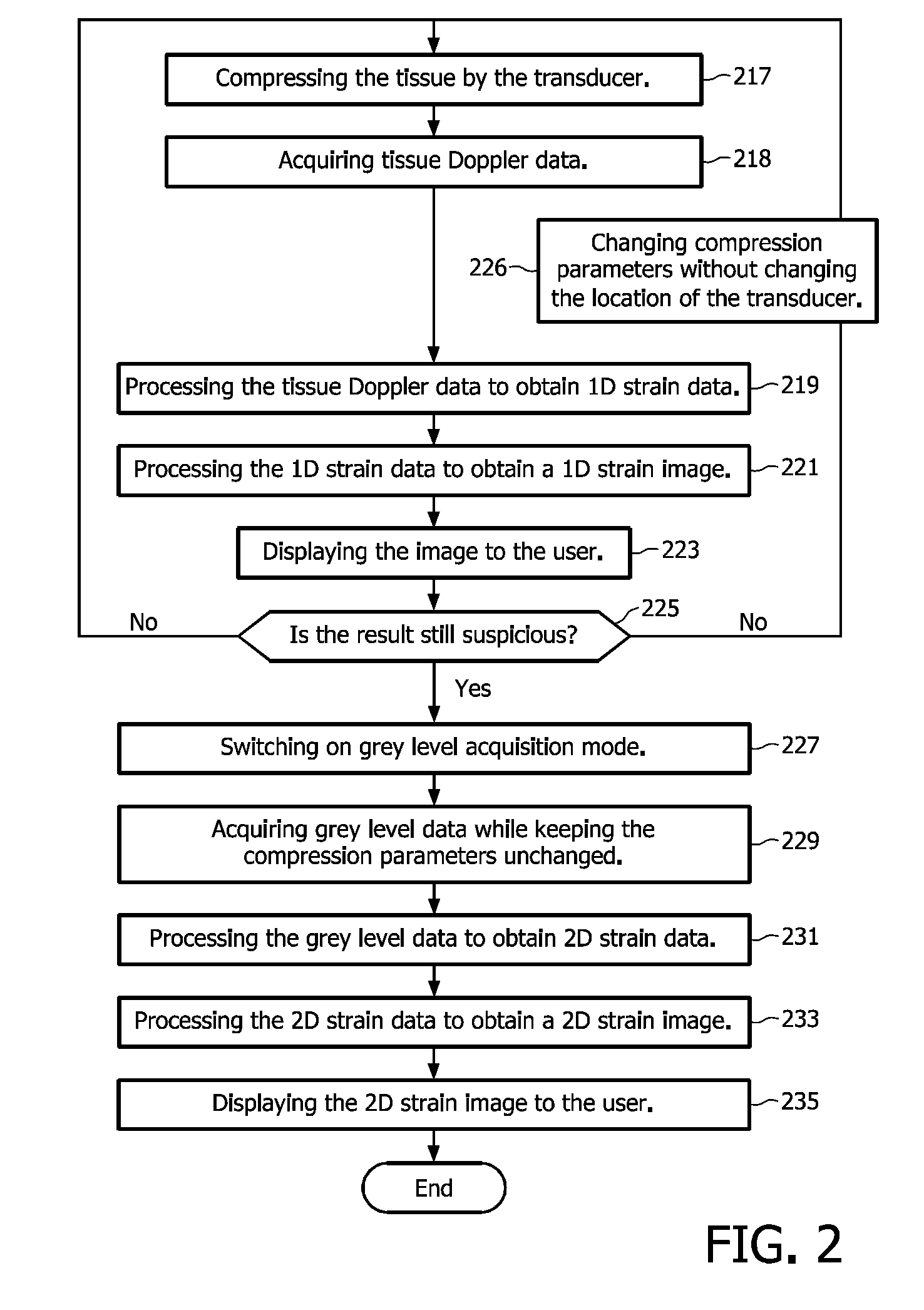
[0013]As will become clear in the following, this dual-mode method allows in an embodiment an early identification and fair assessment of pathology from elastographic analysis for obtaining the best possible result for elastography assessment of a tissue. The invention thereby provides a method capable of quickly providing high quality tissue strain images.

Problems solved by technology

This compression is created by the operator and thus it is virtually impossible to control the speed and extent of the exerted force.
In any case, despite optimal operating conditions, the final quality of the strain image is often less than satisfactory today.
The computations are however too slow to be able to run in real-time and thus they need to be performed off-line.
Accordingly, despite the 2D algorithms have the capacity to achieve good performance, the results cannot be easily obtained.
This thereby limits interoperability.

Method used


Image

  • Tissue strain analysis
  • Tissue strain analysis
  • Tissue strain analysis

Examples


Embodiment Construction

[0025]In the following description a non-limiting exemplary embodiment of the invention will be described in more detail. In this embodiment the invention is applied in breast elastography where the obtained elastogram can help to discriminate between benign and malignant tumours. It is to be noted that the invention is not limited to this application, but the invention can also be applied to echocardiography, where real-time inspection of TDI information would assess the value of acquisition and offline speckle tracking methods will further bring fine computations of relevant clinical parameters.

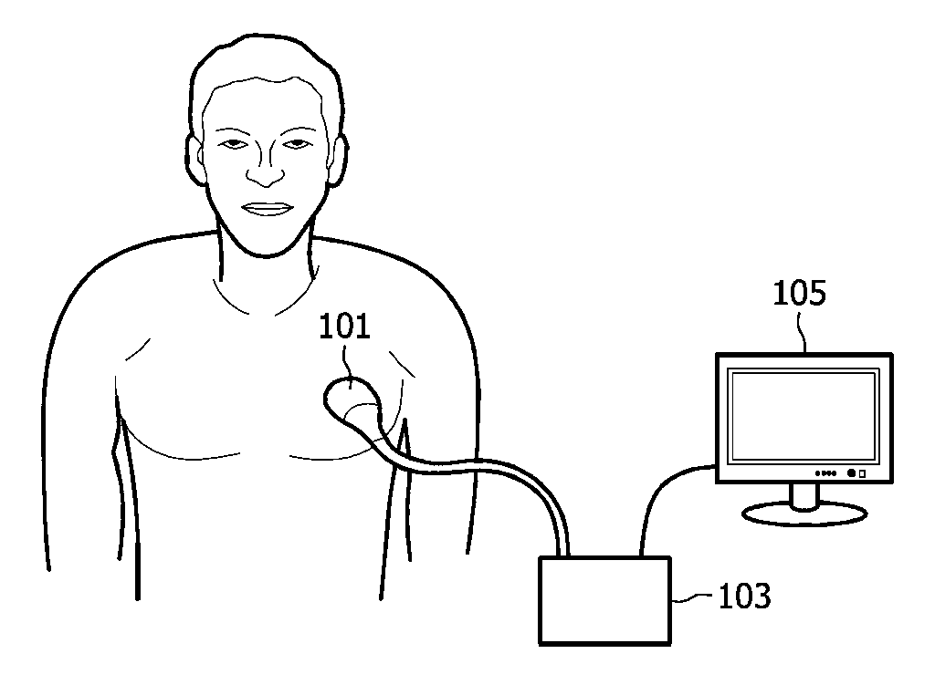
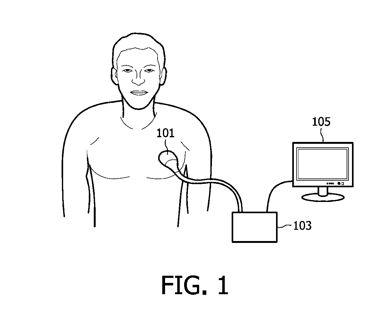
[0026]FIG. 1 shows an ultrasonic sensor probe, also known as a transducer 101 that is placed on the chest of a patient for obtaining ultrasonic signals from tissues of the patient. An ultrasonic transducer is a device that converts energy into ultrasonic or sound waves above the normal range of human hearing. The transducer 101 is connected to a processing unit 103 which is further connecte...


PUM


Abstract

Elasticity variation within biological tissue is often correlated with its pathology. Variation of elasticity can be assessed by specific ultrasound acquisition sequences during which pressure is applied. The tissue motion and deformation during those sequences is correlated to the tissue stiffness. The present invention describes a hybrid method that combines a real-time monitoring mode during acquisition and a non-real-time fine analysis after acquisition. This method allows early identification and fair assessment of pathology from elastographic analysis to obtain the best possible result for elastography assessment.

Description

FIELD OF THE INVENTION[0001]The present invention relates to a method of obtaining tissue strain data. This strain data can be obtained by use of an ultrasonic probe also known as a transducer. The invention also relates to a corresponding computer program product and measurement apparatus.BACKGROUND OF THE INVENTION[0002]Elasticity variation within biological tissue is often correlated with its pathology. Variation of elasticity can be assessed by specific ultrasound acquisition sequences during which pressure is applied. The tissue motion and deformation during those sequences is correlated to the tissue stiffness. Breast cancer tumours for instance present more stiffness against surrounding tissue.[0003]Elastographic analysis relates to elasticity measurement as biological tissues are submitted to an external constraint; the compression from an ultrasound probe for instance. This compression is created by the operator and thus it is virtually impossible to control the speed and e...

Claims


Application Information

Patent Timeline
Patent Type & Authority Applications(United States)
IPC IPC(8): A61B8/00
CPCA61B8/08A61B8/488A61B8/485A61B8/0825
Inventor GERARD, OLIVIERGAUTHIERA, THOMASDUFOUR, CECILE
Owner KONINKLIJKE PHILIPS ELECTRONICS NV
Learn More