Integrated portable digital x-ray imaging system

a portable, digital technology, applied in the field of medical imaging, can solve the problems of inability to perform on-site interpretation of obtained images, inability to meet patient care requirements, and inability to adapt to patient car

Active Publication Date: 2011-05-12
CARESTREAM HEALTH INC
View PDF16 Cites 33 Cited by
  • Summary
  • Abstract
  • Description
  • Claims
  • Application Information

AI Technical Summary

Benefits of technology

[0014]It is an advantage of the present invention that it provides a portable integrated solution for diagnostic imaging in ICU and other urgent care clinical environments.
[0015]It is a further advantage of the present invention that it provides a mechanism for alerting an operator to a detected condition related to image suitability and offers information on possible corrective action according to the condition detected.
[0016]These objects are given only by way of illustrative example, and such objects may be exemplary of one or more embodiments of the invention. Other desirable objectives and advantages inherently achieved by the disclosed invention may occur or become apparent to those skilled in the art. The invention is defined by the appended claims.

Problems solved by technology

In general, however, existing portable x-ray systems are self contained, tending to be somewhat bulky and inflexible.
Unfortunately, this conventional workflow pattern can sometimes be poorly suited to the requirements of patient care.
The need to upload image data to the PACS or other archive system means that interpretation of the obtained images cannot be performed on-site, but requires coordination with off-site diagnosticians.
It can be difficult for the clinical staff to determine the status of a worklist request until some time after the image is obtained.
There can be an undesirable delay in obtaining response information for problems of tube and line placement.
In addition, quality control (QC) suffers, since the technologist must wait for off-site processing and response in order to determine whether or not an obtained image is usable for diagnostic purposes.
With existing solutions, for example, the technologist does not have ready access to some types of information that can help to provide images that are more acceptable and useful to the diagnostician.
Workflow remains cumbersome and assignment of technologist tasks still remains a largely manual process in many ICU environments.
It can be difficult for the technologist to determine what is needed for each patient and to prioritize the sequence of images that are needed accordingly.
Overall, the need for improved support for bedside personnel has not been addressed by conventional solutions, so that considerable back-and-forth communications with off-site staff is often necessary in order to detect problems, such as tube positioning problems.
Conventional approaches have failed to take advantage of the full range of information about the patient and about patient images that can be made available to the imaging technologist and to the ICU staff.
Yet another shortcoming of mobile DR systems relates to lack of information to the technician on the clinical condition of the patient and diagnostic suitability of the image obtained.

Method used


Image

  • Integrated portable digital x-ray imaging system
  • Integrated portable digital x-ray imaging system
  • Integrated portable digital x-ray imaging system

Examples


Embodiment Construction

[0041]The following is a detailed description of the preferred embodiments of the invention, reference being made to the drawings in which the same reference numerals identify the same elements of structure in each of the several figures.

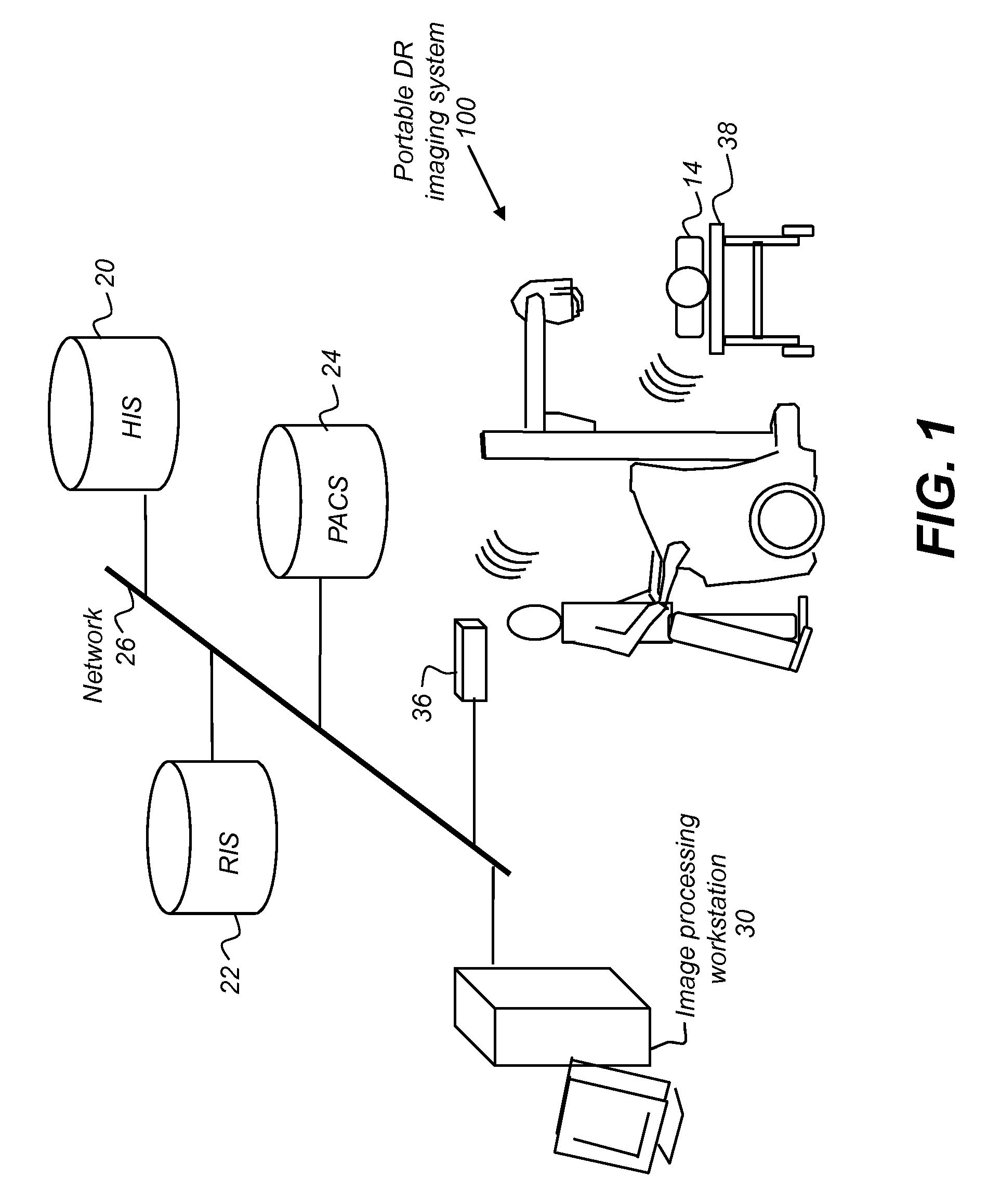
[0042]The schematic block diagram of FIG. 1 shows a mobile digital radiography system 100 that obtains images of a patient 14 in an ICU or other facility and communicates with a number of medical archiving and radiology databases over a network 26. Among the databases that communicate over network 26 are a Hospital Information System (HIS) 20, a Radiologist Information System 22, and a PACS 24. In addition, one or more optional image processing workstations 30 also receive and process images from mobile digital radiography system 100. Mobile digital radiography system 100 has a wireless interface 36 to network 26, typically connecting to a wireless hub or similar data communications interface device. The use of a wireless interface, while not essent...


PUM


Abstract

A method for processing a radiographic image of a patient, executed at least in part on a host processor, initiates exposure in response to an instruction entered from an operator console and obtains radiographic image data from a digital detector that is subjected to the exposure. The obtained radiographic image data is analyzed according to a set of predefined criteria for diagnostic suitability of the image. One or more results of the diagnostic suitability analysis at the operator console is indicated and a listing of one or more corrective actions at the operator console according to the indicated results is provided.

Description

CROSS REFERENCE TO RELATED APPLICATIONS[0001]This is a Continuation-in-Part of commonly assigned U.S. patent application Ser. No. 12 / 581,912 entitled “INTEGRATED PORTABLE DIGITAL X-RAY IMAGING SYSTEM”, in the names of David H. Foos et al., filed Dec. 20, 2009, which claims priority to U.S. Ser. No. 61 / 108,630, provisionally filed on Oct. 27, 2008, entitled “INTEGRATED PORTABLE DIGITAL X-RAY IMAGING SYSTEM”, in the names of David H. Foos et al., both of which are incorporated herein by reference.FIELD OF THE INVENTION[0002]The invention relates generally to medical imaging and in particular to portable X-ray imaging systems. More specifically, the invention relates to a digital X-ray imaging system that integrates imaging, quality control, and data access functions for improved delivery of diagnostic imaging services.BACKGROUND OF THE INVENTION[0003]Portable chest radiography is a widely performed radiographic exam in the intensive care unit (ICU). Radiographs of patients in the ICU ...

Claims


Application Information

Patent Timeline
Patent Type & Authority Applications(United States)
IPC IPC(8): H05G1/64G16H30/40G16H50/20
CPCA61B6/4405A61B6/462A61B6/463G06F19/3406A61B6/482A61B6/563G06F19/321A61B6/465G16H40/63G16H30/40G16H50/20A61B6/5252G16H30/20
Inventor FOOS, DAVID H.SEHNERT, WILLIAM J.HUO, ZHIMINLUO, HUIWANG, XIAOHUI
Owner CARESTREAM HEALTH INC
Why Eureka
Try Eureka