Mammography-Apparatus and Method for Screening the Occurrence of Malignant Cells

Inactive Publication Date: 2011-10-06
ACADEMISCH MEDISCH CENT BIJ DE UNIV VAN
View PDF5 Cites 6 Cited by
  • Summary
  • Abstract
  • Description
  • Claims
  • Application Information

AI Technical Summary

Benefits of technology

[0019]With the mammography-apparatus according to the invention it is possible to apply a method for executing a screening program to quickly detect the occurrence of malignant cells in a breast by using simultaneously and in combination an X-ray mammography detection method and a near-infrared contact-free photoacoustic detection method in which non-focussed pulsed laser-light is aimed at the breast and the induced ultrasound in the breast is measured with a detector and without applying a contact-gel between the detector and the breast. Contrary to all expectations this method and apparatus has proven effective for screening the occurrence of malignant cells and its feeding vessels in the breast.
[0022]A further aspect of the invention is that by using several non-contact ultrasound detectors a coarse localization of the abnormality that produces the acoustic signal can be made. For this purpose use is made of the different times of arrival of the said acoustic signal in the different non-contact ultrasound detectors. The times of arrival in the different non-contact ultrasound detectors are determined by the different distances travelled by the said ultrasound signal through the breast tissue and air on its way to the different detectors. Because of the synchronous x-ray imaging, this coarse localization can be easily related to this image.

Problems solved by technology

X-ray imaging is known to have a poor discriminatory nature when comparing malignant cells with the surrounding cells of normal glandular-tissue.
It is further known that invasive breast tumors in more dense breasts (which are overrepresented in lower age groups, yet also occur with older women) are difficult to depict with X-ray imaging.
Currently some 100 million women are being screened every year, consequently only very little time is available under screening circumstances.
Research shows that in screening programs a significant number of cancers are missed, which is due to the limited diagnostic value of the known mammography-test, and the related necessarily limited amount of time and facilities that are available for a screening.
This known system is unsuitable for reliable screening operations on large numbers of people.
The necessity to apply an opening portion in the compression paddle causes that the image data obtained with this known apparatus are unreliable in view of their lack of repeatability and technical considerations pertaining to the quality of the applied X-ray-imaging.
Evidently the technology disclosed in this article is aiming at imaging the detected abnormalities and is unsuited for purposes of screening large numbers of people in which the time available for any single screening will not be more than 5-10 minutes.
Clearly also the article does not aim to provide a solution for screening, yet investigates the quality of photoacoustic imaging in comparison with X-ray imaging.

Method used


Image

  • Mammography-Apparatus and Method for Screening the Occurrence of Malignant Cells
  • Mammography-Apparatus and Method for Screening the Occurrence of Malignant Cells
  • Mammography-Apparatus and Method for Screening the Occurrence of Malignant Cells

Examples


Embodiment Construction

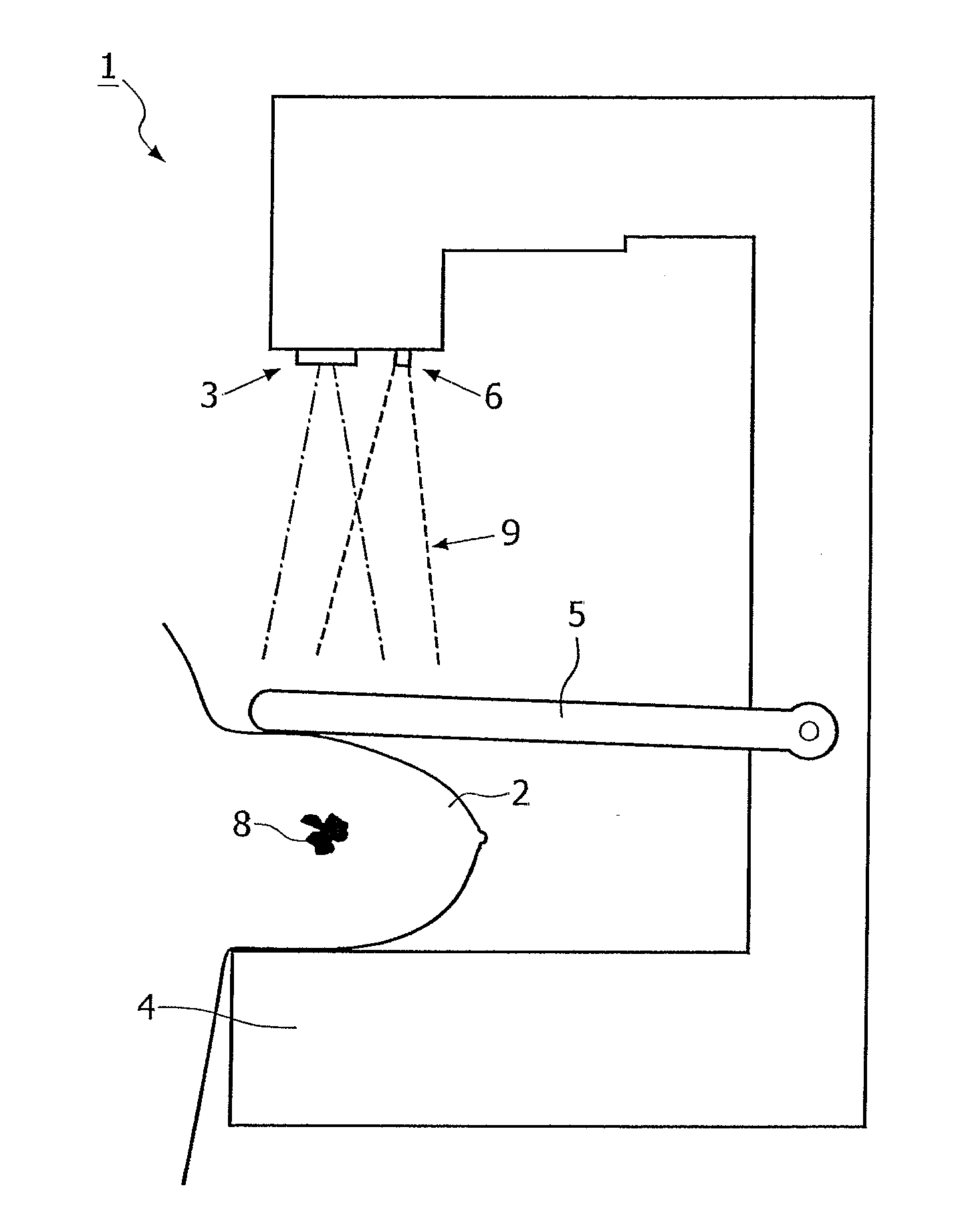
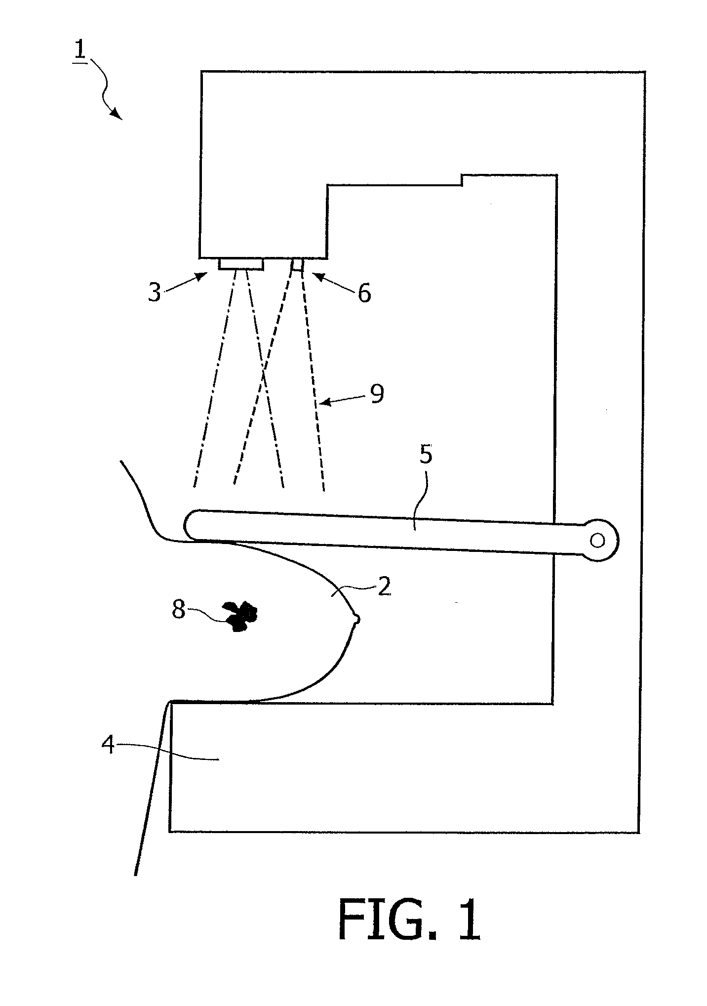
[0031]With reference first to FIG. 1 the mammography-apparatus 1 of the invention is shown for screening malignant cells 8 in a breast 2.

[0032]The mammography-apparatus 1 comprises an X-ray source 3, an X-ray detector 4 and a paddle 5 for pressing the breast 2 against said X-ray detector 4.

[0033]In accordance with the invention the mammography-apparatus 1 further comprises a laser-light-source 6 for transmitting pulsed defocussed laser-light 9 which during use is aimed at the breast 2 in a pre-determined direction which direction is monitored.

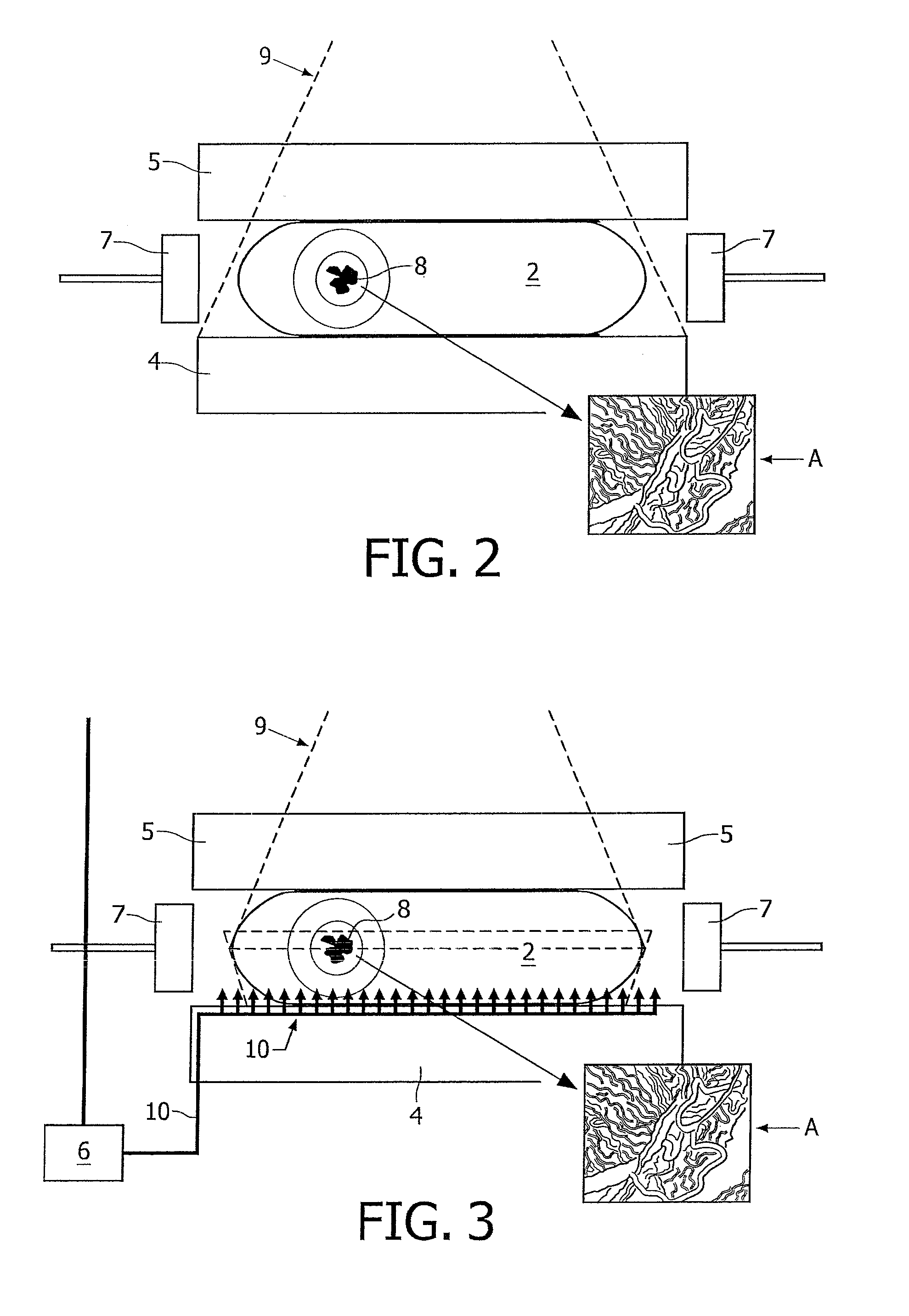
[0034]With reference to FIGS. 2, 3 and 4 showing first, second and third embodiments of the apparatus 1 of the invention, it is shown that the apparatus 1 is also provided with at least one, and most commonly several non-contact ultrasound detectors 7 for detection of ultrasound that originates from said breast 2, which is induced by an excitation stemming from the said defocussed laser-light 9. Thus, it is possible to generally screen the occu...


PUM


Abstract

A mammography-apparatus for detecting malignant cells in a breast comprising an X-ray source, an X-ray detector and a paddle for pressing the breast against said X-ray detector, wherein the mammography-apparatus further comprises a non-focussed laser-beam source for pulsed laser-light, which during use is aimed at the breast, and said apparatus comprises at least one non-contact ultrasound detector for contact-free detection of an ultrasound originating from said breast so as to screen the occurrence or non-occurrence of said malignant cells.

Description

CROSS-REFERENCE TO RELATED APPLICATIONS [0001]This application is a continuation application of International Patent Application Serial No. PCT / NL2009 / 050601, entitled “Mammography-Apparatus and Method for Screening the Occurrence of Malignant Cells”, to Academisch Medisch Centrum van de Universiteit van Amsterdam, filed on Oct. 7, 2009, and the specification and claims thereof are incorporated herein by reference.[0002]This application is a continuation of Netherlands Patent Application Serial No. 2002092, entitled “Mammography-Apparatus and Method for Screening the Occurrence of Malignant Cells”, filed on Oct. 13, 2008 and Netherlands Patent Application Serial No. 2003122, entitled “Mammography-Apparatus and Method for Screening the Occurrence of Malignant Cells”, filed on Jul. 3, 2009, and the specifications and claims thereof are incorporated herein by reference.STATEMENT REGARDING FEDERALLY SPONSORED RESEARCH OR DEVELOPMENT [0003]Not Applicable.INCORPORATION BY REFERENCE OF MAT...

Claims


Application Information

Patent Timeline
IPC IPC(8): A61B8/00A61B6/00
CPCA61B5/0095A61B5/4312A61B6/4417A61B6/502A61B8/0825A61B6/0414A61B6/4441
Inventor DEN HEETEN, GERHARD JOHANGRIMBERGEN, CORNELIS ANTONIUS
Owner ACADEMISCH MEDISCH CENT BIJ DE UNIV VAN
Why Eureka
Try Eureka