Ultrasonic diagnostic apparatus

Active Publication Date: 2011-10-06
FUJIFILM CORP
View PDF1 Cites 56 Cited by
  • Summary
  • Abstract
  • Description
  • Claims
  • Application Information

AI Technical Summary

Benefits of technology

[0019]According to the present invention, the cross sections of the 2D ultrasonic image can be easily switched according to the orientation of the head mounted display. Owing to this, the insertion path of the biopsy needle can be easily conformed even if the operator's hands are occupied for the operations of the ultrasonic probe and the treatment tools when the ultrasonic diagnos

Problems solved by technology

However, in inserting or puncturing a biopsy needle into such a small target, it is difficult to confirm whether the biopsy needle is accurately punctured into the tumor mass by observing the common 2D tomographic image.
To display the planar ultrasonic image (hereinafter, referred to as the 2D ultrasonic image) in arbitrary cross section of the patient's body, setting for generating or displaying the 2D ultrasonic image in which (what types of) cross section requires complex operations, and therefore the usability of the apparatus is not necessarily good.
Even in the case where the 2D ultrasonic image is generated usi

Method used


Image

  • Ultrasonic diagnostic apparatus
  • Ultrasonic diagnostic apparatus
  • Ultrasonic diagnostic apparatus

Examples


Embodiment Construction

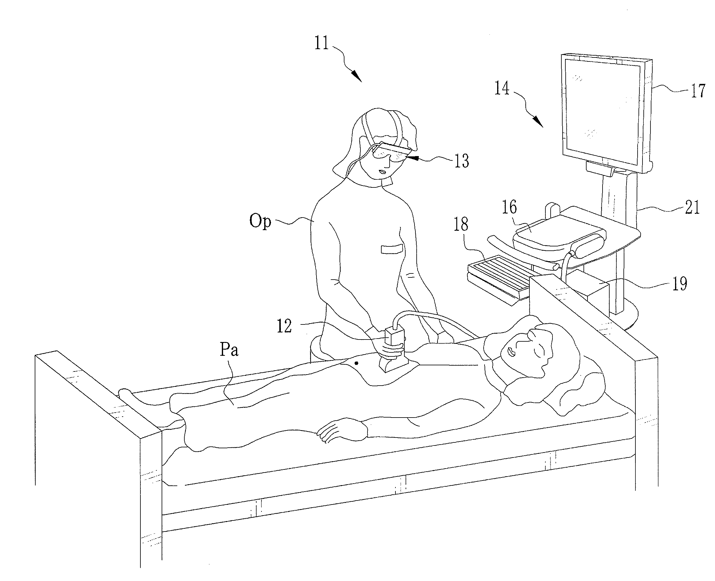
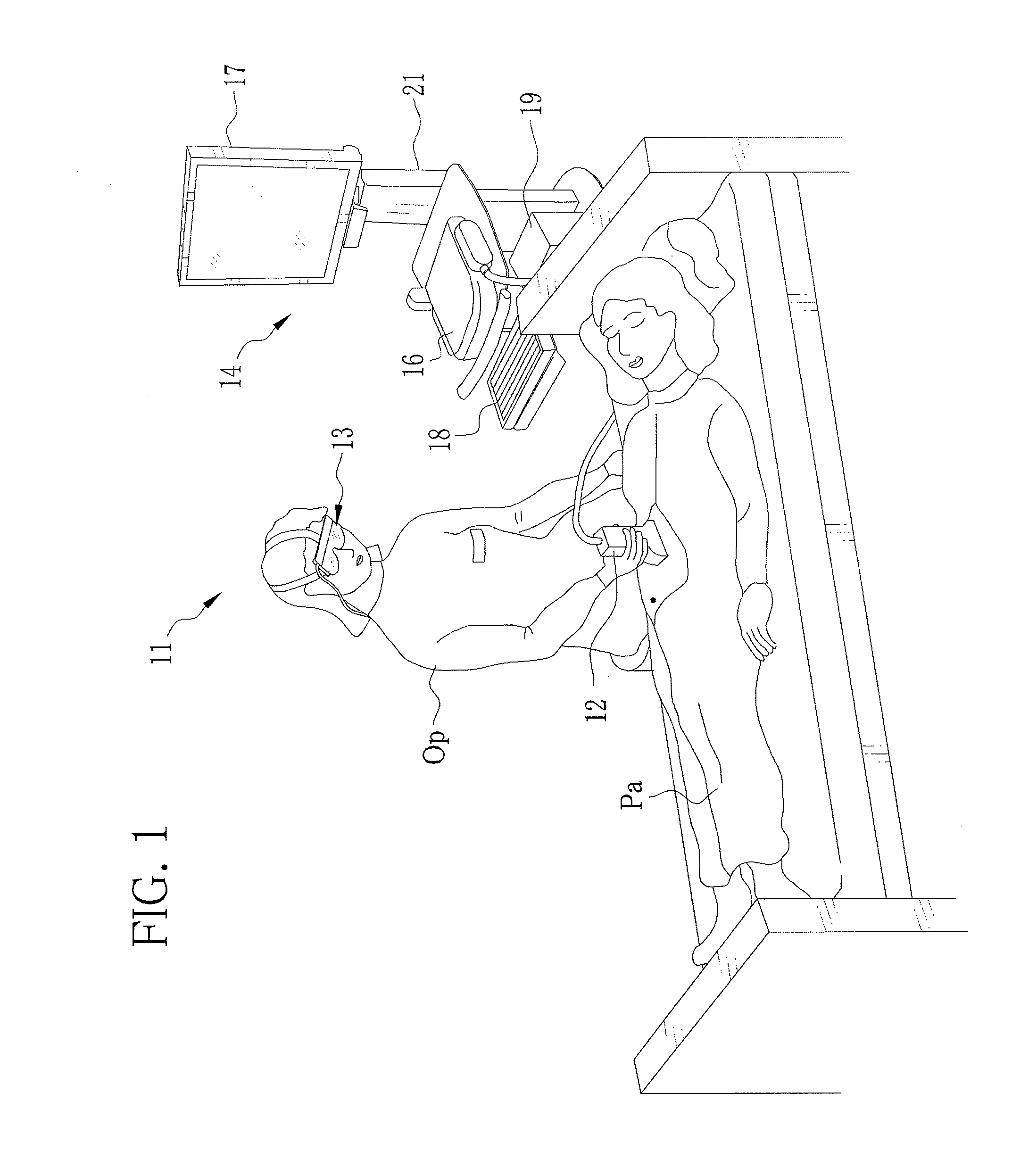
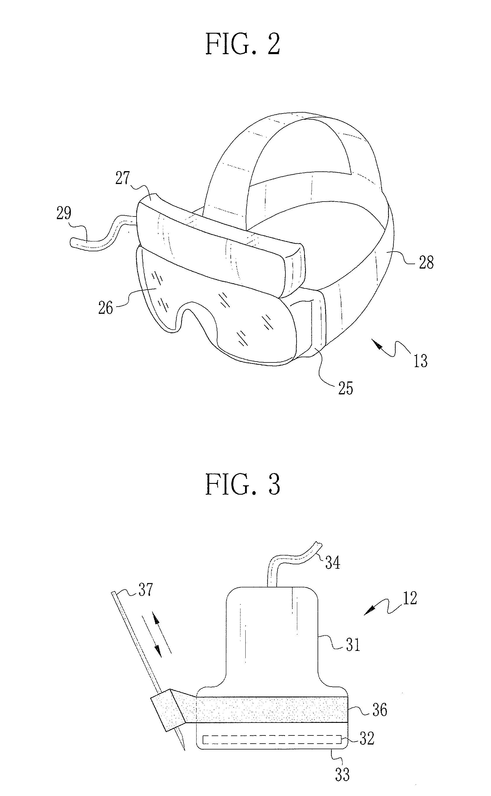
[0034]As shown in FIG. 1, an ultrasonic diagnostic apparatus 11 is an apparatus in which ultrasonic waves are transmitted to the inside of a body of a patient Pa laid quietly on a bed or the like, and an ultrasonic image which is a tomographic image of the inside of the patient's body is generated based on the echoes of the ultrasonic waves and displayed. The ultrasonic diagnostic apparatus 11 is constituted of an ultrasonic probe 12, a head mounted display (HMD) 13, and a main body 14.

[0035]The ultrasonic probe 12 for transmitting the ultrasonic waves and receiving the echoes of the ultrasonic waves is formed to such a size that an operator Op can hold with one hand. The ultrasonic probe 12 is used with being pressed against a surface of the patient Pa's body. The ultrasonic probe 12 is connected to the main body 14 through a flexible communication cable, and its position and angle to be pressed against the patient Pa can be freely adjusted within predetermined ranges. The ultrason...


PUM


Abstract

An ultrasonic diagnostic apparatus is provided with an ultrasonic probe, an ultrasonic image generating section, a head mounted display (HMD), and an orientation measurement section. The ultrasonic probe has two-dimensionally arranged ultrasonic transducers. The ultrasonic image generating section generates a 2D ultrasonic image representing a cross section of a three-dimensional area inside of a patient's body. The HMD has an orientation sensor for outputting signals corresponding to motion of the HMD and a projector for projecting images and the like within the view of an operator Op. The orientation measurement section measures rotation direction and rotation angle of the operator Op's head (HMD). A plurality of cross sections of the three-dimensional area inside of the patient's body for which the 2D image is generated is preliminary set, and the generated 2D ultrasonic image is switched according to the rotation direction and the rotation angle of the HMD.

Description

BACKGROUND OF THE INVENTION[0001]1. Field of the Invention[0002]The present invention relates to an ultrasonic diagnostic apparatus that transmits ultrasonic waves to the inside of a patient's body to obtain tomographic images of the patient's body, and more particularly relates to an ultrasonic diagnostic apparatus that is provided with a head mounted display.[0003]2. Description of the Related Art[0004]An ultrasonic diagnostic apparatus has conventionally been used widely for diagnoses and examinations in the medical field. The ultrasonic diagnostic apparatus transmits ultrasonic waves to the inside of a patient's body, and generates ultrasonic tomographic images (hereinafter, referred to as the ultrasonic images) of the patient's body from the reflected echoes, and display them on a monitor. The ultrasonic diagnostic apparatus is basically constituted of an ultrasonic probe, a processor, and the like. The ultrasonic probe, which is used with being pressed against a surface of the...

Claims


Application Information

Patent Timeline
IPC IPC(8): A61B8/14
CPCG01S7/52084A61B8/461A61B8/462A61B8/483A61B8/54G01S15/899G01S7/52074A61B8/467A61B8/463A61B8/466
Inventor TASHIRO, RIKANISHINO, TOMOHIROTANABE, TSUYOSHI
Owner FUJIFILM CORP
Why Eureka
Try Eureka