Automatic Cardiac Functional Assessment Using Ultrasonic Cardiac Images

a functional assessment and ultrasonic technology, applied in ultrasonic/sonic/infrasonic diagnostics, image enhancement, instruments, etc., can solve the problem of reducing the accuracy of the assessed or quantified cardiac parameters

Inactive Publication Date: 2011-10-27
MINDTREE LTD
View PDF6 Cites 30 Cited by
  • Summary
  • Abstract
  • Description
  • Claims
  • Application Information

AI Technical Summary

Problems solved by technology

The phenomenon of “drift” caused due to the movement artifacts in the ultrasonic cardiac images, which in turn reduces th

Method used


Image

  • Automatic Cardiac Functional Assessment Using Ultrasonic Cardiac Images
  • Automatic Cardiac Functional Assessment Using Ultrasonic Cardiac Images
  • Automatic Cardiac Functional Assessment Using Ultrasonic Cardiac Images

Examples


Embodiment Construction

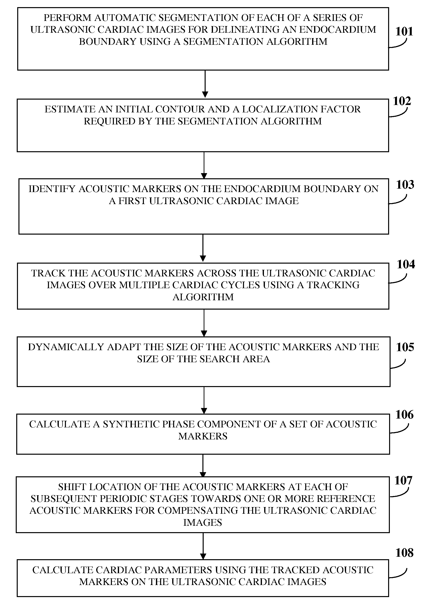
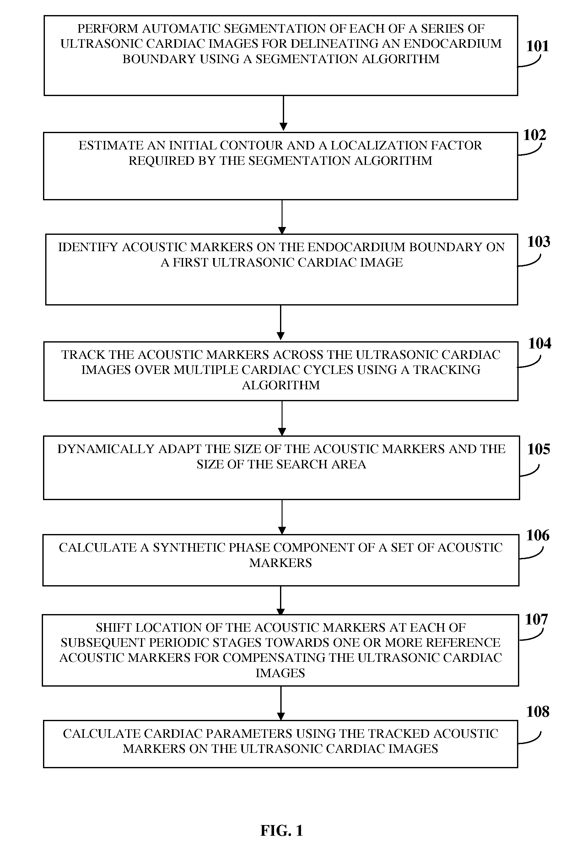
[0049]FIG. 1 illustrates a computer implemented method for performing automatic cardiac functional assessment using a series of ultrasonic cardiac images. A series of ultrasonic cardiac images, for example, echocardiograms are obtained from an echocardiogram database, for example, in an offline mode. Automatic segmentation of each of the ultrasonic cardiac images is performed 101 for delineating an endocardium boundary in each of the ultrasonic cardiac images using a segmentation algorithm. The segmentation algorithm is, for example, based on a region-based active contour algorithm. The segmentation algorithm is configured to automatically delineate the endocardium boundary using localized image statistics. The computer implemented method and system disclosed herein automatically estimates 102 an initial contour and a localization factor that constitute inputs to the segmentation algorithm for delineating the endocardium boundary. One or more periodic stages of each of the cardiac c...


PUM


Abstract

A computer implemented method and system for fully-automatic cardiac functional assessment are provided. Automatic segmentation of a series of ultrasonic cardiac images is performed for delineating an endocardium boundary and an epicardium boundary, in each of the ultrasonic cardiac images using a segmentation algorithm. Multiple acoustic markers are identified on the endocardium boundary on the ultrasonic cardiac images. The acoustic markers are tracked across the ultrasonic cardiac images over multiple cardiac cycles using a tracking algorithm. Multiple cardiac parameters are calculated using the tracked acoustic markers on drift compensated ultrasonic cardiac images. The computer implemented method and system for cardiac functional assessment is fully-automatic without requiring user intervention or inputs.

Description

CROSS REFERENCE TO RELATED APPLICATIONS[0001]This application claims the benefit of non-provisional patent application number 1160 / CHE / 2010 titled “Automatic Cardiac Functional Assessment Using Ultrasonic Cardiac Images”, filed on Apr. 27, 2010 in the Indian Patent Office.[0002]The specification of the above referenced patent application is incorporated herein by reference in its entirety.BACKGROUND[0003]Cardiac functional assessment refers to the measurement and analysis of cardiac parameters, where specific cardiac parameters such as left ventricular volume, ejection fraction, tissue strain, etc. are assessed and used by cardiologists and echocardiographers to diagnose cardiac diseases. Much of the present day cardiac functional assessment is manual and subjective, and has a high dependency on the interpretation of echocardiograms by a medical practitioner, which can vary from practitioner to practitioner.[0004]Cardiac functional assessment referred herein as cardiac computer-aide...

Claims


Application Information

Patent Timeline
IPC IPC(8): G06K9/00
CPCG06T7/0012G06T7/0083A61B8/0883G06T2207/10132G06T2207/30048G06T7/0097G06T7/12G06T7/174
Inventor KUMAR, ANURAGRAO, JAIDEEP HARIKUMAR, PRASHANTAYERRAGUDI VENUGOPALACHARYULU, SAMPATH KRISHNANKANTH, SUDHAKUMAR, RAKSHAN
Owner MINDTREE LTD
Why Eureka
Try Eureka