
Sacroiliac joint fixation system

a fusion system and sacroiliac joint technology, applied in the field of sacroiliac joint fixation fusion system, can solve the problems of increased operative time, increased hospitalization, pain, recovery time,

Active Publication Date: 2011-10-27
JCBD
View PDF7 Cites 175 Cited by
  • Summary
  • Abstract
  • Description
  • Claims
  • Application Information

AI Technical Summary

Benefits of technology

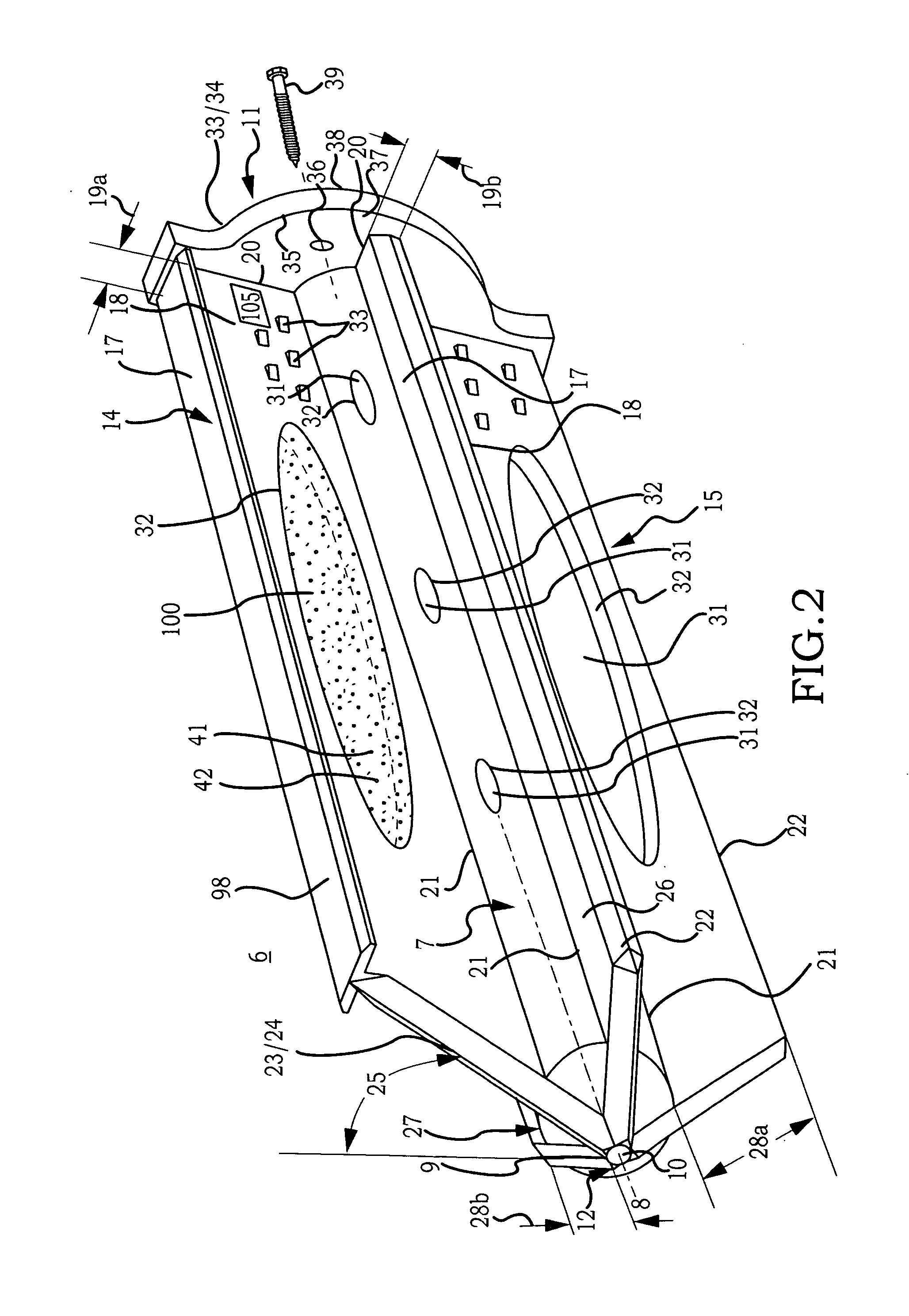
[0011]Accordingly, a broad object of the invention can be to provide an inventive sacroiliac joint implant for fixation and fusion of the sacroiliac joint. Embodiments of the sacroiliac joint implant can provide an elongate body, which can further include at least one radial member, or a pair of members which extend distance radially outward from the longitudinal axis of the elongate body adapted for non-transverse placement between the articular surfaces of the sacroiliac joint and as to certain embodiments can further provide a third radial member and additionally a fourth radial member each adapted to extend a distance radially outward from the longitudinal axis of the elongate body into the bone of the sacrum or the ilium.
[0012]Another broad object of the invention can be to provide an inventive method for fixation and fusion of the sacroiliac joint which utilizes the inventive sacroiliac implant. The inventive method comprising the steps of performing a minimally invasive posterior surgery that allows access to the posterior aspect of the sacroiliac joint for removing a sufficient portion of the articular cartilage or tissue between the articular surfaces of the sacroiliac joint which can be replaced by embodiments of the above described sacroiliac implant. A portion of the subcondral bone of the sacroiliac joint can be removed to provide an implant receiving space in the plane of the sacroiliac joint (non-trans-sacroiliac) configured to allow interference fitting of the elongate member, or the elongate member with at least a first radial member between the opposed surfaces of the implant receiving space. The inventive method can further include the step of providing the implant receiving space with one or more radial member receiving channels cut into the bone (including one or more of the subchondral, cortical, or cancellous) and thereby locating one or more radial members in the bone of the sacrum or the bone of the ilium. The inventive method avoids conventional trans-sacroiliac placement of fixation elements or S1 pedicle and intrailiac screws joined by a rod while providing immediate fixation of the sacroiliac joint.
[0014]Another broad object of the invention can be to provide particular embodiments of the inventive fixation fusion implant with an amount of curvature along the length of the implant which allows placement of embodiments of the fixation fusion implant which have an increased surface area which remain within or substantially within the articular portion of the sacroiliac joint.

Problems solved by technology

However, while each of these methods have been utilized for fixation and fusion of the sacroiliac joint over the past several decades, substantial problems with respect to the fixation and fusion of the sacroiliac joint remain unresolved.
A significant problem with certain conventional methods for fixation and fusion of the sacroiliac joint including the anterior approach, posterior approach, or lateral approach may be that the surgeon has to make a substantial incision in the skin and tissues for direct access to the sacroiliac joint involved.
Often referred to as an “open surgery”, these procedures have the attendant disadvantages of requiring general anesthesia and can involve increased operative time, hospitalization, pain, and recovery time due to the extensive soft tissue damage resulting from the open surgery.
A danger to open surgery using the anterior approach can be damage to the L5 nerve root which lies approximately two centimeters medial to the sacroiliac joint or damage to the major blood vessels.
Use of trans-sacroiliac and S1 pedicle-iliac bone implants can also involve the risk of damage to the lumbosacral neurovascular elements.
Damage to the lumbosacral neurovascular elements as well as delayed union or non-union of the sacroiliac joint by use of these procedures may require revision surgery to remove all or a portion of the implants or repeat surgery as to these complications.
Another significant problem with conventional procedures utilizing minimally invasive small opening procedures can be that the procedures are technically difficult requiring biplanar fluoroscopy of the articular surfaces of the sacroiliac joint and extensive surgical training and experience.
Despite the level of surgical training and experience, there is a substantial incidence of damage to the lumbosacral neurovascular elements.
Additionally, sacral anomalies can further lead to mal-placement of implants leading to damage of surrounding structures.
Additionally, these procedures are often performed with out fusion of the sacroiliac joint which does not remove the degenerative joint surface and thereby does not address the degenerative condition of the sacroiliac joint which may lead to continued or recurrent sacroiliac joint pain.
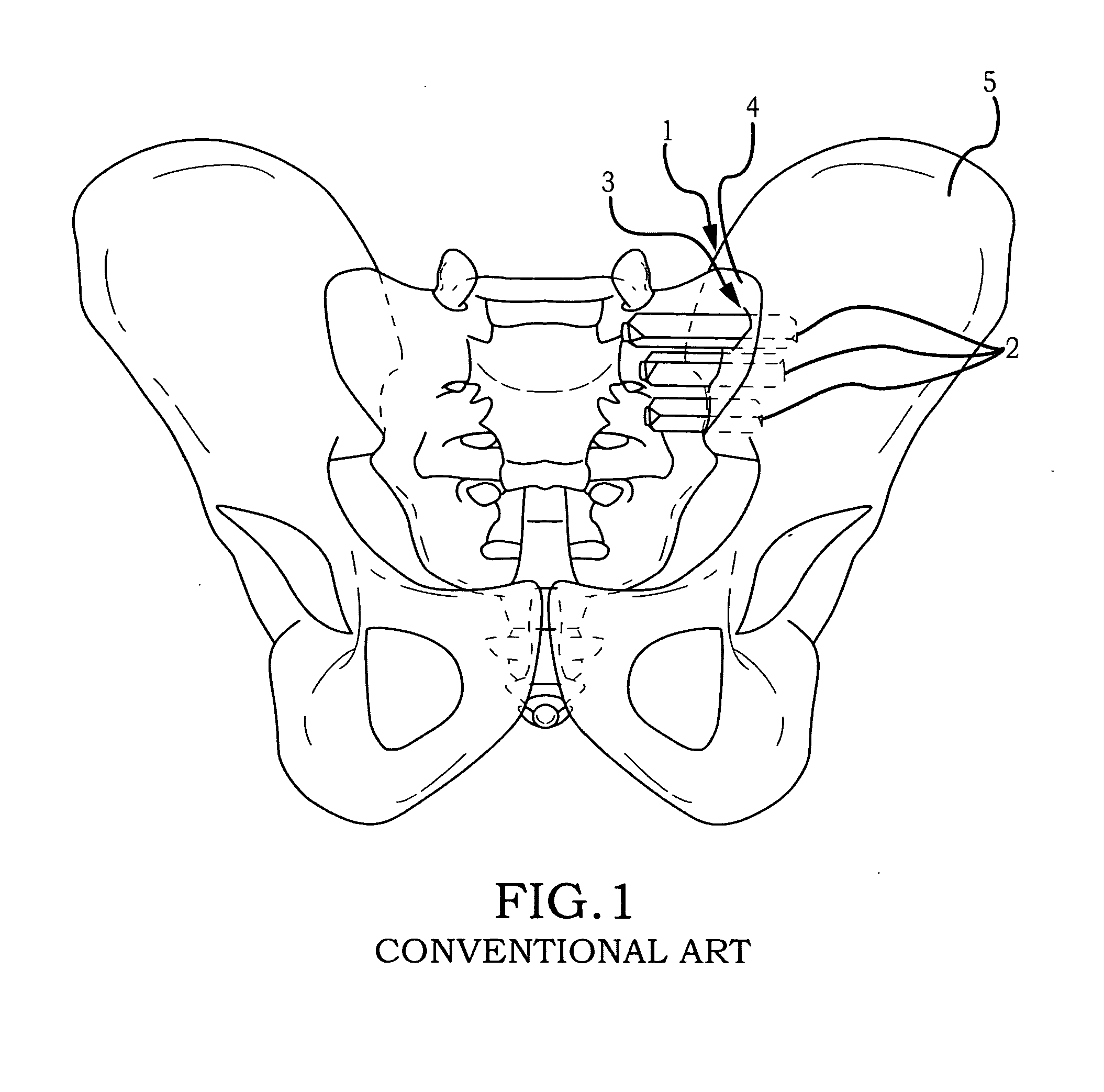
Another significant problem with conventional procedures can be the utilization of multiple trans-sacroiliac elongate implants, which do not include a threaded surface.
This approach requires the creation of trans-sacroiliac bores in the pelvis and nearby sacral foramen which can be of relatively large dimension and which are subsequently broached with instruments which can result in bone being impacted into the pelvis and neuroforamen.
The creation of the trans-sacroiliac bores and subsequent broaching of the bores requires a guide pin which may be inadvertently advanced into the pelvis or sacral foramen resulting in damage to other structures.
Additionally, producing the trans-sacroiliac bores, broaching, or placement of the elongate implants may result in damage to the lumbosacral neurovascular elements, as above discussed.
Additionally, there may be no actual fusion of the articular portion of the sacroiliac joint which may result in continued or recurrent pain requiring additional surgery.
Another substantial problem with conventional procedures can be that placement of posterior extra-articular distracting fusion implants and bone grafts as described for example in U.S. patent application Ser. No. 10 / 797,481 of Stark may be inadequate with respect to removal of the articular surface or preparation of cortical bone, the implant structure and fixation of the sacroiliac joint.
The method may not remove sufficient amounts of the articular surfaces or cortical surfaces of the sacroiliac joint to relieve pain in the sacroiliac joint.
The implant structures described may have insufficient or avoid engagement with the articular surfaces or cortical bone of the sacroiliac joint for adequate fixation or fusion.
The failure to sufficiently stabilize and fuse the sacroiliac joint with the implant structures and methods described by the Stark application may result in a failure to relieve the condition of sacroiliac joint being treated.
Additionally, the method of driving apart a sacrum and ilium as described by Stark may lead to mal-alignment of the sacroiliac joint and increased pain.

Method used


Image

  • Sacroiliac joint fixation system
  • Sacroiliac joint fixation system
  • Sacroiliac joint fixation system

Examples


example 1

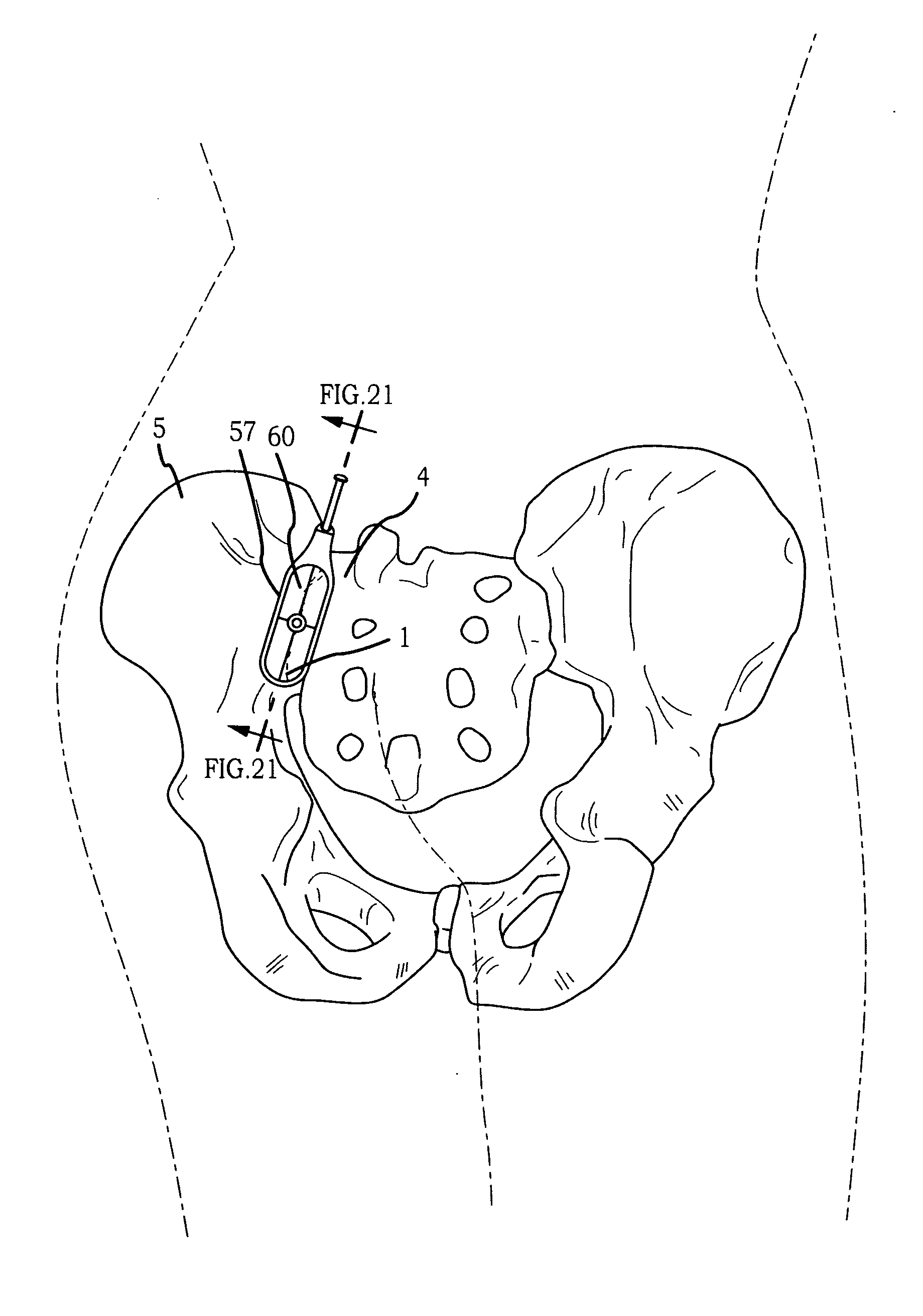
[0104]An embodiment of the inventive sacroiliac joint implant having a configuration substantially as shown by FIGS. 3-6 and as above-described was inserted into a patient under direct visualization and with assisted lateral fluoroscopy. The procedure was performed for the purpose of assessing in an actual reduction to practice the ability of the inventive sacroiliac joint implant to be safely implanted between inferior or caudal portion the articular surfaces of the sacroiliac joint substantially as shown in FIG. 25 to confirm the that implantation of the sacroiliac joint implant into a implant receiving space configured substantially as above-described acts to immobilize the sacroiliac joint. The sacroiliac joint implant as above-described implanted into the inferior (caudal) portion of the sacroiliac joint proved to immediately immobilize the sacroiliac joint.

[0105]As can be easily understood from the foregoing, the basic concepts of the present invention may be embodied in a var...


PUM


Abstract

A sacroiliac joint fixation fusion system that provides a method of fixation and fusion of the sacroiliac joint and a sacroiliac joint implant which upon placement within the articular region of the sacroiliac joint facilitates stability and fusion of the sacroiliac joint.

Description

[0001]This Application is a continuation of U.S. patent application Ser. No. 12 / 998,712, filed May 23, 2011, which is the National Stage of International Patent Cooperation Treaty Patent Application No. PCT / US11 / 00070, filed Jan. 13, 2011, which claims the benefit of U.S. Provisional Patent Application No. 61 / 335,947, filed Jan. 13, 2010, each hereby incorporated by reference herein.I. TECHNICAL FIELD[0002]Generally, a sacroiliac joint fixation fusion system that provides a method of fixation and fusion of the sacroiliac joint and a sacroiliac joint implant which upon placement within the articular space of the sacroiliac joint facilitates stability and fusion of the sacroiliac joint.II. BACKGROUND[0003]The sacroiliac joint is the joint between the sacrum and the ilium of the pelvis, which are joined by ligaments. In humans, the sacrum supports the spine and is supported in turn by an ilium on each side. The sacroiliac joint is a synovial joint with articular cartilage and irregular...

Claims


Application Information

Patent Timeline
Patent Type & Authority Applications(United States)
IPC IPC(8): A61F2/30
CPCA61B17/025A61B17/7043A61F2/30988A61F2002/30622A61B17/8645A61B17/1739A61B17/7055A61B17/1757A61F2002/30995A61B17/1626A61B2017/0046A61F2/4455A61F2/4611A61F2002/4687A61F2310/00017A61F2310/00023A61F2310/00179A61F2310/00359A61F2002/30179A61B17/1742A61B17/7074A61F2/30749
Inventor DONNER, EDWARD JEFFREY
Owner JCBD
Learn More