Anti-fgfr2 antibodies
a technology of fgfr2 and antibodies, applied in the field of molecular biology, immunology and oncology, can solve the problems of difficult generation of specific antibodies specific for human fgfr2, and achieve the effect of reducing or eliminating an immune respons
- Summary
- Abstract
- Description
- Claims
- Application Information
AI Technical Summary
Benefits of technology
Problems solved by technology
Method used
Image

Examples
example 1
Cell Lines and Reagents
[0078]KATO III, HEC-1-A, AN3 CA, SNU-16, and human lung cancer cell lines were acquired from the American Type Culture Collection (Rockville, Md.). FDCP-1 and Ba / F3, MFM-223, MFE-296, MFE-280, MFE-319 and ESS-1 cells were obtained from the German Collection of Microorganisms and Cell Cultures. All human cell lines were cultured according to the instructions specified by the suppliers, at 37° C., in an atmosphere containing 5% CO2. All FGFs were purchased from R&D Systems, Inc. (Minneapolis, Minn.).
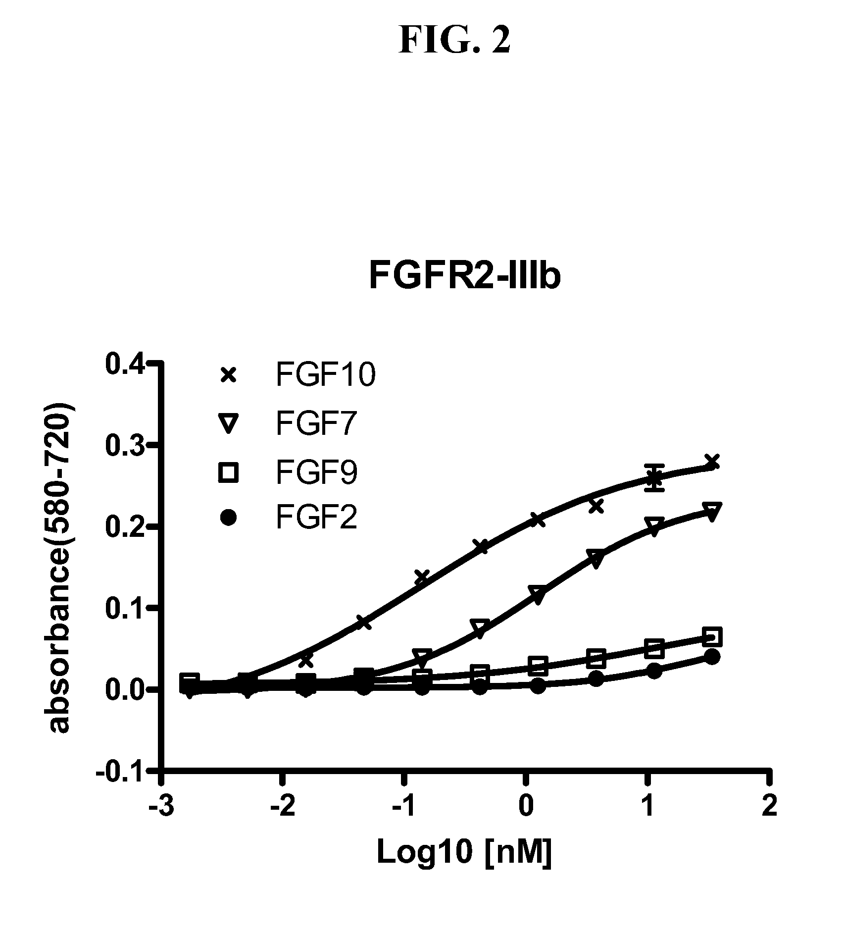
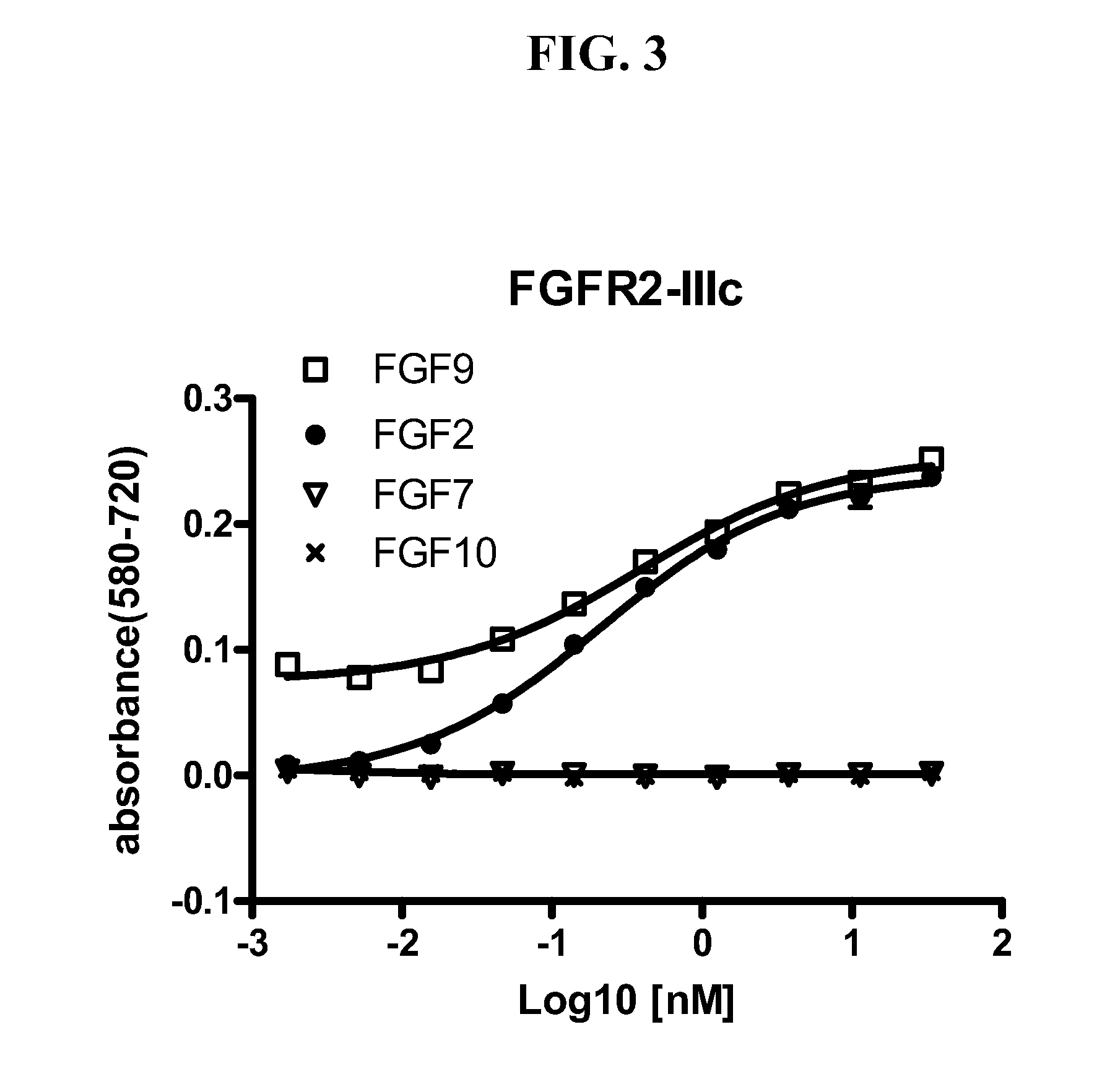
[0079]To establish cell-based assays to screen for functional FGFR2 antibodies, we first engineered Ba / F3 and FDCP-1 cells to express wild type FGFR2 and cancer-associated mutants or variants of FGFR2. FGFR-driven FDCP cells and Ba / F3 cells were obtained by the following methods. FDCP-1 cells were transfected by electroporation with plasmids encoding the IIIb, IIIc isoform or C-terminally truncated variant of human FGFR2 as well as cancer-associated FGFR2-IIIb S252W,...
example 2
Production of Anti-FGFR2 Monoclonal Antibodies
[0083]Mice were immunized with a 1:1 mixture of human FGFR2 IgD2-IgD3 (IIIb) and human FGFR2 IgD2-IgD3 (IIIc) fused with a human Fc moiety at their C-termini. Mouse immunizations and cell fusions were performed by a commercial vendor (Precision Antibody, Columbia, Md.).
[0084]In a primary screen, hybridoma supernatants were screened to detect binding to human FGFR2 IgD2-IgD3, using an ELISA format. Antibodies that passed the primary screen were subjected to a secondary screen, which was a cell-based proliferation assay described in Example 3 (below).
[0085]The primary screen was performed using the supernatants of the murine hybridoma clones yielded from the splenic fusion of the mice immunized with the extracellular domain of human FGFR2. Assay plates were coated with 100 ng / well of recombinant soluble FGFR2 extracellular domain and then blocked with 5% milk in PBS for one hour at room temperature. Then 50 μA of hybridoma supernatant was ...
example 3
Identification of FGFR2 Antagonist Antibodies
[0086]To screen for FGFR2 antagonist antibodies, hybridoma supernatants containing FGFR2 antibodies were added to FDCP cells ectopically expressing one of the following five forms of FGFR2: (1) wild type FGFR2-IIIb; (2) wild type FGFR2-IIIc; (3) FGFR2-III(b) S252W; (4) FGFR2-III(b) N550K; and (5) FGFR2-III(b) with C-terminal truncation. The supernatants were added to the FGFR2-expressing cells at a 1:1 ratio (volume) in a flat-bottomed 96-well plate (70,000 cells / well) with heparin (5 μg / ml)±FGF1 (8 ng / ml). After incubation at 37° C. for 2 days, MTT assays were conducted as described above.
[0087]The supernatant of clone 4B9 demonstrated potent and selective inhibition of the FDCP-1 proliferation driven by the IIIb-isoform of FGFR2. Antibody 4B9 (also referred to as antibody GP369), produced by clone 4B9, was purified by conventional techniques for further characterization. Surface plasmon resonance analysis indicated that antibody 4B9 exh...
PUM
| Property | Measurement | Unit |
|---|---|---|
| Molar density | aaaaa | aaaaa |
| Temperature | aaaaa | aaaaa |
| Molar density | aaaaa | aaaaa |
Abstract
Description
Claims
Application Information
 - R&D
- Intellectual Property
- Life Sciences
- Materials
- Tech Scout
- Unparalleled Data Quality
- Higher Quality Content
- 60% Fewer Hallucinations
Browse by: Latest US Patents, China's latest patents, Technical Efficacy Thesaurus, Application Domain, Technology Topic, Popular Technical Reports.
© 2025 PatSnap. All rights reserved.Legal|Privacy policy|Modern Slavery Act Transparency Statement|Sitemap|About US| Contact US: help@patsnap.com