

Method for diagnosis of cancer and monitoring of cancer treatments
a cancer and treatment technology, applied in the field of cancer diagnosis and cancer treatment monitoring based on the analysis of circulating dna, can solve problems such as hammering its clinical application in cancer diagnosis
- Summary
- Abstract
- Description
- Claims
- Application Information
AI Technical Summary
Benefits of technology
Problems solved by technology
Method used
Image

Examples
example 1
Methods of Sample Analysis and DNA Fragment Pattern Selection
[0065]Overall, sample analysis and fragment pattern selection is carried out in five subsequent steps. Step (1) isolation of circulating DNA, step (2) quantification of isolated DNA, step (3) quantification of DNA fragments by real time PCR, step (4) data evaluation and fragment pattern selection and finally step (5) computation of the DNA fragmentation index (DFI).
[0066]Step (1): Isolation of Circulating DNA
[0067]Circulating DNA is isolated from 1 to 2.5 ml frozen (−20° C.) or fresh serum samples using the Qiagen MinElute Virus Vacuum kit (Cat. No. 57714, Qiagen, Hilden, Germany) following the instructions of the manufacturer (Qiagen kit manual, 3rd edition, March 2007, page 23-25). Frozen plasma samples (2-2.5 ml) are processed using the Qiagen MinElute Virus Spin kit (Cat. No. 57704, Qiagen, Hilden, Germany) following the instructions of the manufacturer (Qiagen kit manual, 3rd edition, February 2007, page 19-22).
[0068]...
example 2
Relapse Diagnosis of Metastatic Breast Cancer by DNA-Fragmentation Analysis
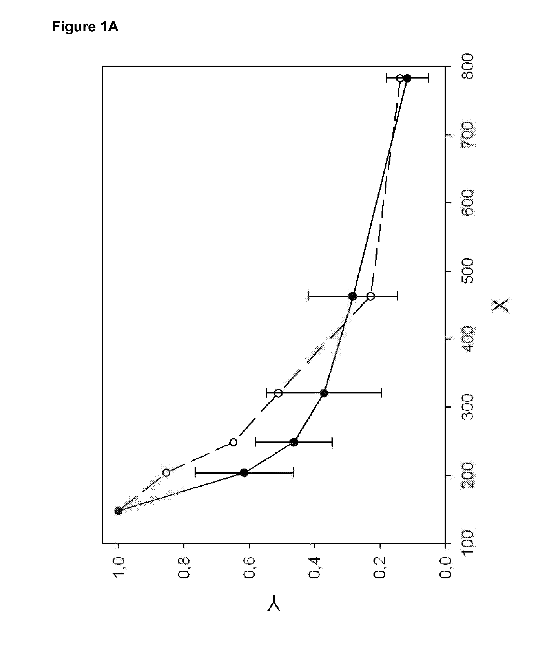
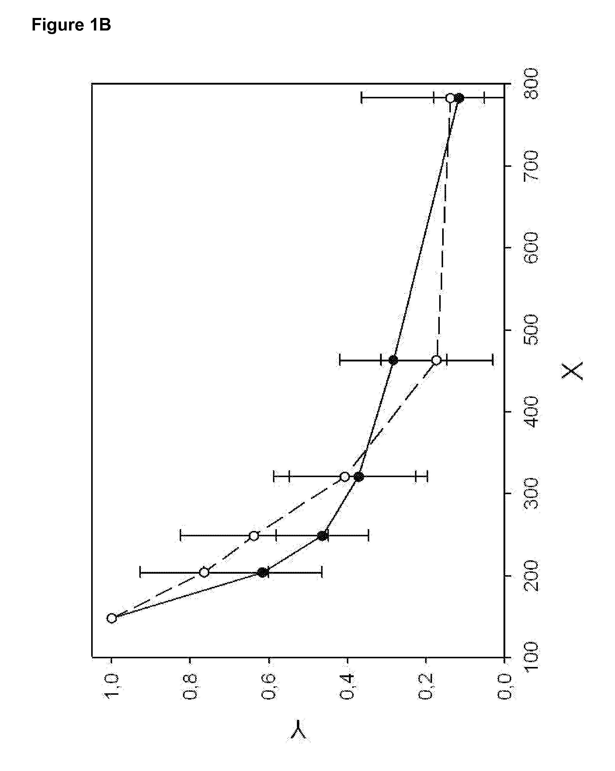
[0098]The DNA fragmentation pattern of healthy donors (n=17) and a pool of patients (n=10) with metastatic breast cancer at various clinical stages is recorded (Example 1). LINE1 fragments with indicated by length are analyzed (see Table 2), normalized, and plotted (see FIG. 1B). The DNA fragmentation pattern (see FIG. 1A) of pooled breast cancer sera indicates an increased level of DNA fragments between 200 and 400 bp as compared to healthy controls.
[0099]The DNA fragmentation index (DFI) is computed for each patient / pool according to equation [2] (using LINE1-B, LINE1-C, LINE1-E and LINE1-F, see Table 2) as well as equation [4] (using LINE1-B, LINE1-C, LINE1-D, LINE1-E and LINE1-F, see Table 2).
[0100]The mean DFI4 (equation [2]) for healthy control is 0.02±0.01 compared to 0.64 for the breast cancer pool; thus indicating a potential cut off around 0.1.
[0101]Similar results are obtained using equation [4]. T...
example 3
Diagnosis of Hepatocellular Carcinoma by DNA-Fragmentation Analysis
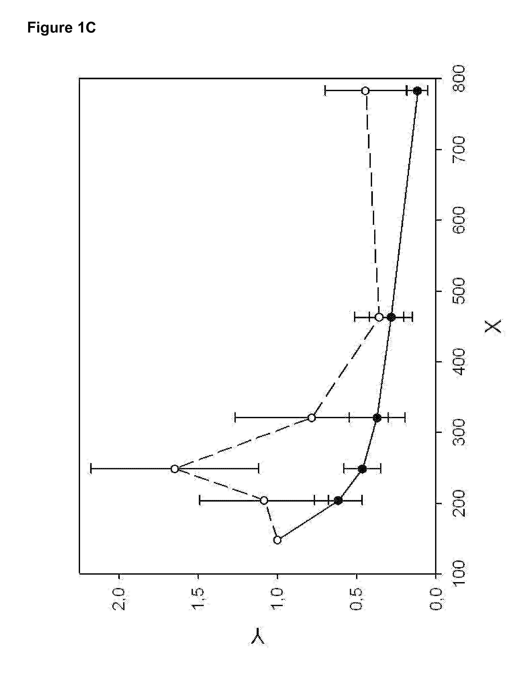
[0110]The DNA fragmentation pattern of patients with liver cirrhosis (at-risk patients) and patients with HCC is recorded as indicated in Example 1). LINE1 fragments with indicated sizes are analyzed (see Table 2), normalized, and plotted (see FIG. 2). FIG. 2 shows a significantly different curve of the DNA fragmentation pattern of HCC patients as compared to at risk patients suffering from liver cirrhosis. Therefore, the diagnosis of HCC may be obtained by recording the DNA fragmentation pattern in the indicated range of 148 to 783 bp, but at least between 148 and 463 bp and comparison with the DNA-fragmentation pattern of patients with liver cirrhosis and / or chronic hepatitis C. The DNA-fragmentation pattern of HCC patients differs most significantly from the control group between 204 and 463 bp.
[0111]Computation of DFI is performed according to equation [1], [2] and [4.1]. In order to optimize the LINE1 pattern de...
PUM
| Property | Measurement | Unit |
|---|---|---|
| concentrations | aaaaa | aaaaa |
| volume | aaaaa | aaaaa |
| volume | aaaaa | aaaaa |
Abstract
Description
Claims
Application Information