
Method and system for non-invasive monitoring of patient parameters

Active Publication Date: 2012-03-29
GENERAL ELECTRIC CO
View PDF3 Cites 83 Cited by
  • Summary
  • Abstract
  • Description
  • Claims
  • Application Information

AI Technical Summary

Benefits of technology

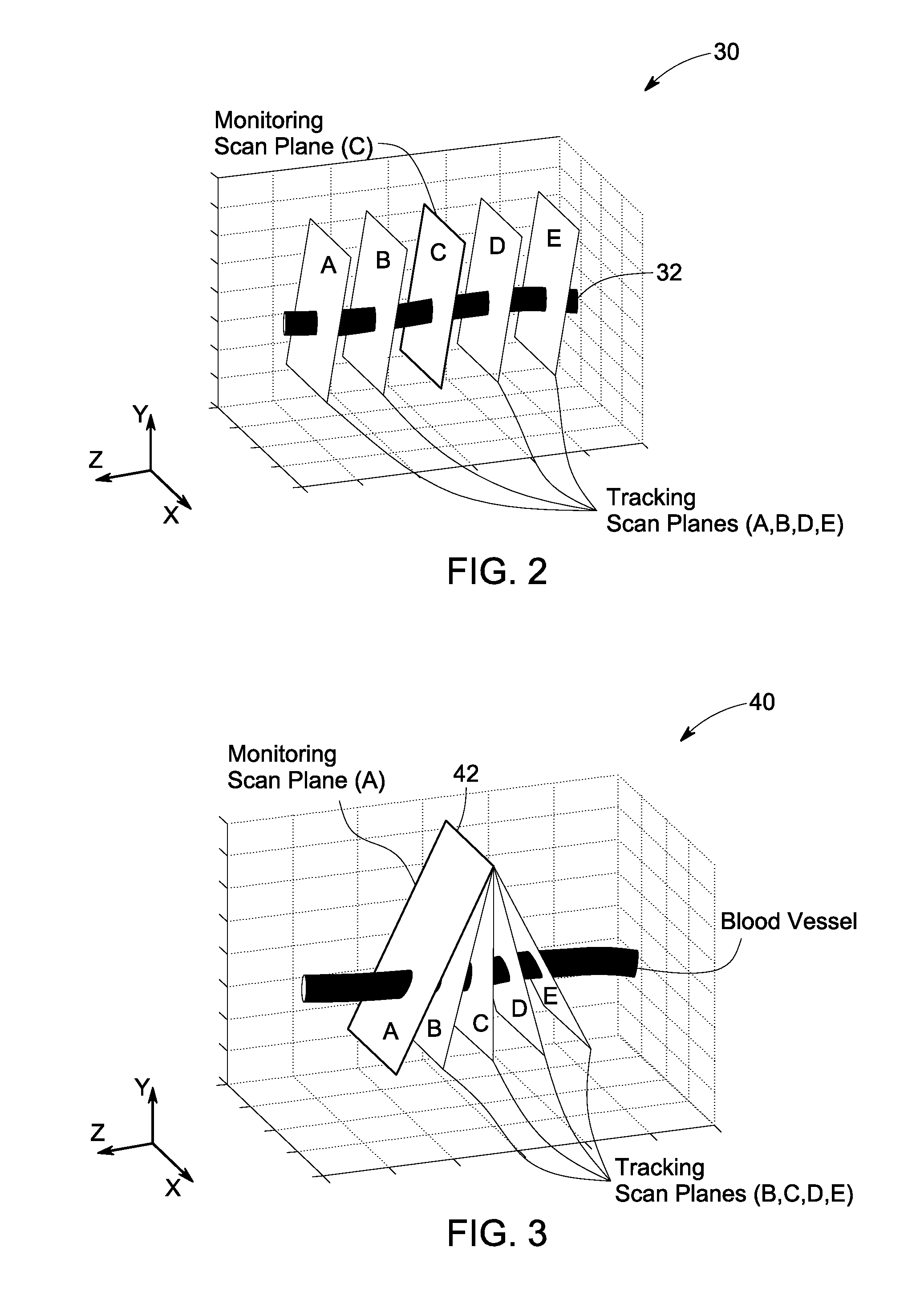
[0009]In accordance with an embodiment of the invention, a system for continuous non-invasive monitoring of multiple arterial parameters of a patient is provided. The system includes an ultrasound imager and an ultrasound transducer acoustically coupled to the patient for acquiring a plurality of ultrasound data. The system also includes an ultrasound processor coupled to the ultrasound transducer. The ultrasound processor is configured to detect a blood vessel through a short-axis view and using a color flow processing within a monitoring scan plane, determine a centerline of the blood vessel through a volume using data from a plurality of tracking scan planes, determine a blood flow direction along the centerline, estimate a Doppler angle and a flow angle for the monitoring scan plane, check if the Doppler angle is above or below a threshold value and adjust the tilt of the monitoring scan plane, estimate a contour of the blood vessel within the monitoring scan plane using a B-mode image, estimate a cross-sectional area of the blood vessel based on the estimated contour and the centerline of the blood vessel. The ultrasound processor is also configured to reduce noise in two-dimensional blood velocity estimates obtained from the color flow processing, estimate a volumetric flow rate of blood from the two dimensional blood velocity estimates within the monitoring scan plane, the centerline and the contour of the blood vessel. The ultrasound processor is further configured to update the centerline within a volume of the blood vessel using data of the plurality of tracking scan planes. Finally the system includes a display coupled to the processor and said display is configured to output a continuous quantitative waveforms based on the cross-sectional area of the blood vessel and the volumetric flow rate of blood.
[0010]In accordance with another embodiment of the invention, a method of processing continuously measured ultrasound data of a patient is provided. The method includes receiving the continuously measured ultrasound data acquired via an ultrasound transducer attached to the patient for detecting a blood vessel through a short-axis view and using color flow processing within a monitoring scan plane. The method also includes determining a centerline of the blood vessel through a volume using data from multiple tracking scan planes. Further, the method includes determining a blood flow direction along the centerline, estimating a Doppler angle and a flow angle for the monitoring scan plane, checking if the Doppler angle is above or below a threshold value and adjusting tilt of the monitoring scan plane, estimating a contour of the blood vessel within the monitoring scan plane using a B-mode image, estimating a cross-sectional area of the blood vessel based on the estimated contour and the centerline of the blood vessel and reducing noise in the two-dimensional blood velocity estimates obtained from the color flow processing. The method includes estimating a volumetric flow rate of blood from the two dimensional blood velocity estimates within the monitoring scan plane, the centerline and the contour of the blood vessel. Further, the method includes updating the centerline within a volume of the blood vessel using data of the plurality of tracking scan planes and finally generating continuous quantitative waveforms based on the cross-sectional area of the blood vessel and the volumetric flow rate of blood through the vessel.

Problems solved by technology

However, continuous ultrasound measurements of cross-sectional area and volumetric flow rate of blood vessels using two-dimensional imaging are difficult to perform and typically only provide qualitative measures because of the inability to verify proper alignment of the ultrasound probe and determine the orientation of the ultrasound beam relative to the vessel.
This technique requires several manual steps by a sonographer and is thus, impractical for continuous vascular monitoring.
This approach requires a sonographer and is prone to alignment errors thereby, making the process impractical for continuous vascular monitoring applications.
Neither of these methods that utilize volumetric acquisition are suitable for producing continuous waveforms over the cardiac period because of the long acquisition time for the volumetric color flow frames and the potential for significant motion over the frame time.

Method used


Image

  • Method and system for non-invasive monitoring of patient parameters
  • Method and system for non-invasive monitoring of patient parameters
  • Method and system for non-invasive monitoring of patient parameters

Examples


Embodiment Construction

[0024]When introducing elements of various embodiments of the present invention, the articles “a,”“an,”“the,” and “said” are intended to mean that there are one or more of the elements. The terms “comprising,”“including,” and “having” are intended to be inclusive and mean that there may be additional elements other than the listed elements. Further, the term ‘processing’ may refer to reading or recording or rewriting or retrieving of data from a data storage system. Any examples of operating parameters are not exclusive of other parameters of the disclosed embodiments.

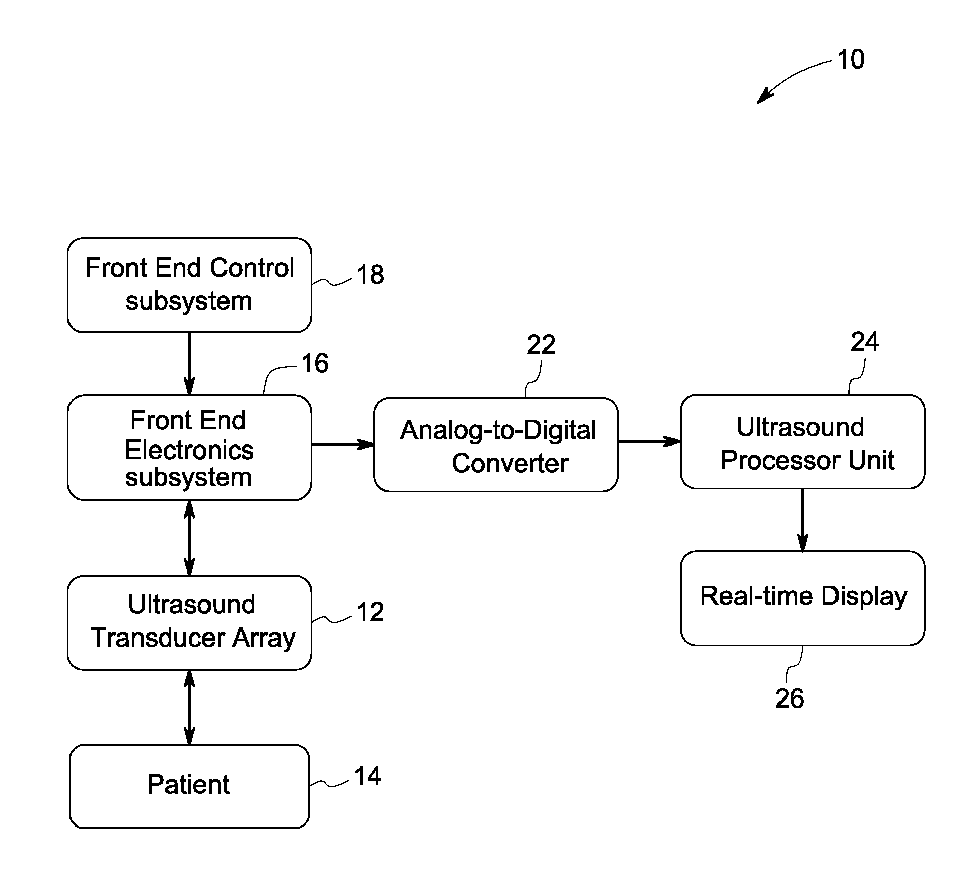
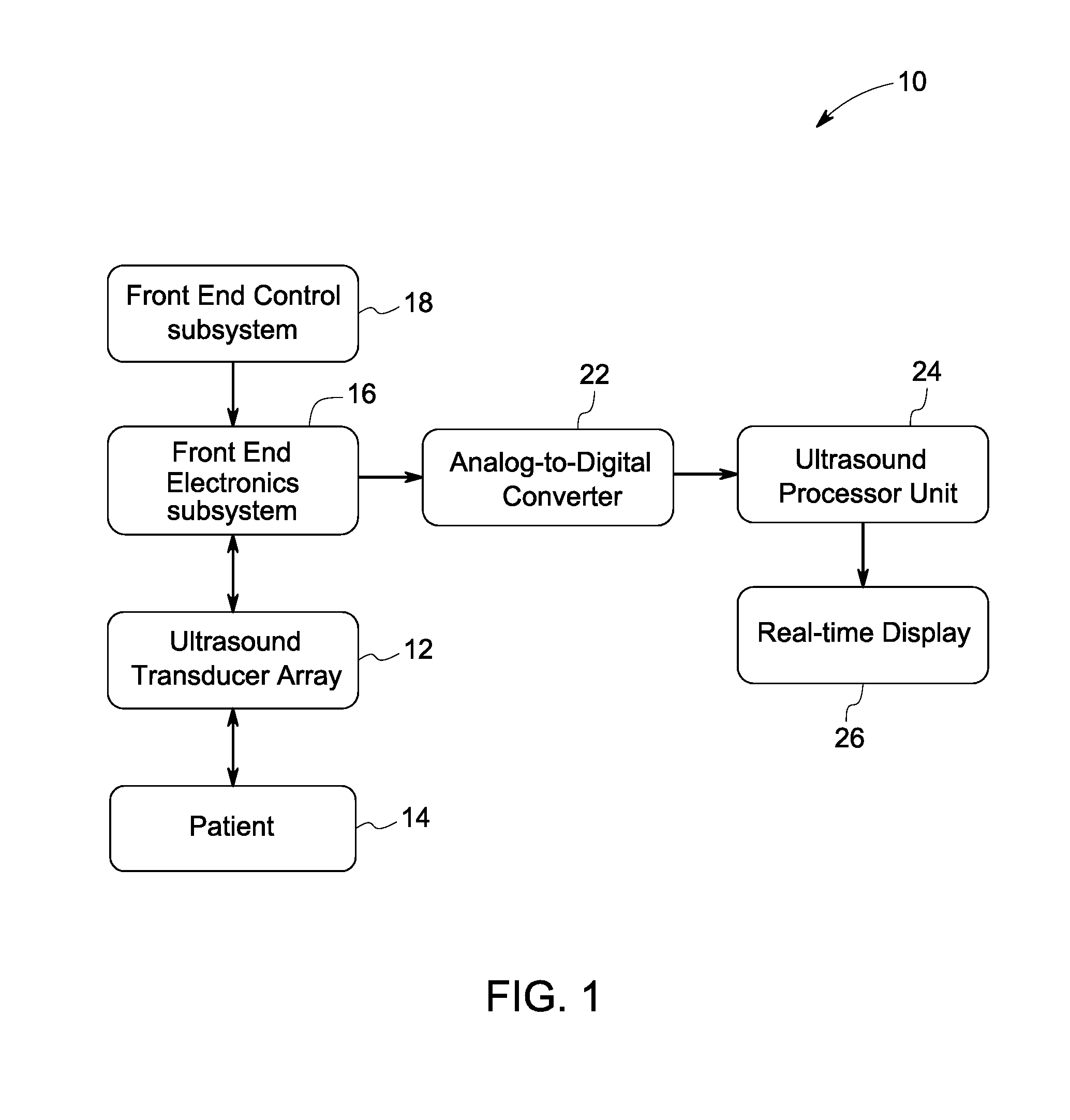
[0025]FIG. 1 is a block diagram illustrating various components of an ultrasound-based patient monitoring system 10 in accordance with an embodiment of the invention. The system 10 includes an ultrasound transducer array 12 that is in contact with a patient 14 and acoustically coupled using an ultrasound gel for continuously acquiring ultrasound data. Further, the ultrasound transducer array 12 is connected to an elect...


PUM


Abstract

A method for continuous non-invasive monitoring of multiple arterial parameters of a patient is provided. The method includes continuously acquiring ultrasound data via an ultrasound transducer attached to the patient for detecting a blood vessel using color flow processing within a monitoring scan plane. Further, the method includes processing the continuously acquired ultrasound data to generate continuous quantitative waveforms based on an estimated cross-sectional area of the blood vessel and an estimated volumetric flow rate of blood through the vessel and displaying the generated continuous quantitative waveforms for monitoring the arterial parameters of the patient in real-time.

Description

[0001]This invention was made with Government support under contract number R01HL094487 awarded by the National Institute of Health (NIH). The Government has certain rights in the invention.BACKGROUND[0002]The present invention relates generally to methods and system for monitoring of arterial parameters and more particularly to a method and system for continuous non-invasive monitoring of patient parameters.[0003]Generally, non-invasive monitoring of arterial parameters is done using ultrasound measurement systems. However, continuous ultrasound measurements of cross-sectional area and volumetric flow rate of blood vessels using two-dimensional imaging are difficult to perform and typically only provide qualitative measures because of the inability to verify proper alignment of the ultrasound probe and determine the orientation of the ultrasound beam relative to the vessel. Current quantitative flow measurements use the long-axis (longitudinal) view of the vessel along with interac...

Claims


Application Information

Patent Timeline
IPC IPC(8): A61B8/06
CPCA61B8/06A61B8/5223A61B8/488A61B8/08G16H50/30
Inventor DENTINGER, AARON MARKPATWARDHAN, KEDAR ANILHOCTOR, RALPH THOMAS
Owner GENERAL ELECTRIC CO
Learn More