Device for collapsing and loading a heart valve into a minimally invasive delivery system

a delivery system and heart valve technology, applied in the field of aortic heart valve disease, can solve the problems of undue stress on the stented valve, difficult mastery, and unsatisfactory crimping devices, and achieve the effect of minimally invasive and undue stress

Active Publication Date: 2012-04-05
ST JUDE MEDICAL CARDILOGY DIV INC
View PDF13 Cites 37 Cited by
  • Summary
  • Abstract
  • Description
  • Claims
  • Application Information

AI Technical Summary

Benefits of technology

[0005]Present crimping devices and methods for collapsing a stented valve have proven to be unsatisfactory as they include bulky devices, are difficult to master, are time consuming, impart undue stress on the stented valve, or include other undesirable qualities. Such devices include direct radial devices, as are well known in the art. It would therefore be beneficial to provide devices and methods for collapsing a stented bioprosthetic heart valve using apparatuses and techniques that overcome the deficiencies of conventional devices. Preferably, such devices and methods may also assist with loading of the collapsed stented valve onto a minimally invasive delivery device.

Problems solved by technology

Present crimping devices and methods for collapsing a stented valve have proven to be unsatisfactory as they include bulky devices, are difficult to master, are time consuming, impart undue stress on the stented valve, or include other undesirable qualities.

Method used


Image

  • Device for collapsing and loading a heart valve into a minimally invasive delivery system
  • Device for collapsing and loading a heart valve into a minimally invasive delivery system
  • Device for collapsing and loading a heart valve into a minimally invasive delivery system

Examples


Embodiment Construction

[0056]In the following detailed description, preferred embodiments of a device for collapsing and loading a stented bioprosthetic valve (also referred to as a stented valve) onto a minimally invasive delivery system are described in accordance with the present invention. In describing the embodiments illustrated in the drawings, specific terminology may be used for the sake of clarity. However, the invention is not intended to be limited to the specific terms so selected, and it is to be understood that specific terms may also include technical equivalents that operate in a similar manner to accomplish a similar purpose. Where like elements have been depicted in multiple embodiments, identical reference numerals have been used in the multiple embodiments for ease of understanding.

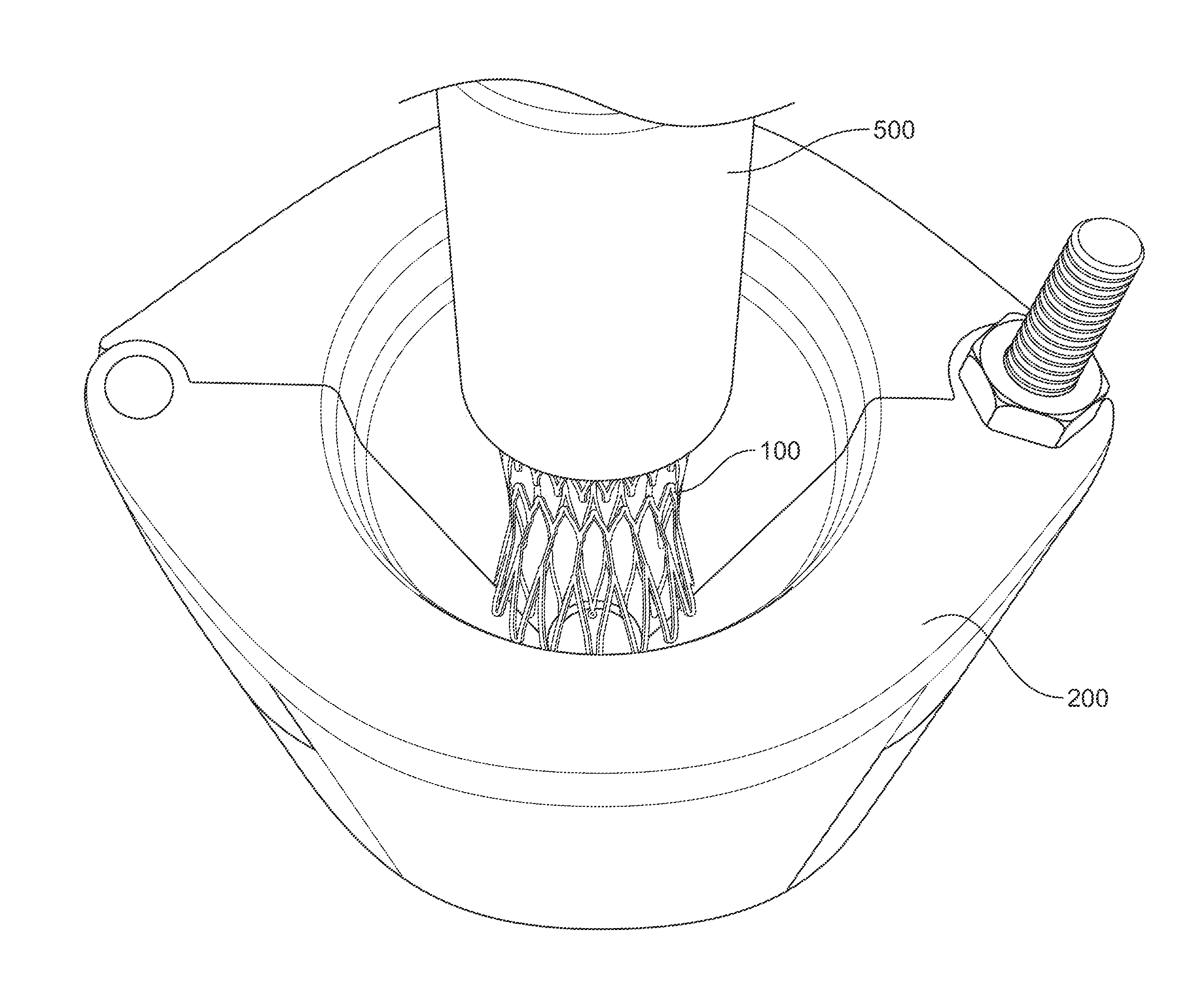
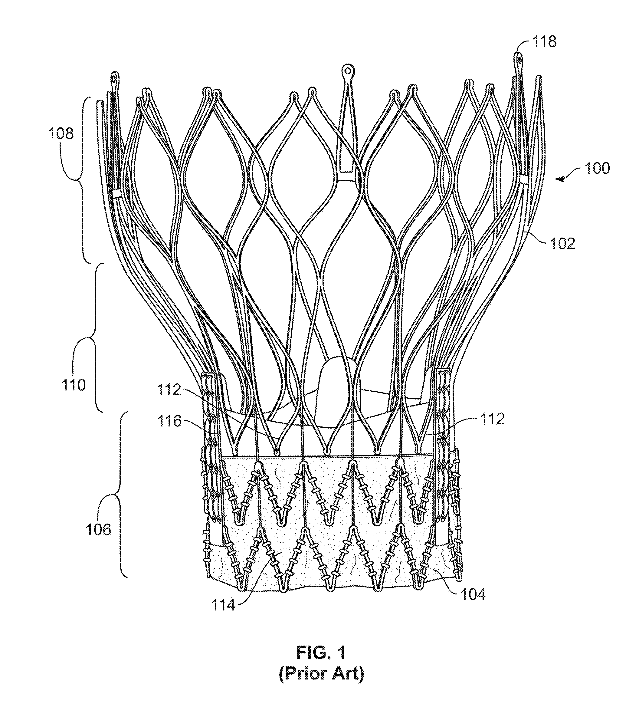
[0057]Referring to FIG. 1, there is shown a conventional bioprosthetic stented valve 100. When installed in the heart, the stented valve 100 is positioned at the annulus of the compromised native valve such...


PUM

PropertyMeasurementUnit
temperatureaaaaaaaaaa
dimensionaaaaaaaaaa

Abstract

A device is provided for collapsing a stented bioprosthetic valve, including first section and second sections, each spanning between first and second ends of the device. The second section of the device is associated with the first section to at least partially enclose an internal cavity formed by the first and second sections, the internal cavity tapering from an open insertion portion at a first end of the device to an open exit portion at a second end of the device. The insertion portion has a larger dimension than the exit portion. When the first section and second section are substantially enclosing the internal cavity, a stented bioprosthetic valve may be inserted into the insertion portion and collapsed as it is moved toward and through the exit portion. The valve may then be loaded on an apparatus for insertion into the body.

Description

CROSS-REFERENCE TO RELATED APPLICATIONS[0001]The present application claims the benefit of the filing date of U.S. Provisional application No. 61 / 374,429, filed Aug. 17, 2010, the disclosure of which is hereby incorporated by reference herein.BACKGROUND OF THE INVENTION[0002]Heart valve disease, and particularly aortic heart valve disease, is a medical problem afflicting many individuals throughout the world. Over time, natural heart valves may leak causing aortic insufficiency or regurgitation. They may also become blocked or exhibit limited function due to aortic stenosis. Among the treatment options for either condition is the destruction of the native valve and insertion of a prosthetic valve through a transapical approach using a minimally invasive delivery device.[0003]Prosthetic valves may be formed from biological materials such as harvested bovine valves or pericardium tissue. Such valves are typically fitted within, and form a portion of, a biologically compatible stent, w...

Claims


Application Information

Patent Timeline
Patent Type & Authority Applications(United States)
IPC IPC(8): A61F2/24B23P11/00
CPCA61F2/2427Y10T29/49927A61F2002/9522A61F2/9522A61F2/9525Y10T29/53657Y10T29/53796Y10T29/53843Y10T29/53852Y10T29/539A61F2/2418
Inventor DALE, THEODORE PAULCHALEKIAN, AARON J.GLAZIER, VALERIE J.WANG, HUISUN
Owner ST JUDE MEDICAL CARDILOGY DIV INC
Why Eureka
Try Eureka