Method and system for brain tumor segmentation in multi-parameter 3D mr images via robust statistic information propagation

a statistical information propagation and multi-parameter technology, applied in image enhancement, image analysis, instruments, etc., can solve the problems of time-consuming, labor-intensive and subjective manual segmentation of brain tumor images commonly used in clinics, and achieve the effect of reducing performance degradation

Active Publication Date: 2012-04-19
INST OF AUTOMATION CHINESE ACAD OF SCI
View PDF5 Cites 29 Cited by
  • Summary
  • Abstract
  • Description
  • Claims
  • Application Information

AI Technical Summary

Benefits of technology

[0010]This method tries to exploit the local and global consistency of the image to be segmented for the tumor segmentation, therefore, it can ...

Problems solved by technology

However, manual segmentation of brain tumor images commonly used in clinics is time-consuming, labor-intensive and subjective to considerable variation in intra- and inter-rater performance.
It however remains a challenging task to achieve robust segmentation as brain tumors differ much in appearance, location, size, and shape.
However, the supervised PBT classifier relies on the image consistency between training images and the image to be segmented, and it...

Method used


Image

  • Method and system for brain tumor segmentation in multi-parameter 3D mr images via robust statistic information propagation
  • Method and system for brain tumor segmentation in multi-parameter 3D mr images via robust statistic information propagation
  • Method and system for brain tumor segmentation in multi-parameter 3D mr images via robust statistic information propagation

Examples


Embodiment Construction

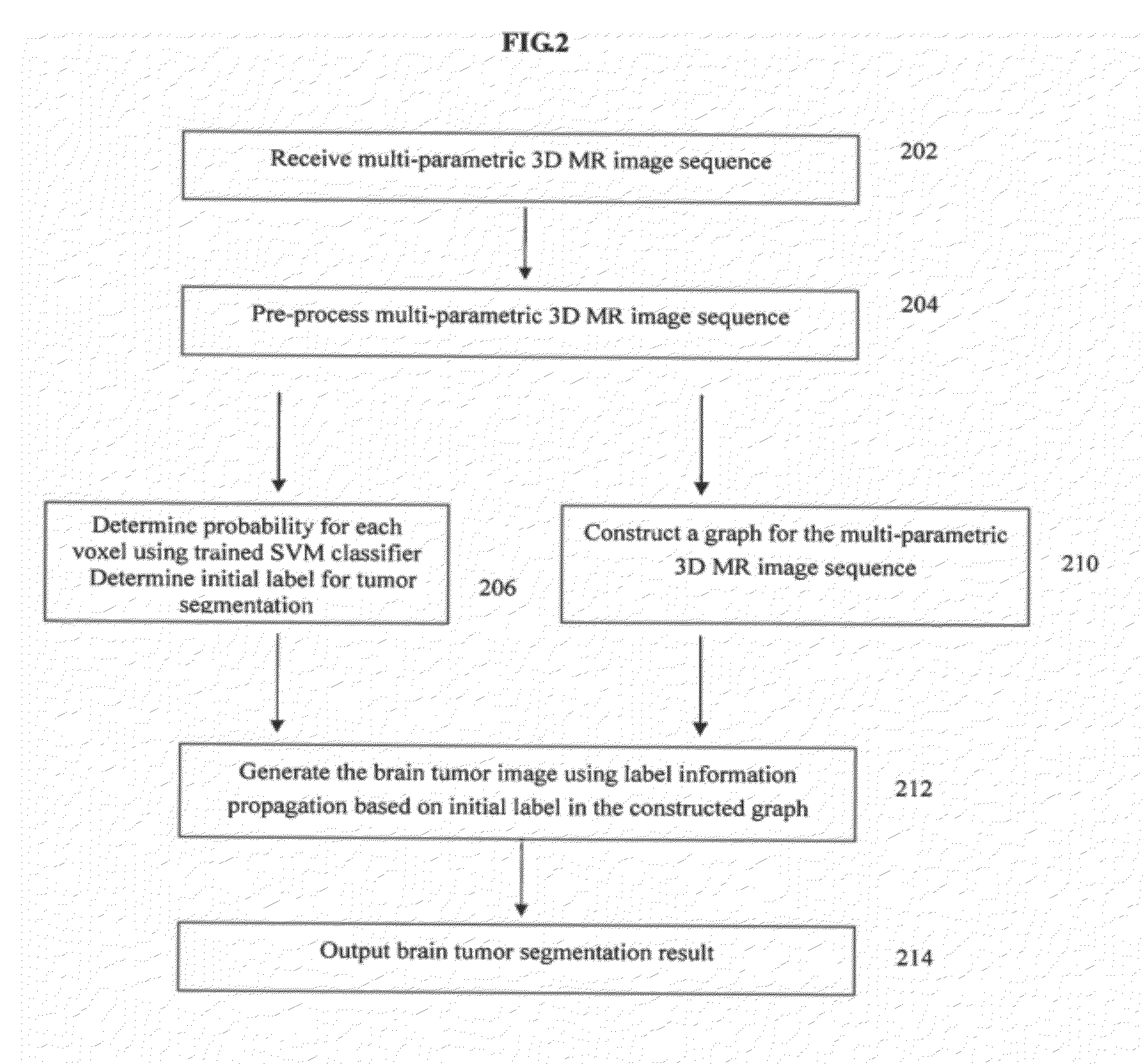
[0014]The present invention is directed to a method for automatic brain tumor segmentation in 3D MR images. Embodiments of the present invention are described herein to give a demonstration of this brain tumor segmentation method.

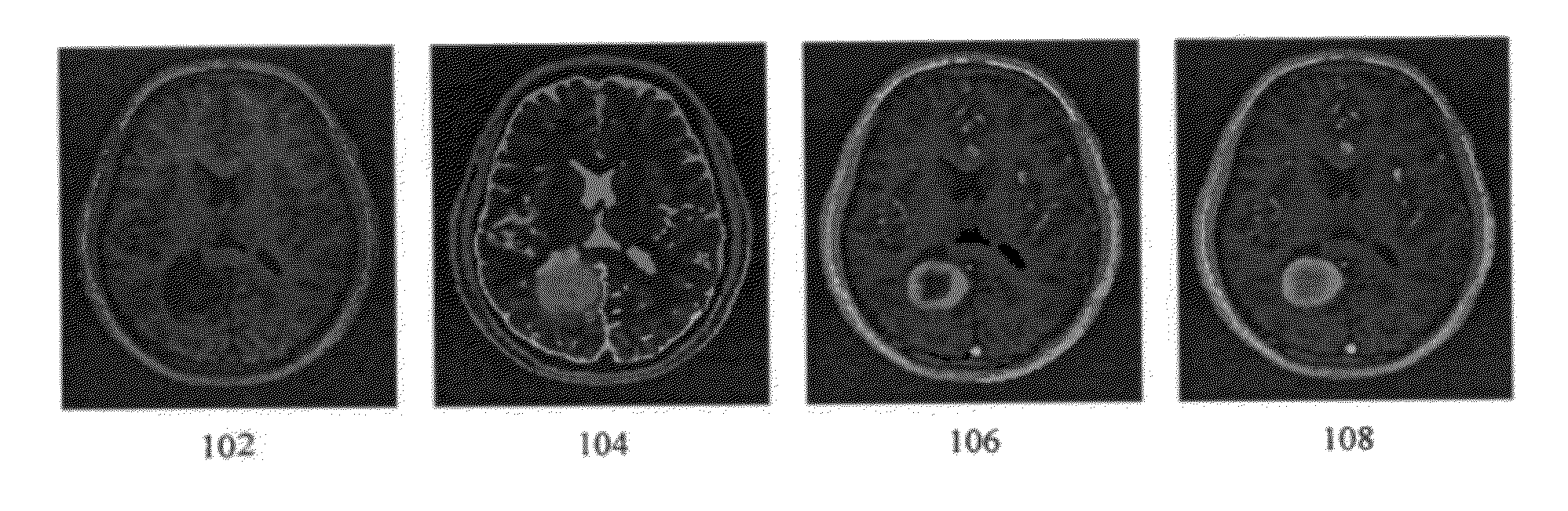
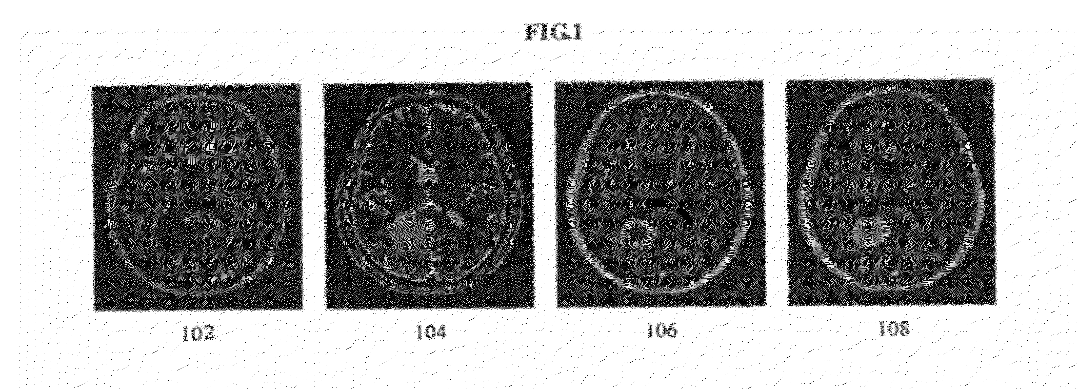
[0015]Embodiments of the present invention are directed to fully automatic brain tumor segmentation in multi-parametric 3D MR images. A multi-parametric 3D image sequence can include T1 weighted MR volume, T2 weighted MR volume, the contrast enhanced T1 weighted MR volume, just to name a few. FIG. 1 illustrates exemplary MR images showing a brain tumor. As illustrated in FIG. 1, images 102, 104, 106 show an axial slice of a multi-parametric 3D MR image sequence. In particular, image 102 shows a T1 weighted image, image 104 shows a T2 weighted image, image 106 shows a contrast enhanced T1 weighted image, and image 108 shows an annotated gro and truth of a brain tumor overlaid with the contrast enhanced T1 weighted image.

[0016]Embodiments of the present inven...


PUM


Abstract

A method for brain tumor segmentation in multi-parametric 3D MR images. The method comprises: pre-processing an input multi-parametric 3D MR image; classifying each voxel in the pre-processed multi-parametric 3D MR image, determining the probability that the voxel is part of a brain tumor, and obtaining an initial label information for the image segmentation based on the classification probability; constructing a graph based representation for the pre-processed image to be segmented; and generating the segmented brain tumor image using the initial label information and graph based representation. This method tries to exploit the local and global consistency of the image to be segmented for the tumor segmentation and can alleviate partially the performance degradation caused by the inter-subject image variability and insufficient statistical information from training.

Description

PRIORITY CLAIM[0001]The present application is a National Phase entry of PCT Application No. PCT / CN2010 / 074517, filed Jun. 25, 2010, the disclosure of which is hereby incorporated by reference herein in its entirety.TECHNICAL FIELD[0002]The present invention relates to medical image processes, and particularly to fully automatic segmentation of brain tumors in multi-parametric 3D MR images.BACKGROUND ART[0003]Detection and segmentation of brain tumors in magnetic resonance images is of great importance in medical diagnosis. Reliable and precise tumor segmentation provides helpful information for surgical planning and therapy accessing. The tumor segmentation can also be used for general modeling of a pathological brain and the construction of pathological brain atlases. However, manual segmentation of brain tumor images commonly used in clinics is time-consuming, labor-intensive and subjective to considerable variation in intra- and inter-rater performance. Accordingly, a method for...

Claims


Application Information

Patent Timeline
IPC IPC(8): G06K9/00
CPCG06T7/0081G06T7/0087G06T7/0093G06T2207/30096G06T2207/20081G06T2207/30016G06T2207/10088G06T7/11G06T7/143G06T7/162
Inventor FAN, YONGLI, HONGMING
Owner INST OF AUTOMATION CHINESE ACAD OF SCI
Why Eureka
Try Eureka