Spatio-Temporal Analysis for Automatic Contrast Injection Detection on Angiography During Trans-Catheter Aortic Valve Implantation

a technology of automatic contrast injection and angiography, which is applied in the field of spatial analysis of automatic contrast injection detection on angiography during transcatheter aortic valve implantation, can solve the problems of not normalizing and subsequent classification tasks are difficult, and achieve good volume of contrast injection

Active Publication Date: 2012-05-31
SIEMENS MEDICAL SOLUTIONS USA INC
View PDF4 Cites 11 Cited by
  • Summary
  • Abstract
  • Description
  • Claims
  • Application Information

AI Technical Summary

Problems solved by technology

This simple feature, however, is not normalized and its value can change significantly across different sequences and / or patients, making the subsequent classification task difficult.

Method used


Image

  • Spatio-Temporal Analysis for Automatic Contrast Injection Detection on Angiography During Trans-Catheter Aortic Valve Implantation
  • Spatio-Temporal Analysis for Automatic Contrast Injection Detection on Angiography During Trans-Catheter Aortic Valve Implantation
  • Spatio-Temporal Analysis for Automatic Contrast Injection Detection on Angiography During Trans-Catheter Aortic Valve Implantation

Examples


Embodiment Construction

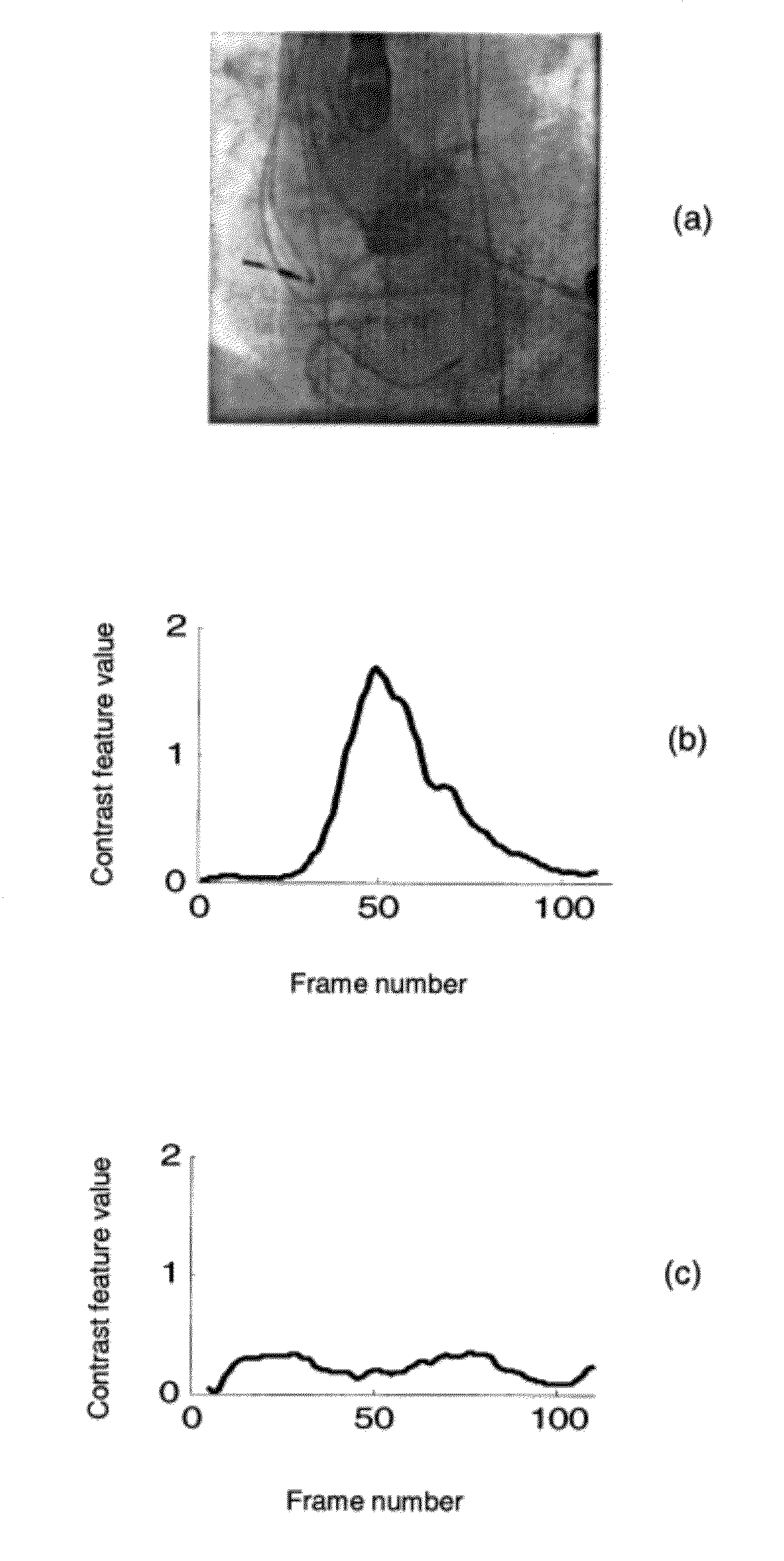
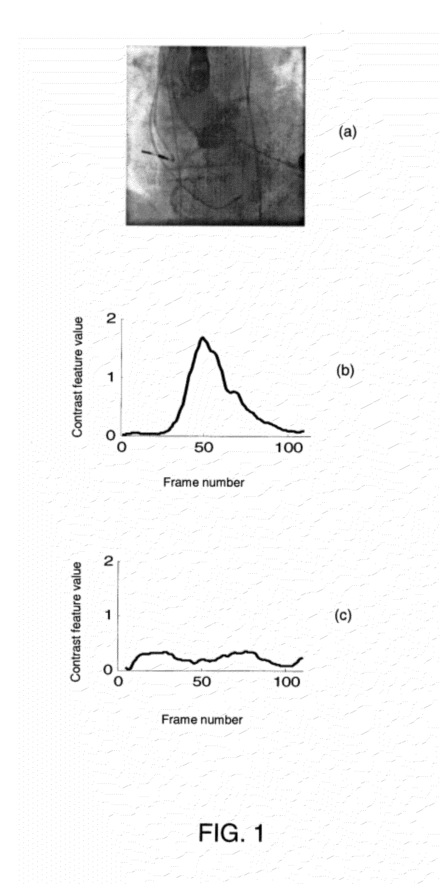
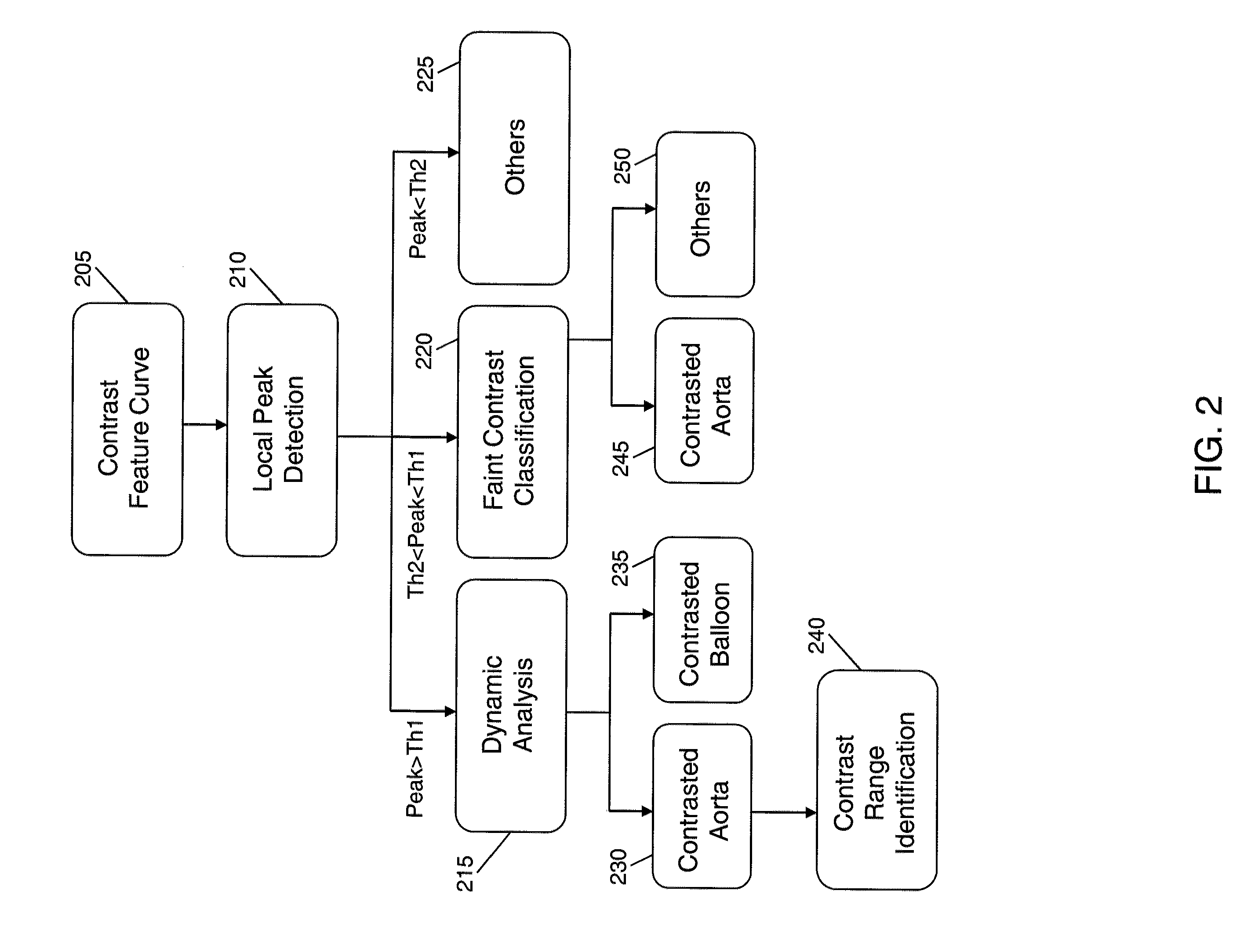
[0039]Disclosed herein, in accordance with an exemplary embodiment of the present invention, is a method for detecting contrast agent injection in fluoroscopic and / or angiographic sequences by an integrated temporal and spatial analysis. This method aims at solving difficult cases that are associated with the contrast feature curve developed in R. Liao et al., “Automatic Detection of Contrast Injection on Fluoroscopy and Angiography for Image guided Trans-Catheter Aortic Valve Implantations (TAVI),” SPIE, 2011. Briefly, in the method disclosed herein, a cascaded classifier removes contrasted balloons from a contrasted aorta by combining a spatio-temporal feature with shape features. A local classifier is trained using temporal information provided by the contrast feature curve to identify a range of contrasted frames. Further, for sequences with faint contrast, frames with a high contrast feature value may be tested with a classifier and features may be selected therefrom using a re...


PUM


Abstract

A method that includes generating a contrast feature curve for a medical image sequence including a plurality of frames, where the contrast feature curve represents contrast feature values of the frames. The method further includes detecting a peak in the contrast feature curve, and determining whether the peak corresponds to at least one of contrast injection in an aortic root, contrast injection in a balloon, and a non-contrast injected region.

Description

CROSS-REFERENCE TO RELATED APPLICATIONS[0001]This application claims priority under 35 U.S.C. §119 to U.S. provisional application No. 61 / 409,641, filed Nov. 3, 2010, the disclosure of which is incorporated by reference herein in its entirety.[0002]This application is related to commonly assigned U.S. non-provisional application entitled “AUTOMATIC DETECTION OF CONTRAST INJECTION,” attorney docket no. 2010P19699US (8706-1333), which claims priority under 35 U.S.C. §119 to U.S. provisional application No. 61 / 385,594, filed Sep. 23, 2010, each of which is incorporated by reference herein in its entirety.BACKGROUND OF THE INVENTION[0003]1. Technical Field[0004]The present invention relates to a method for automatically detecting whether there is contrast injection in the aortic root on a fluoroscopy or angiography sequence.[0005]2. Discussion of the Related Art[0006]Aortic valve disease leads to 60,000 surgical aortic valve replacements every year in Europe and even more in the United ...

Claims


Application Information

Patent Timeline
Patent Type & Authority Applications(United States)
IPC IPC(8): G06K9/00
CPCG06K2209/05G06T2207/30104G06T2207/10121G06T7/0016G06V2201/03
Inventor LIAO, RUIYAN, MICHELLE XIAOHONGYOU, WEI
Owner SIEMENS MEDICAL SOLUTIONS USA INC
Why Eureka
Try Eureka