Graphene-encapsulated nanoparticle-based biosensor for the selective detection of biomarkers

a biosensor and nanoparticle technology, applied in the field of graphene-encapsulated nanoparticle-based biosensors for selective detection of biomarkers, can solve the problem that their potential as biosensors has not been fully explored

Active Publication Date: 2012-08-30
RUTGERS THE STATE UNIV
View PDF1 Cites 95 Cited by
  • Summary
  • Abstract
  • Description
  • Claims
  • Application Information

AI Technical Summary

Benefits of technology

[0006]In one embodiment, the present invention provides a field effect transistor (FET) in which a source electrode and a drain electrode are formed apart from each other on a semi-conductor substrate and a gate electrode incorporating a uniform layer of reduced graphene oxide encapsulated semiconductor nanoparticles (rGO-NPs) is disposed between and contacts both the source and drain electrodes. In another embodiment, the gate electrode is functionalized with a linker molecule having the structure:

Problems solved by technology

Nevertheless, there have been only a few reports on developing graphene FET-based biosensors, and their potential as biosensors has not been fully explored.

Method used


Image

  • Graphene-encapsulated nanoparticle-based biosensor for the selective detection of biomarkers
  • Graphene-encapsulated nanoparticle-based biosensor for the selective detection of biomarkers
  • Graphene-encapsulated nanoparticle-based biosensor for the selective detection of biomarkers

Examples


example 1

Materials and Methods

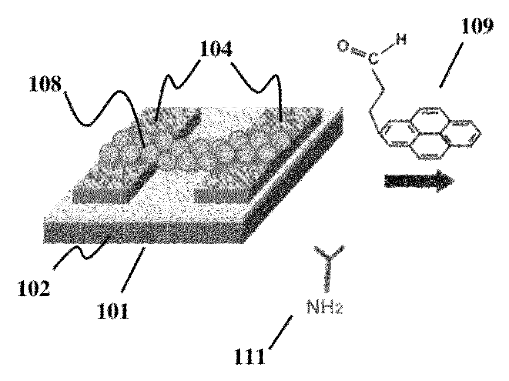
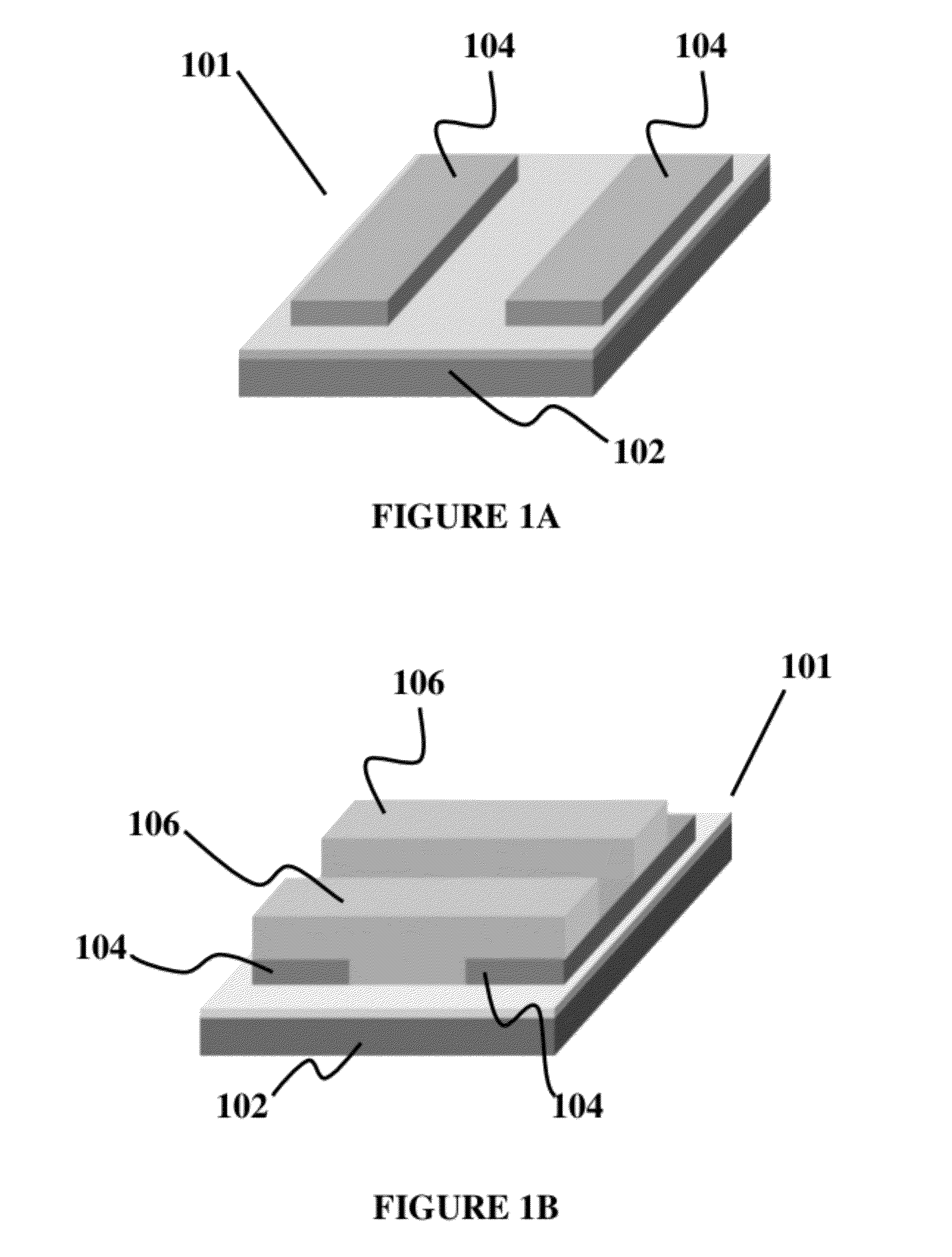
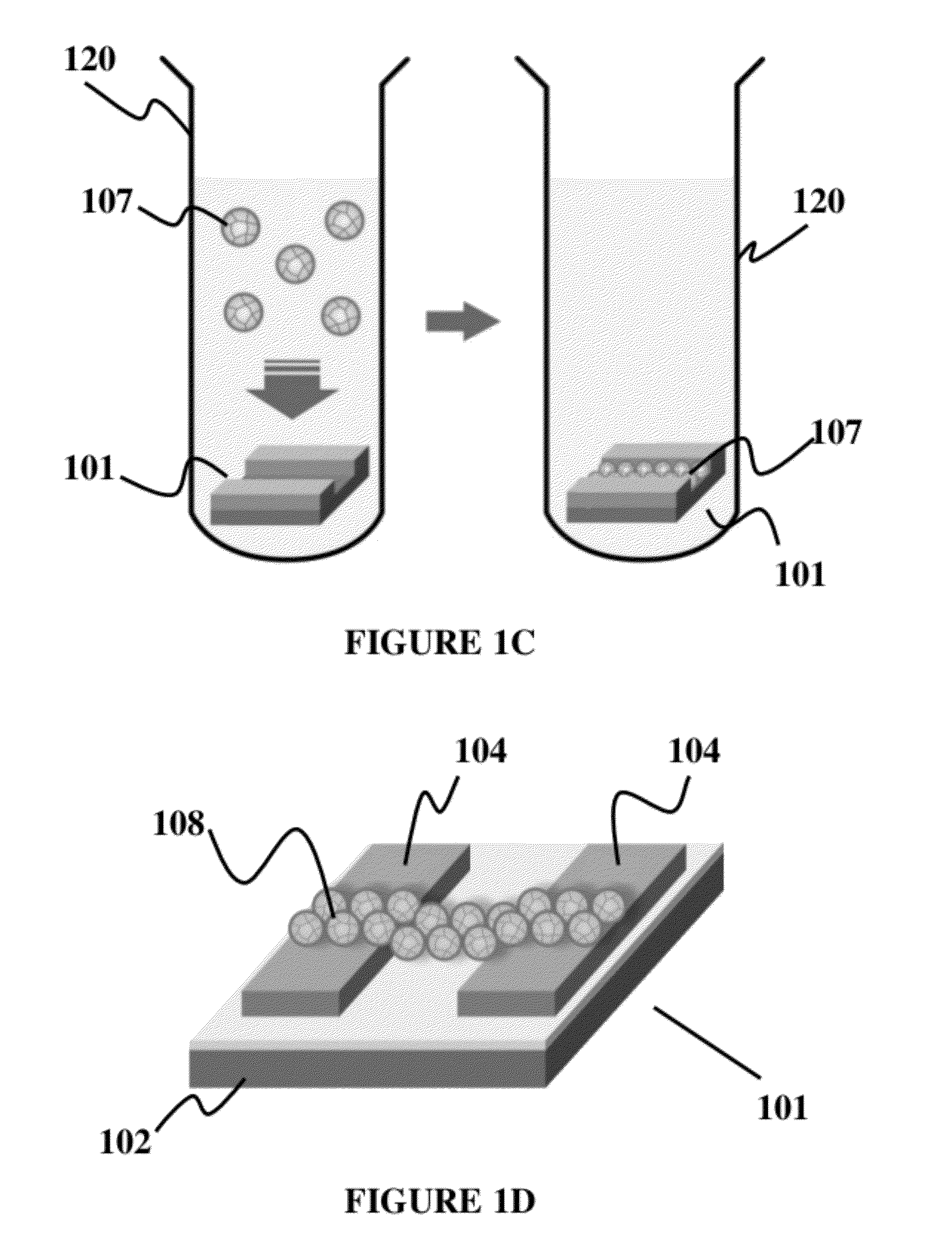
[0038]Preparation of reduced graphene oxide and SiO2 NPs: GO was obtained from SP-1 graphite utilizing the modified Hummer method. See, e.g., Eda et al., Nat. Nanotechnol. 2008, 3, 270. For the GO assembly on the surface of NPs, GO suspension was injected into the nanoparticle solution for 10 minutes, and GO-NPs were separated from GO solution using a centrifuge. SiO2 NP (100 nm) solution was purchased from Corpuscular Inc. For GO-NP assembly, the photo resist-patterned substrate was placed in the NP solution, and GO-NPs were assembled on the substrate by applying centrifugal force for three minutes. After the deposition of GO-NPs on the substrate, the GO on the NP surface, having the low conductance, was reduced to graphene by exposure to hydrazine vapor overnight.

[0039]Surface molecular pattering: 3-Aminopropyltriethoxysilane (APTES) and cysteamine molecules were used in forming SAMs. For the patterning of APTES SAM on SiO2, the photoresist (AZ5214) was first ...

example 2

[0045]The following example illustrates the fabrication and application of a reduced graphene oxide (rGO) encapsulated nanoparticle (NP)-based FET biosensor for selective and sensitive detection of key biomarker proteins for breast cancer. This example demonstrates the use of Human Epidermal growth factor Receptor 2 (HER2) and Epidermal Growth Factor Receptor (EGFR), which are known to be over-expressed in breast cancers, as examples to demonstrate the high sensitivity and selectivity of the graphene-encapsulated NP biosensor. This biosensor could be used to detect any biomarkers with relative ease. In typical experiments for fabricating the graphene encapsulated NPs-based biosensors, individual silicon oxide NPs (100 nm), functionalized with 3-aminopropyltriethoxysilane (APTES), were first coated with thin layers of graphene oxide (GO), which prevent aggregation and maintain high electrical conductivity (FIG. 1). This was mainly achieved via the electrostatic interaction between th...


PUM

PropertyMeasurementUnit
semiconductoraaaaaaaaaa
ring structureaaaaaaaaaa
conductiveaaaaaaaaaa

Abstract

A field effect transistor (FET) with a source electrode and a drain electrode distanced apart from each other on a semi-conductor substrate, and a gate electrode consisting of a uniform layer of reduced graphene oxide encapsulated semiconductor nanoparticles (rGO-NPs), wherein the gate electrode is disposed between and contacts both the source and drain electrodes. Methods of making and assay methods using the FETs are also disclosed, including methods in which the rGO-NPs are functionalized with binding partners for biomarkers.

Description

CROSS-REFERENCE TO RELATED APPLICATION[0001]This application claims the benefit of U.S. provisional application Ser. No. 61 / 443,950 filed Feb. 17, 2011, the disclosure of which is incorporated herein by reference in its entirety.STATEMENT REGARDING FEDERALLY FUNDED RESEARCH[0002]The research leading to the present invention was supported in part by NIH New Innovator Award, 2009, Grant No. NIH-1DP20D00646201. Accordingly, the U.S. Government has certain rights in this invention.BACKGROUND OF THE INVENTION[0003]Nanomaterials, such as silicon nanowires (SiNWs), carbon nanotubes (CNTs), and graphene, have gained much attention for use in electrical biosensors due to their nanoscopic and electrical properties. See, for example, Cui et al., Science 2001, 293, 1289; Patolsky et al. Protoc. 2006, 1, 1711; Bradley et al. Nano Lett. 2004, 4, 253; Chen et al. J. Am. Chem. Soc. 2004, 126, 1563; Star et al., Nano Lett. 2004, 4, 1587; Tang et al., Nano Lett. 2006, 6, 1632; Lu et al., ACS Nano 200...

Claims


Application Information

Patent Timeline
Patent Type & Authority Applications(United States)
IPC IPC(8): G01N27/00G01N27/414H01L21/335B82Y40/00B82Y99/00
CPCH01L29/4908H01L29/7869G01N27/4146B82Y30/00G01N27/4145H01L29/78696
Inventor LEE, KI-BUMMYUNG, SUNGSOLANKI, ANIRUDDH
Owner RUTGERS THE STATE UNIV
Why Eureka
Try Eureka