

Retinal neuroprotection by ion channel blockers regulated by the sur subunit
a technology of ion channel blockers and sur subunits, applied in the direction of biocide, amide active ingredients, drug compositions, etc., can solve the problems of insufficient efficacy at clinically feasible doses, cell damage and death, retinal cell degeneration leading to visual impairment,
- Summary
- Abstract
- Description
- Claims
- Application Information
AI Technical Summary
Benefits of technology
Problems solved by technology
Method used
Image

Examples
example 1
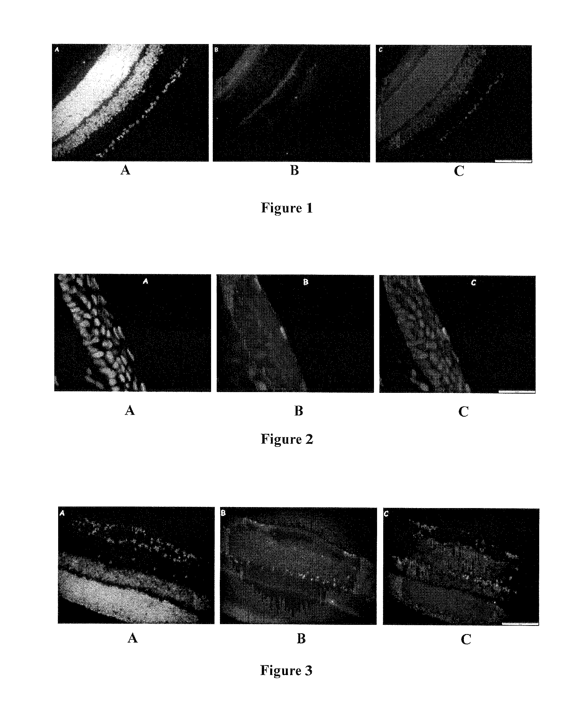
Effect of Glibenclamide on NMDA-Induced Retinal Degeneration
[0090]Experimental Protocol
[0091]Dimethylsulfoxide (DMSO) was needed to solubilize the glibenclamide. To reduce the number of intravitreal injections, NMDA and glibenclamide (5 g, MPBIO® Europe, Illkirch, France) previously dissolved in DMSO were co-injected in a same solution after checking that the mixture did not precipitate at the concentrations used. DMSO was used at the lowest concentration required to dissolve glibenclamide (260 mM).
[0092]Three glibenclamide solutions containing 1, 10 or 100 ng glibenclamide per 3 μL of solution were prepared. For this, a stock solution containing 9 mg of glibenclamide powder dissolved in 900 μL DMSO (10 μg / μL glibenclamide concentration) was diluted in 1× PBS to obtain the desired concentrations.
[0093]As per the experimental protocol described below, the test groups received a co-injection of glibenclamide and NMDA prepared by mixing 3 μL of glibenclamide solution (comprising 1, 10 ...
example 2
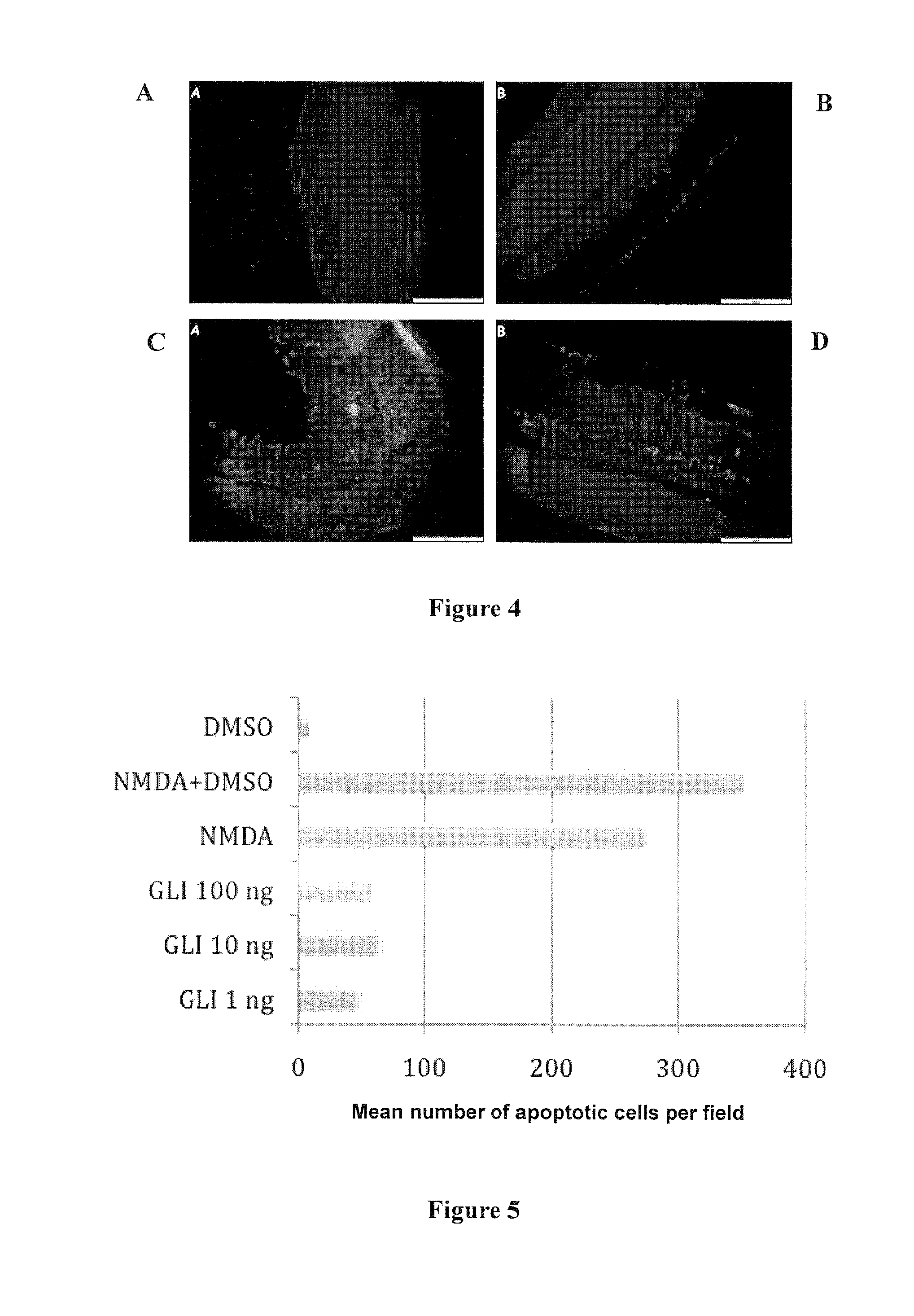
Effect of Different Routes of Administration of Glibenclamide on Retinal Degeneration
[0117]Experimental Protocol
[0118]Eight groups of 2 rats were used. Both eyes of each animal received identical treatment. Three routes of administration of glibenclamide were tested: intravitreal route (1 ng or 0.01 ng injected in each eye), oral route (10 mg / kg body weight) and subconjunctival route (1 ng glibenclamide injected in each eye).
[0119]The glibenclamide stock solution for the intravitreal and subconjunctival injections was obtained by diluting 4.5 mg glibenclamide in 300 μL of pure DMSO. This solution was then frozen. The glibenclamide solutions at different concentrations used for the intravitreal and subconjunctival injections were obtained by dilution in BSS® buffer (Alcon) immediately before use.
[0120]The glibenclamide solutions for oral administration (10 mg / kg) were obtained by dissolving 8 mg glibenclamide in 300 μM DMSO and 1030 μL of water to give a 6 mg / ml glibenclamide solutio...
example 3
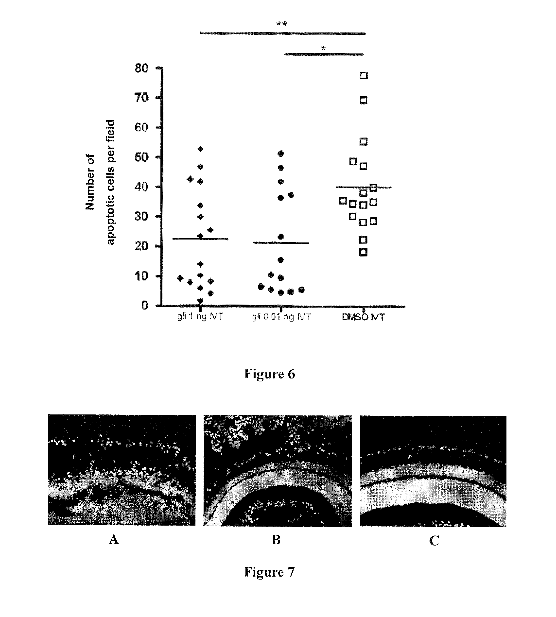
Preventive Effect of Glibenclamide on NMDA-Induced Retinal Degeneration
[0144]Experimental Protocol
[0145]Three groups of 2 rats were used. Both eyes of each animal received identical treatment.
[0146]The preventive effect of glibenclamide was evaluated by injecting into the vitreous body of each eye 100 ng or 100 μg glibenclamide diluted in BSS® buffer, 5 days before intravitreal injection of NMDA. Glibenclamide was diluted in BSS buffer and not in DMSO so as to obtain a prolonged-effect suspension, resulting from the low solubility of glibenclamide.
[0147]Administration of 100 μg glibenclamide allowed a test of the potential toxicity of a high dose.
[0148]The glibenclamide stock solution for intravitreal injections was obtained by diluting 4.5 mg glibenclamide in 300 μL of pure DMSO. This solution was then frozen. The glibenclamide solutions at different concentrations used for the intravitreal injections were obtained by dilution in BSS® buffer (Alcon) immediately before use.
[0149]NMD...
PUM
 Abstract
Description
Claims
Application Information