Obtaining data for automatic glaucoma screening, and screening and diagnostic techniques and systems using the data

a technology of automatic glaucoma and data acquisition, applied in image data processing, medical data mining, othalmoscopes, etc., can solve the problems of poor sensitivity of current tests, low cost effectiveness of routine glaucoma screening in the whole population, and loss of peripheral vision

Active Publication Date: 2012-09-13
SINGAPORE HEALTH SERVICES PTE +1
View PDF1 Cites 24 Cited by
  • Summary
  • Abstract
  • Description
  • Claims
  • Application Information

AI Technical Summary

Benefits of technology

[0019]The present invention aims, in some aspects, to provide new and useful methods and systems for using images to extract data useful for detecting glaucoma from fundus images (e.g. a single fundus image per eye).

Problems solved by technology

Progression of the disease results in loss of peripheral vision, so patients may complain of “tunnel vision” (only being able to see the centre).
They are useful in finding the molecular pathways of disease, but usually not useful in finding genes that predict risks of disease.
However, routine screening for glaucoma in the whole population is not cost effective and limited by poor sensitivity of current tests.
So far no technique employing genetic information has been employed in screening patients or assessing risk factors, and indeed it may not be possible to extract sufficient information from genetic data alone to make a screening operation possible.
Furthermore, currently, there is no systematic way to detect and manage early glaucoma.
Treatment at this stage is limited to surgery, is expensive, requires skilled personnel, and does not restore vision.
IOP measurement is neither specific nor sensitive enough to be an effective screening tool.
Furthermore, optic disc assessment by an ophthalmologist is subjective and the availability of HRT is very limited because of the cost involved as well as a shortage of trained operators.
In some cases, where the gradient values are gradual, difficulties in the correct cup identification can occur.
This technique has the weakness that the color information may not be accurate.
Although this method is non-reliant on color or pallor, some of the challenges include correct identification of kinks, as well as the absence of kinks in some retinal images.
However, no results were presented on the accuracy of results compared to clinical ground truth.
While some of the results presented are promising, the key challenge lies in the use of stereoscopic photography as compared to monocular (“non-stereo”) photography.
Stereoscopic photography demands specific hardware and requires specialized training, both of which may be unsuitable for the needs for mass screening.

Method used


Image

  • Obtaining data for automatic glaucoma screening, and screening and diagnostic techniques and systems using the data
  • Obtaining data for automatic glaucoma screening, and screening and diagnostic techniques and systems using the data
  • Obtaining data for automatic glaucoma screening, and screening and diagnostic techniques and systems using the data

Examples


Embodiment Construction

[0055]Section 1 of the following text describes an embodiment of the invention (“AGLAIA”). In section 2 it describes four optional features of the embodiment, the first three of which have application to other systems for studying fundus images.

1. The Embodiment

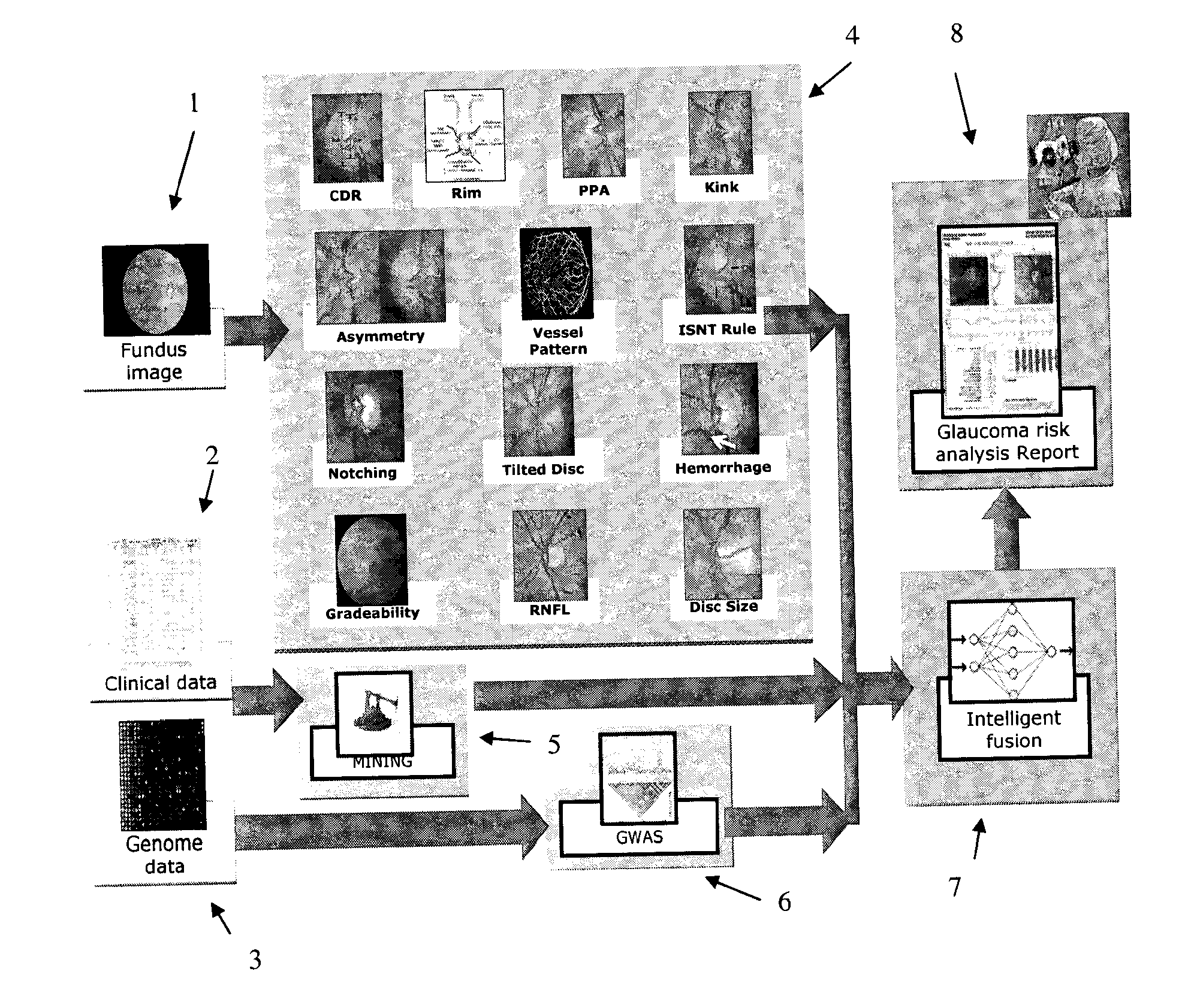
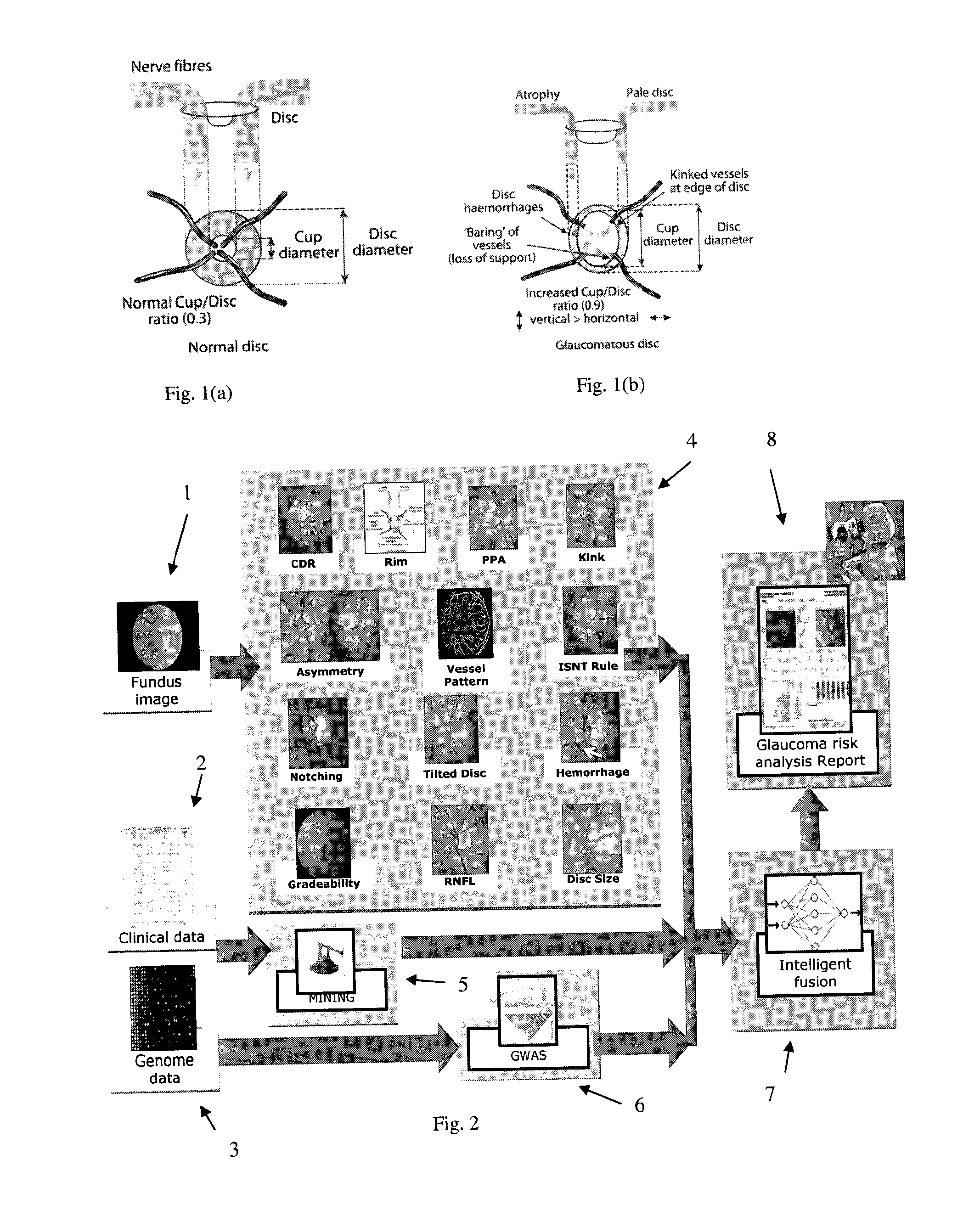
[0056]The structure of an embodiment of the invention is illustrated schematically in FIG. 2. The embodiment is referred to as “AGLAIA” (Architecture for automatic glaucoma diagnosis and its genetic association study through medical image informatics). There are three inputs to the system. A first input is at least one (typically, exactly one) non-stereo fundus image 1 of each eye of a subject. A second input is clinical data 2 describing the medical history of the subject. The third input is genome data 3 (genetic data), for example data obtained from a DNA microarray. The DNA microarray typically measures the respective levels of expression in the subject of each of a large number of genes, or detects single nucleotide poly...


PUM


Abstract

A non-stereo fundus image is used to obtain a plurality of glaucoma indicators. Additionally, genome data for the subject is used to obtain genetic marker data relating to one or more genes and/or SNPs associated with glaucoma. The glaucoma indicators indicators and genetic marker data are input into an adaptive model operative to generate an output indicative of a risk of glaucoma in the subject. In combination, the genetic indicators and genome data are more informative about the risk of glaucoma than either of the two in isolation. The adaptive model may be a two-stage model, having a first stage in which individual genetic indicators are combined with respective portions of the genome data by first adaptive model modules to form respective first outputs, and a second stage in which the first outputs are combined by a second adaptive mode. Texture analysis is performed on the fundus images to classify them based on their quality, and only images which are determined to meet a quality criterion are subjected to an analysis to determine if they exhibit glaucoma indicators. Also, the images are put into a standard format. The system may include estimating the position of the optic cup by combining results from multiple optic cup segmentation techniques. The system may include estimating the position of the optic disc by applying edge detection to the funds image, excluding edge points that are unlikely to be optic disc boundary points, and estimating the position of an optic disc by fitting an ellipse to the remaining edge points.

Description

FIELD OF THE INVENTION[0001]The present invention relates to methods and systems for obtaining data useful in detecting glaucoma in a human subject. It further relates to methods and systems for generating an output indicative of a high likelihood of glaucoma. The output may be used in screening subjects for glaucoma, or even performing a glaucoma diagnosis for a subject.BACKGROUND OF THE INVENTION[0002]Glaucoma is a chronic eye condition in which the nerve that connects the eye to the brain (optic nerve) is progressively damaged. Patients with early glaucoma do not have visual symptoms. Progression of the disease results in loss of peripheral vision, so patients may complain of “tunnel vision” (only being able to see the centre). Advanced glaucoma is associated with total blindness. Worldwide, it is the second leading cause of blindness, affecting 60 million people by 2010, and responsible for approximately 5.2 million cases of blindness (15% of the total burden of world blindness)...

Claims


Application Information

Patent Timeline
Patent Type & Authority Applications(United States)
IPC IPC(8): G06K9/46
CPCA61B3/12A61B5/7267A61B5/7264A61B5/02007A61B3/0025G06T7/0012G06T2207/30041G16H50/20G16H50/70A61B5/7275G06T7/40
Inventor LIU, JIANGZHANG, ZHUOWONG, WING KEE DAMONTAN, NGAN MENGYIN, FENGSHOULEE, BENG HAILI, HUIQILIM, JOO HWEECHEUNG, CAROLAUNG, TINWONG, TIEN YINLIANG, ZIYANGCHENG, JUNMANI, BASKARAN
Owner SINGAPORE HEALTH SERVICES PTE
Why Eureka
Try Eureka