

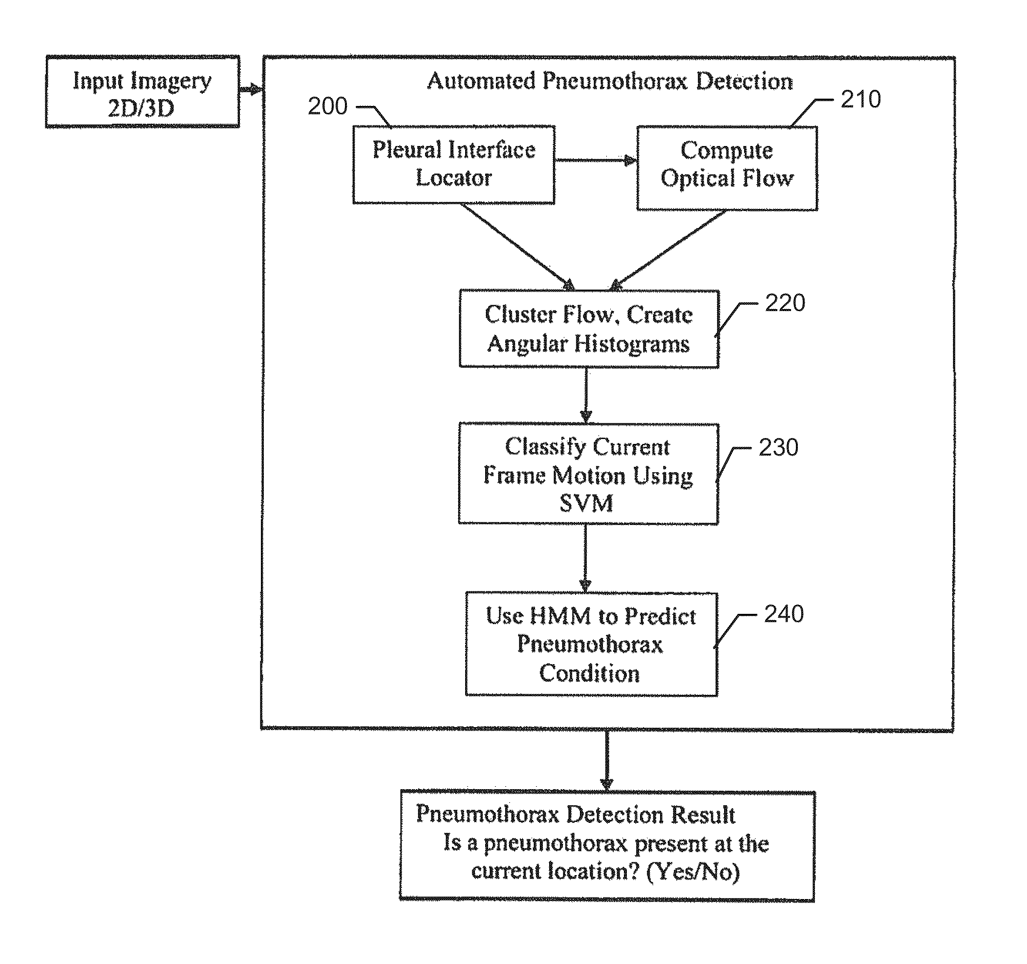
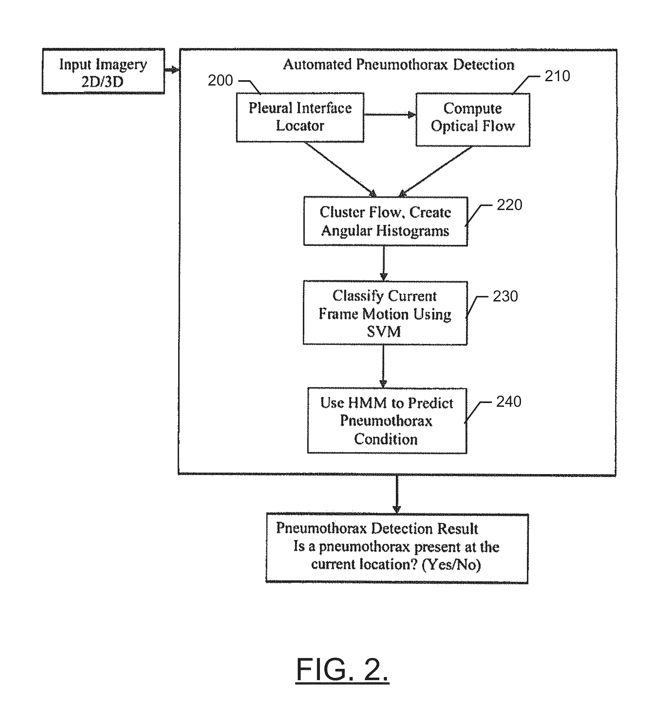
Automated Pneumothorax Detection
a pneumothorax and automatic technology, applied in the field of automatic detection of lung related ailments or conditions, can solve problems such as hemodynamic instability and/or death
- Summary
- Abstract
- Description
- Claims
- Application Information
AI Technical Summary
Benefits of technology
Problems solved by technology
Method used
Image

Examples
Embodiment Construction
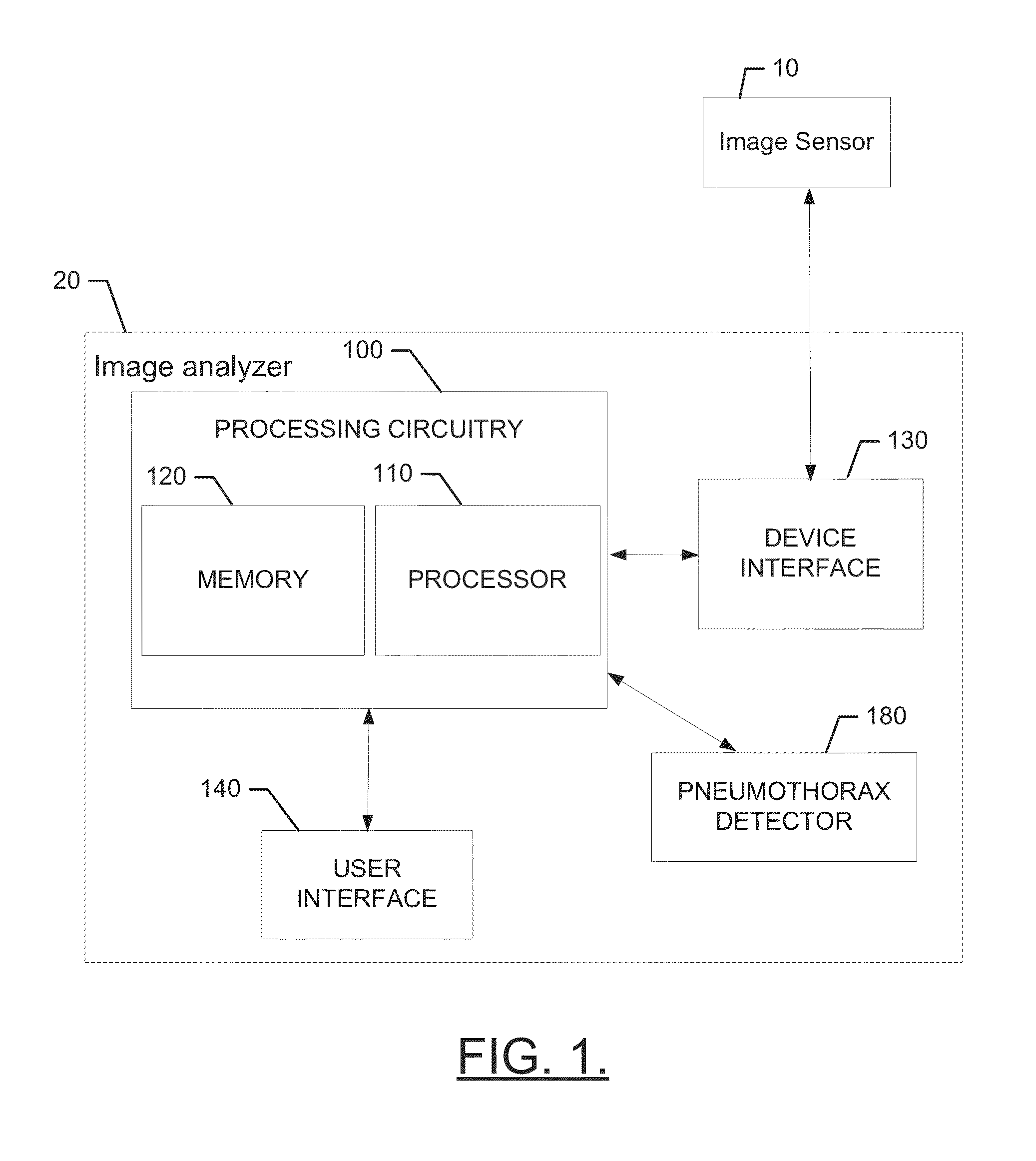
[0017]Some example embodiments now will be described more fully hereinafter with reference to the accompanying drawings, in which some, but not all example embodiments are shown. Indeed, the examples described and pictured herein should not be construed as being limiting as to the scope, applicability or configuration of the present disclosure. Rather, these example embodiments are provided so that this disclosure will satisfy applicable legal requirements. Like reference numerals refer to like elements throughout.
[0018]As indicated above, some example embodiments may enable the provision of a mechanism by which to diagnose pneumothorax automatically on the basis of machine executed analysis of image data of lungs. In some cases, the image data may be one-dimensional, two-dimensional or three-dimensional video imagery that may be obtained by time varying imaging modalities such as ultrasound (including Doppler ultrasound), CT or cine-MRI. The image data may be analyzed to identify o...
PUM
 Abstract
Description
Claims
Application Information