System and Method for Controlling Radiation Dose for Radiological Applications

a radiological application and radiation dose technology, applied in the field of medical imaging, can solve the problems of affecting patient safety, affecting the safety of patients, and affecting the accuracy of diagnosis, so as to reduce the local nose level

Inactive Publication Date: 2013-08-08
MAYO FOUND FOR MEDICAL EDUCATION & RES
View PDF3 Cites 25 Cited by
  • Summary
  • Abstract
  • Description
  • Claims
  • Application Information

AI Technical Summary

Benefits of technology

[0011]In accordance with one aspect of the invention, a method for reconstructing an image acquired by delivering an irradiating dose of radiation to a subject is disclosed that includes obtaining a set of medical imaging data acquired using a dose of irradiating radiation delivered to a subject, and selecting at least one of a plurality of mechanisms for reducing the dose of irradiating radiation that could be delivered to the subject to acquire an additional set of medical imaging data. The method also includes inserting noise into the medical imaging data to simulate the at least one of the plurality of mechanisms for reducing the dose of irradiating radiation that could be applied to acquire the additional set of medical imaging data to thereby generate a simulated set of medical imaging data at a reduced dose of irradiating radiation. The method further includes reconstructing a simulated reduced dose image from the simulated set of medical imaging data.

Problems solved by technology

The drastically increased use of CT in modern clinical settings has generated serious public health concerns regarding the cancer risks associated with the radiation exposure from CT.
However, lowering radiation dose alone generally produces a noisier image and may degrade diagnostic performance.
Despite the tremendous effort in the CT community to minimize radiation dose, scanning protocols and radiation doses still vary widely among different CT practices, which poses substantial risks to patient safety.
The substantial variation in protocols and radiation dose is largely attributable to the lack of an efficient and widely available approach to optimizing CT protocols.
This approach requires exploratory low-dose scans on a number of patients, which is tedious and can potentially result in diagnostically compromised image.
The technical details of the tools are not publicly available and the accuracy is usually out of the users' control, which makes their applications rather limited.
However, the effectiveness of denoising mechanisms are widely variable and, more troubling, can make substantial and varied changes to the images.
That is, ineffective or overly aggressive denoising mechanisms can reduce the clinical usefulness and credibility of the images acquired.
In doing so, the difficulty of controlling radiation dose to the patient can be compounded by the need to perform subsequent data acquisitions to replace overly-noisy images.

Method used


Image

  • System and Method for Controlling Radiation Dose for Radiological Applications
  • System and Method for Controlling Radiation Dose for Radiological Applications
  • System and Method for Controlling Radiation Dose for Radiological Applications

Examples


Embodiment Construction

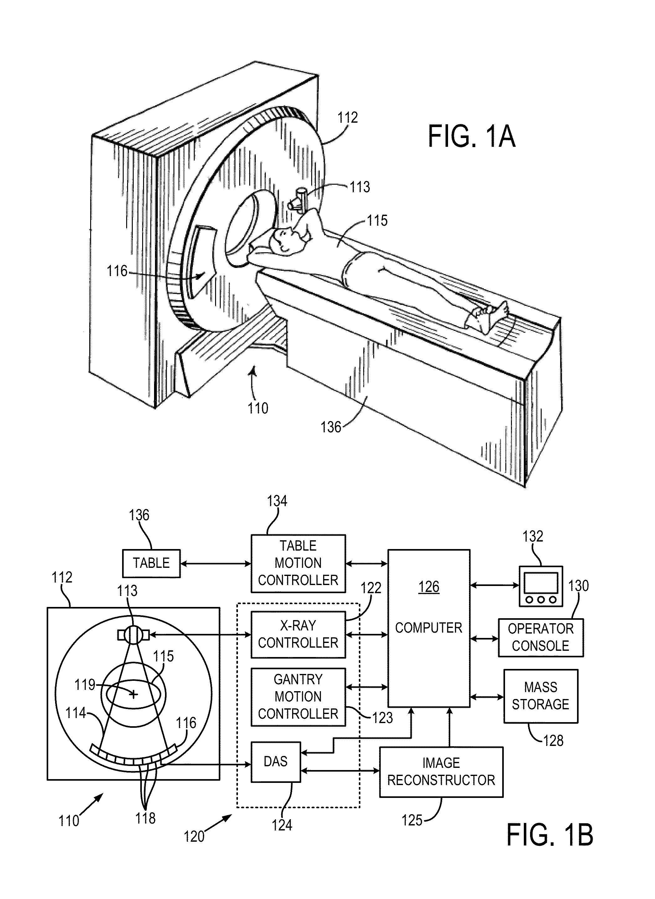
[0027]With initial reference to FIGS. 1A and 1B, a computed tomography (CT) imaging system 110 includes a gantry 112 representative of at least a “third generation” CT scanner. In the illustrated example, the gantry 112 has a pair of x-ray sources 113 that each project a fan beam or cone beam of x-rays 114 toward a detector array 116 on the opposite side of the gantry 112. The detector array 116 is formed by a number of detector elements 118 that together sense the projected x-rays that pass through a medical patient 115. During a scan to acquire x-ray projection data, the gantry 112 and the components mounted thereon rotate about a center of rotation 119 located within the patient 115 to acquire attenuation data.

[0028]The rotation of the gantry 112 and the operation of the x-ray source 113 are governed by a control mechanism 120 of the CT system 110. The control mechanism 120 includes an x-ray controller 122 that provides power and timing signals to the x-ray sources 113 and a gant...


PUM


Abstract

A system and method for reconstructing an image acquired by delivering an irradiating dose of radiation to a subject includes acquiring imaging data using a dose of irradiating radiation and selecting at least one of a plurality of mechanisms for reducing the dose that could be delivered to the subject to acquire additional imaging data. Noise is inserted into the imaging data to simulate the at least one of the plurality of mechanisms for reducing the dose that could be applied to acquire the additional imaging data to thereby generate simulated imaging data at a reduced dose of irradiating radiation. A simulated reduced dose image is reconstructed from the simulated imaging data. A method is provided for utilizing a non-local means filter adapted using a map of local noise to produce denoised medical imaging data reflecting reduced local nose levels from those in originally-acquired medical imaging data.

Description

CROSS-REFERENCE TO RELATED APPLICATIONS[0001]The present application is based on, claims priority to, and incorporates herein by reference in its entirety U.S. Provisional Patent Application No. 61 / 595,999, filed Feb. 7, 2012, and entitled “SYSTEM AND METHOD FOR CONTROLLING RADIATION DOSE FOR RADIOLOGICAL APPLICATIONS.”BACKGROUND OF THE INVENTION[0002]The present invention relates to medical imaging and, more particularly, to systems and methods for controlling radiation doses delivered when performing imaging processes using ionizing radiation.[0003]In a computed tomography system, an x-ray source projects a beam which is collimated to lie within an X-Y plane of a Cartesian coordinate system, termed the “imaging plane.” The x-ray beam passes through the object being imaged, such as a medical patient or other non-medical patient or object, such as in industrial CT imaging, and impinges upon an array of radiation detectors. The intensity of the transmitted radiation is dependent upon...

Claims


Application Information

Patent Timeline
Patent Type & Authority Applications(United States)
IPC IPC(8): G06T11/00A61B6/03
CPCA61B6/5258G06T11/003A61B6/583A61B6/5205A61B6/542A61B6/032
Inventor YU, LIFENGMANDUCA, ARMANDOLI, ZHOUBOFLETCHER, JOEL G.MCCOLLOUGH, CYNTHIA H.
Owner MAYO FOUND FOR MEDICAL EDUCATION & RES
Why Eureka
Try Eureka