Assays for Anti-drug antibodies in the presence of abundant endogenous protein counterpart of the drug
a technology of endogenous protein and anti-drug antibodies, which is applied in the field of kits and methods for detecting anti-drug antibodies in the presence of an abundant endogenous protein counterpart of the drug, can solve the problems of serious side effects, reduced therapeutic protein effectiveness, and changes in drug pharmacokinetics or pharmacodynamics that alter the efficacy of the drug, so as to improve the sensitivity of the assay and improve the sensitivity of the immunogenicity assay
- Summary
- Abstract
- Description
- Claims
- Application Information
AI Technical Summary
Benefits of technology
Problems solved by technology
Method used
Image

Examples
example 1
Sample Preparation
[0055]This Example illustrates the preparation of samples for use in a bridging ELISA, as described in Example 2.
[0056]Removal of HSA and enrichment for immunoglobulin is achieved using a 0.2 mL NAb protein A / G spin column (Thermo Scientific), and buffers provided therewith in the NAb spin kit available from Thermo Scientific. Every reagent and kit component, including columns, and human serum are brought to room temperature. Columns are placed in 2 mL labeled collection tubes and centrifuged at 5,000 g for 1 minute. The flow-through is discarded. Each column is then washed by adding 400 μL of Pierce IgG Binding Buffer (0.2 M, pH 7.2; prepared by dissolving the contents of one pouch provided in the NAb spin kit in a final volume of 500 mL of deionized water), mixing briefly, centrifuging the column at 5,000 g for 1 minute, and discarding the flow-through. The washing step is repeated.
[0057]The bottom of each spin column is capped with the kit-provided rubber cap, a...
example 2
Bridging ELISA
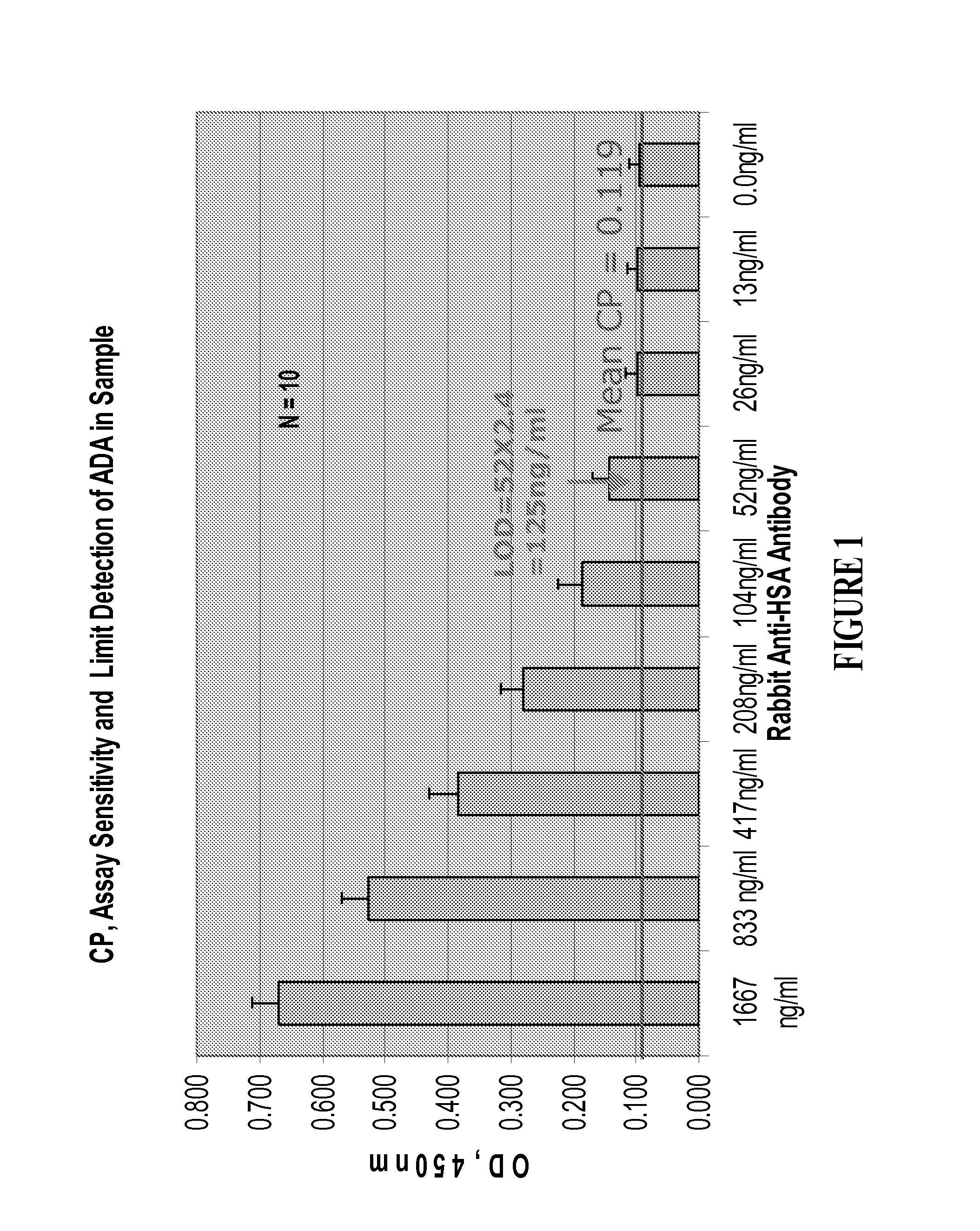
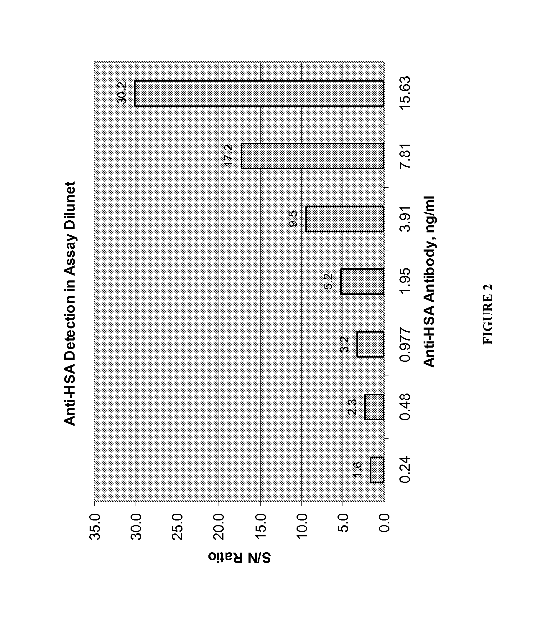
[0059]This Example illustrates the detection of anti-HSA antibodies using a bridging ELISA. Each well of an Immobilizer amine 96-well plate (VWR / NUNC) is coated with 100 μL of a 20 μg / mL solution of HSA in PBS. The plate is incubated at 4° C. overnight. The coating solution is removed without washing, and the plate is blocked by adding 250 μL / well KPL milk block / diluent (prepared by diluting Milk Diluent / Blocking Solution Concentrate (KPL) 1 / 20 with reagent quality water (i.e., 1 mL concentration diluted with 19 mL water)). The plate is incubated for 2 hours at room temperature, at which point the blocking solution is removed without washing and the plate is blotted on a paper towel (TechniCloth, available from ITW Texwipe, Kernersville, N.C.).
[0060]A solution of biotin-conjugated human albumin (Biotin-HSA; Jackson ImmunoResearch, West grove, PA) is prepared at a concentration of 8 μg / mL in KPL milk block / diluent, and 50 μL of this Biotin-HSA solution is added to each ...
example 3
Clinical Application of Sample Preparation and Bridging ELISA Methods
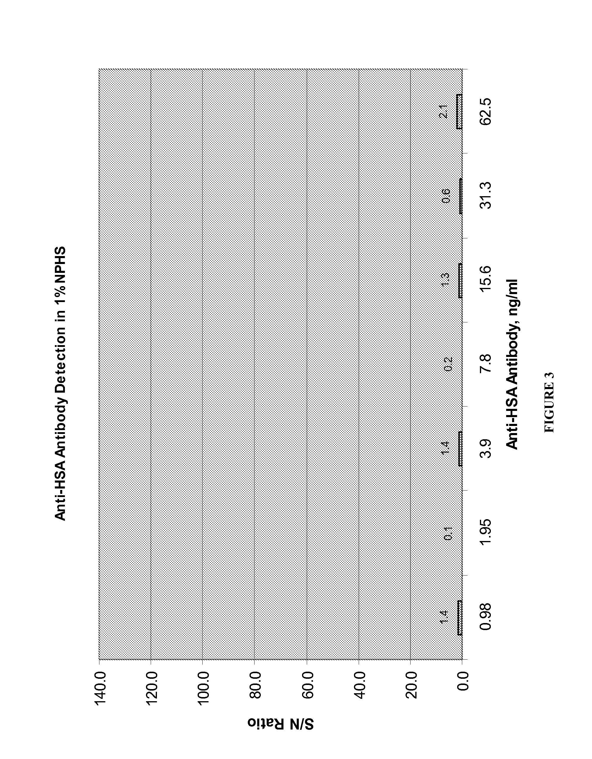
[0064]This Example illustrates the use of the procedures described in Examples 1 and 2 to detect the presence or absence of anti-HSA antibodies in serum obtained from test subjects treated with a fusion protein that comprises a HSA sequence.
[0065]Nine human subjects were treated with B2B3-1, a bispecific antibody that comprises an HSA moiety and is described in detail in US Patent Application No. 2011 / 0059076, which is hereby incorporated by reference.). Serum was obtained from each subject, and samples were prepared and tested as described in Examples 1 and 2. All samples tested negative for the presence of anti-HSA antibodies. These results provide critical safety information for use in assessing candidate drugs undergoing clinical testing.
PUM
 Abstract
Description
Claims
Application Information
 - R&D
- Intellectual Property
- Life Sciences
- Materials
- Tech Scout
- Unparalleled Data Quality
- Higher Quality Content
- 60% Fewer Hallucinations
Browse by: Latest US Patents, China's latest patents, Technical Efficacy Thesaurus, Application Domain, Technology Topic, Popular Technical Reports.
© 2025 PatSnap. All rights reserved.Legal|Privacy policy|Modern Slavery Act Transparency Statement|Sitemap|About US| Contact US: help@patsnap.com