Methods for generating stereoscopic views from monoscopic endoscope images and systems using the same

a technology of endoscope and stereoscopic view, which is applied in the field of methods for generating stereoscopic views and systems using the same, can solve the problems that the monoscopic nature of the imaging system requires surgeons a long and tedious training period before operation

Active Publication Date: 2013-10-03
IND TECH RES INST
View PDF2 Cites 68 Cited by
  • Summary
  • Abstract
  • Description
  • Claims
  • Application Information

AI Technical Summary

Benefits of technology

[0006]In an exemplary embodiment of a method for generating stereoscopic views from monoscopic endoscope images, monoscopic images are obtained by capturing, with an endoscope, images of organs in an operating field. A background depth map of the operating field is obtained for each image corresponding to the organs on which the operation is to be performed. An instrument depth map corresponding to the depth of one or more instruments used during the operation is obtained for each image, wherein the one or more instruments are inserted in the patient's body cavity through a small incision. The background depth map and the instrument depth map are then merged to generate an integrated depth map for each image. Stereoscopic views are generated according to the monoscopic images and the corresponding integrated depth maps.
[0007]An exemplary embodiment of a system for generating stereoscopic views from monoscopic endoscope images includes a storage unit and a processing unit. The storage unit includes monoscopic images, wherein the monoscopic images captured with an endoscope are images of organs in an operating field. The processing unit calculates, for each monoscopic image, a background depth map of the operating field, and calculates an instrument depth map corresponding to one or more instruments used during the operation, wherein the one or more instruments are inserted in the patient's body cavity through a small incision. The processing unit merges the background depth map and the instrument depth map to generate an integrated depth map for each monoscopic image. The processing unit generates stereoscopic views according to the monoscopic images and the corresponding integrated depth map.

Problems solved by technology

The monoscopic nature of the imaging system requires surgeons a long and tedious training period prior to operation, because the sensation of depth is absent, contrary to traditional open surgery where stereopsis naturally occurs to the surgeon.

Method used


Image

  • Methods for generating stereoscopic views from monoscopic endoscope images and systems using the same
  • Methods for generating stereoscopic views from monoscopic endoscope images and systems using the same
  • Methods for generating stereoscopic views from monoscopic endoscope images and systems using the same

Examples


Embodiment Construction

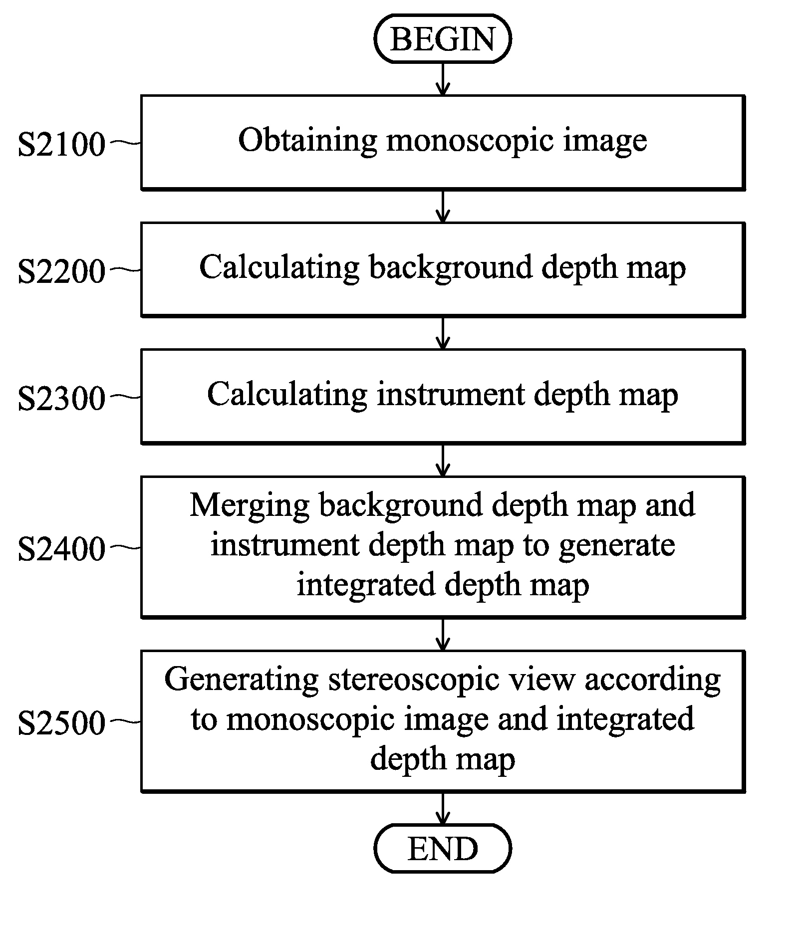
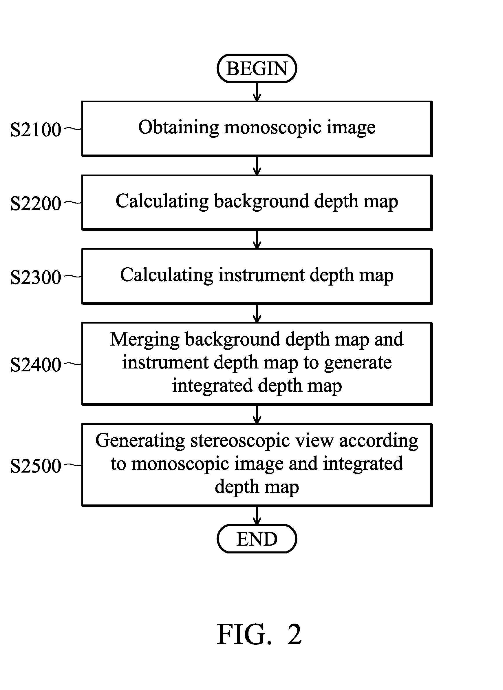
[0021]Methods for generating stereoscopic views from monoscopic endoscope images and systems using the same are provided.

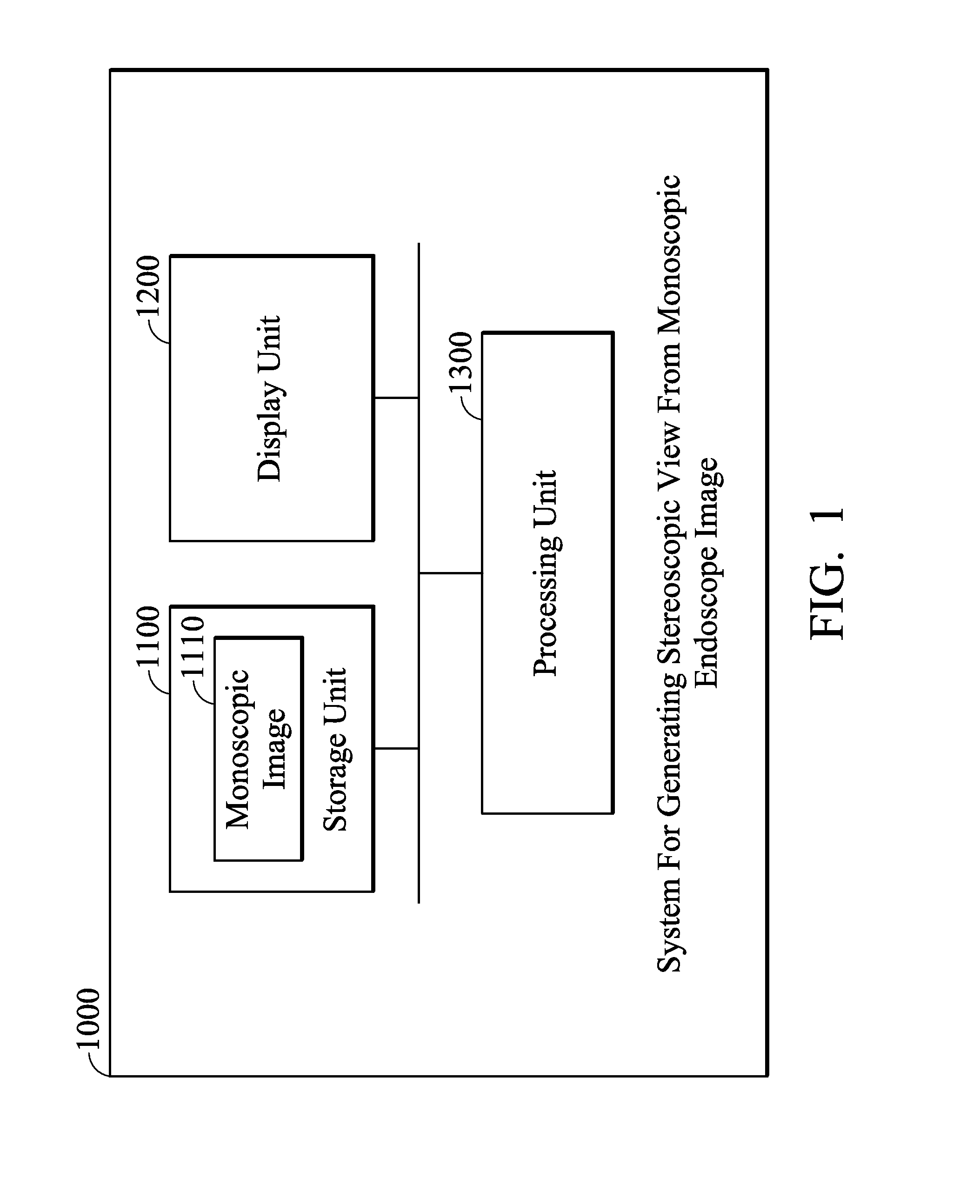
[0022]FIG. 1 is a schematic diagram illustrating an exemplary embodiment of a system for generating stereoscopic views from monoscopic endoscope images of the disclosure. The system for generating stereoscopic views from monoscopic endoscope images 1000 can be used in an electronic device, such as a monoscopic endoscope, a computer system, a display device, a receiving device, a playback device, a capturing device, and others.

[0023]The system for generating stereoscopic views from monoscopic endoscope images 1000 comprises a storage unit 1100, a display unit 1200, and a processing unit 1300. The storage unit 1100 comprises at least one monoscopic image 1110. It is understood that the monoscopic images 1110 can be captured by an image capture unit (not shown) of an endoscope. The monoscopic images 1110 are obtained by capturing images of organs in an operating fiel...


PUM


Abstract

Methods for generating stereoscopic views from monoscopic endoscope images and systems using the same are provided. First, monoscopic images are obtained by capturing images of organs in an operating field via an endoscope. A back-ground depth map of the operating field is obtained for each image. An instrument depth map corresponding to an instrument is obtained for each image, wherein the instrument is inserted in a patient's body cavity. The background depth maps and the instrument depth maps are merged to generate an integrated depth map for each image. Stereoscopic views are generated according to the monoscopic images and the integrated depth maps.

Description

BACKGROUND[0001]1. Technical Field[0002]This disclosure relates generally to methods for generating stereoscopic views and systems using the same, and, more particularly to methods for generating stereoscopic views from monoscopic images captured by an endoscope and systems using the same.[0003]2. Description of the Related Art[0004]Minimal invasive surgery (MIS) consists of using an imaging system and instruments of small diameters which can be introduced in small incisions in the body, in order to reduce the sequels of surgical interventions. The imaging system is in most cases a monoscopic endoscope consisting of an optical system, a camera, a sensor or the like and a display system. The monoscopic nature of the imaging system requires surgeons a long and tedious training period prior to operation, because the sensation of depth is absent, contrary to traditional open surgery where stereopsis naturally occurs to the surgeon. There is therefore a need to provide surgeons with dept...

Claims


Application Information

Patent Timeline
Patent Type & Authority Applications(United States)
IPC IPC(8): H04N13/02G16B45/00
CPCH04N13/026G06T7/0081G06T7/0083H04N2213/003G06T2207/10068G06T2207/20061G06T2207/10028A61B1/00009A61B1/00193G06T7/11G06T7/12H04N13/261
Inventor ANGOT, LUDOVICLIU, KAI-CHEHUANG, WEI-JIA
Owner IND TECH RES INST
Why Eureka
Try Eureka