Image processing apparatus and image processing method

Active Publication Date: 2013-10-03
CANON KK
View PDF41 Cites 4 Cited by
  • Summary
  • Abstract
  • Description
  • Claims
  • Application Information

AI Technical Summary

Benefits of technology

[0008]The present invention has been made in consideration of the above situation, and provides a tec

Problems solved by technology

A DSA image as an image obtained by this DSA has the problem that the subtraction process makes noise larger than that of an image before the su

Method used


Image

  • Image processing apparatus and image processing method
  • Image processing apparatus and image processing method
  • Image processing apparatus and image processing method

Examples


first embodiment

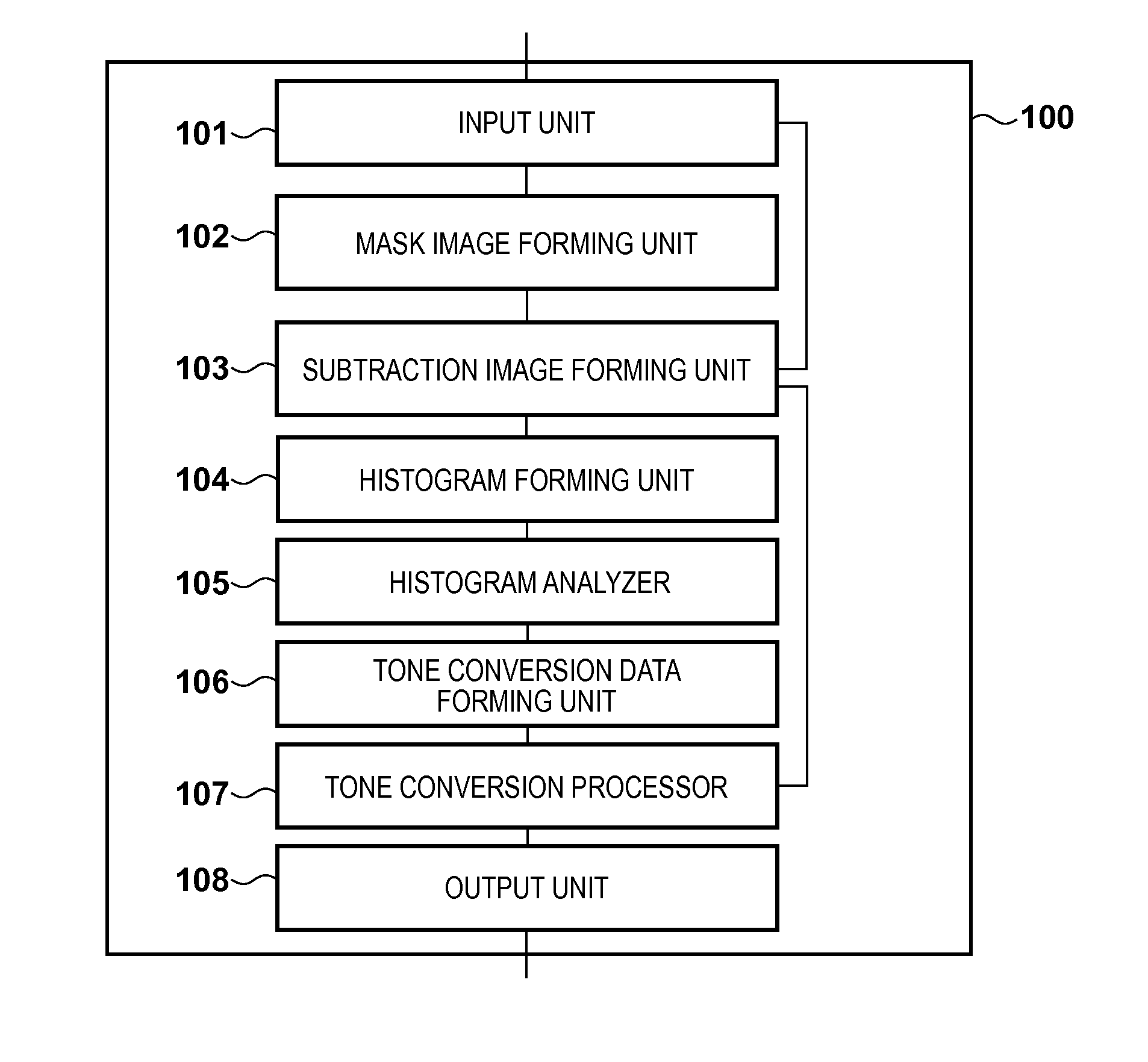
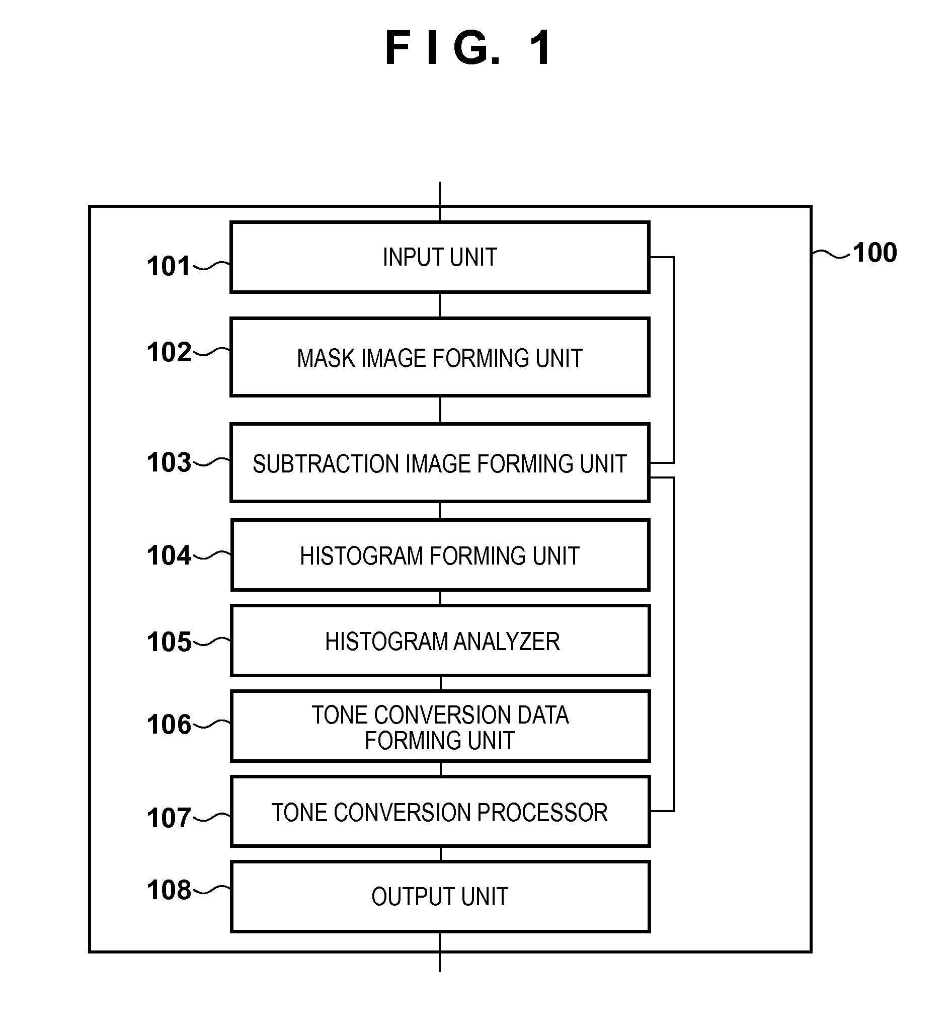
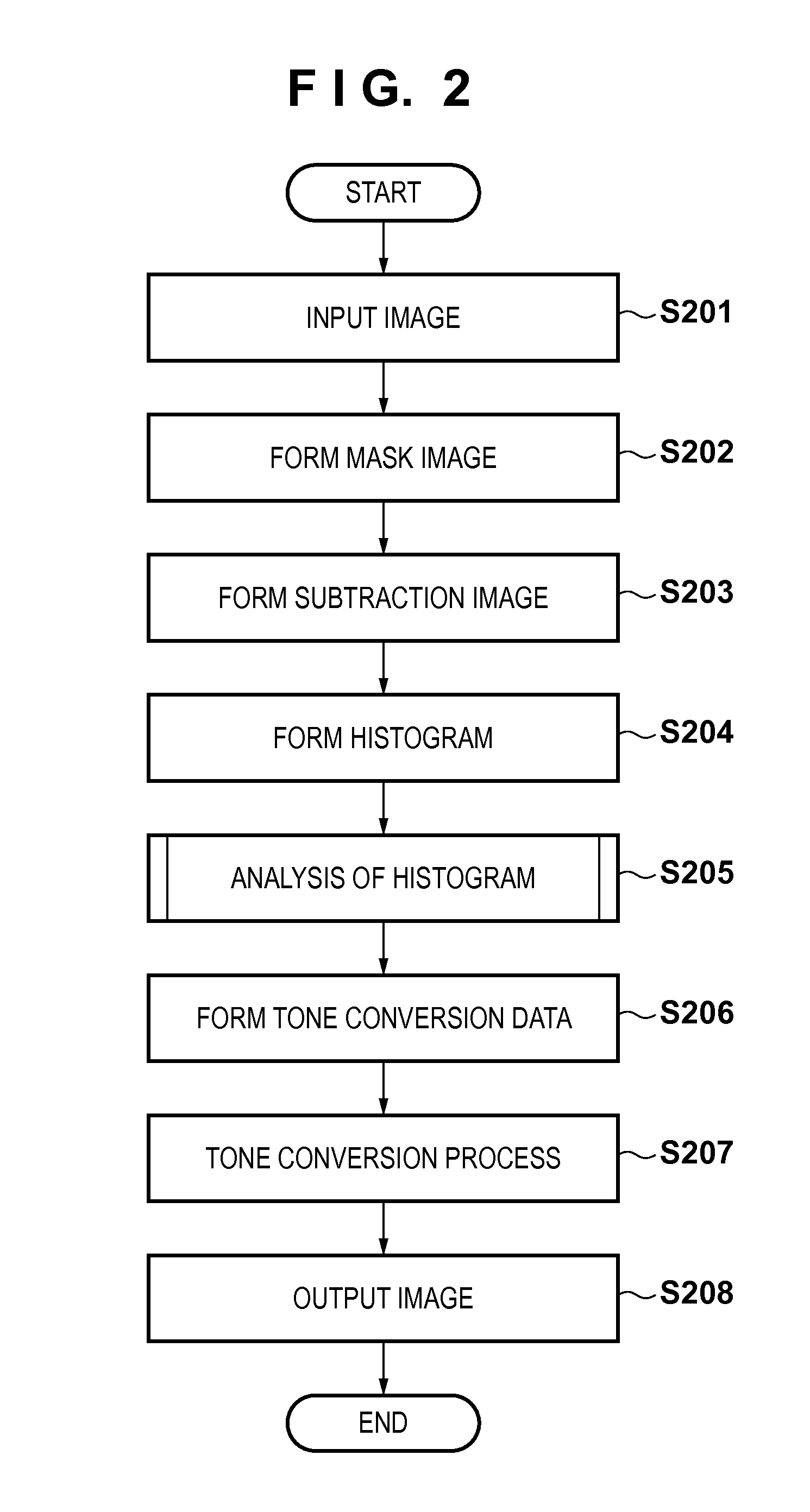
[0022]FIG. 1 shows an example of the functional configuration of an image processing apparatus according to this embodiment. The operation of each unit shown in FIG. 1 will be explained with reference to FIG. 2 showing the flowchart of processing performed by the image processing apparatus.

[0023]In step S201, an input unit 101 receives, from an X-ray image supply apparatus, X-ray images obtained by continuously imaging a target portion into which a radiopaque dye is injected (a radiopaque dye injection target portion) from a state before the radiopaque dye is injected to a state after the radiopaque dye is injected. The X-ray image supply apparatus can be an X-ray radioscopy apparatus for capturing the X-ray images or an image storage device storing the X-ray images. However, the supply form of X-ray images is not limited to any specific supply form.

[0024]Then, the input unit 101 outputs some of sequentially input X-ray images to a mask image forming unit 102, and outputs the rest t...

modification 1

[0046]When the histogram analyzer 105 analyzes the difference histogram, the width of pixel values to be analyzed can also be limited in accordance with, for example, a target portion into which the radiopaque dye is injected, the amount of radiopaque dye to be used, the injection speed, the frame rate, and the imaging conditions. This makes it possible to increase the analytical accuracy.

modification 2

[0047]The tone conversion data formed by the tone conversion data forming unit 106 can be generated for each frame, or tone conversion data generated for a given specific frame can be used for all frames. This makes it possible to form a DSA image having small contrast variations between frames.


PUM


Abstract

A histogram of pixel values in a difference image between a post-injection X-ray image and composite image is generated. A histogram of pixel values in a difference image between a pre-injection X-ray image and the composite image is generated. From the difference between the histograms, information on pixel values in a region where a radiopaque dye is imaged in the difference image between the post-injection X-ray image and composite image is collected. Pixel values in the difference image between the post-injection X-ray image and composite image are converted by using the collected information, and the converted difference image is output.

Description

BACKGROUND OF THE INVENTION[0001]1. Field of the Invention[0002]The present invention relates to an image processing technique for an X-ray image.[0003]2. Description of the Related Art[0004]There is a procedure called angiography for extracting a structure such as a blood vessel that cannot be seen by ordinary X-ray imaging. Angiography is an imaging method of extracting a blood vessel by injecting a medicine called a radiopaque dye into the blood vessel and performing X-ray imaging.[0005]As this angiography, there is an image processing method called digital subtraction angiography (DSA) that extracts only a blood vessel by removing structures except for the blood vessel by performing a subtraction process between an image before a radiopaque dye is injected and an image after the radiopaque dye is injected.[0006]A DSA image as an image obtained by this DSA has the problem that the subtraction process makes noise larger than that of an image before the subtraction. Japanese Patent...

Claims


Application Information

Patent Timeline
IPC IPC(8): A61B6/00
CPCA61B6/5205A61B6/504A61B6/481
Inventor MACHIDA, YOSHIHITO
Owner CANON KK
Why Eureka
Try Eureka