Diagnosis kit and method of using the same

a technology of diagnostic kit and kit, which is applied in the field of diagnostic kit, can solve the problems of difficult to diagnose, difficult to detect only a specific substance among a lot of substances contained in a specimen, and high fever, headache, nausea and the like illness

Inactive Publication Date: 2014-01-02
PANASONIC INTELLECTUAL PROPERTY MANAGEMENT CO LTD
View PDF21 Cites 17 Cited by
  • Summary
  • Abstract
  • Description
  • Claims
  • Application Information

AI Technical Summary

Benefits of technology

[0016]This diagnosis kit can detect extraneous organisms in the red blood cells easily with a small amount of the biological specimen.

Problems solved by technology

However, it is difficult to detect only a specific substance among a lot of substances contained in a specimen especially when a content of the contained substance is small.
After infecting a human via anopheles, the malaria, or plasmodium become parasitic on the red blood cells and increase rapidly in population, and cause high fever, headache, nausea and the like illness.
In conventional methods for detecting chemical compounds, such as antibodies, produced in the blood as a result of the infection, it is difficult to make diagnosis at an early stage since the chemical compounds do not have a detectable density until the population increases to a considerable size.
Since this method can be carried out only by observing the stain, it is easily determinable as to whether or not the red blood cells are parasitized with the malaria protozoa.
This work requires a high level of technique or an expensive apparatus.
In addition, the micrographic observation that stained nucleic acid derived from the white blood cells may be misjudged as the nucleic acid derived from the malaria protozoa, which results in poor accuracy.
Furthermore, it causes a substantial increase in the examination cost when counting is made by highly-skilled examination personnel in the cause of micrographic observation, besides such factors that the examination speed and the accuracy depend largely upon competency of the examination personnel.
In this measurement, erroneous judgment may be made due to differences in density of the biological specimen if spread excessively over a measuring position, which decreases the accuracy.
This method is therefore not suitable for such applications as a simplified test that uses a little amount, e.g. about 1 microliter taken from a finger for the measurement.
Moreover, regions where the examinations for malaria and the like may be necessary, that sufficient supply of electric power is not available, and the infrastructure for maintaining and controlling expensive detecting apparatuses is not established adequately.

Method used


Image

  • Diagnosis kit and method of using the same
  • Diagnosis kit and method of using the same
  • Diagnosis kit and method of using the same

Examples


exemplary embodiment 1

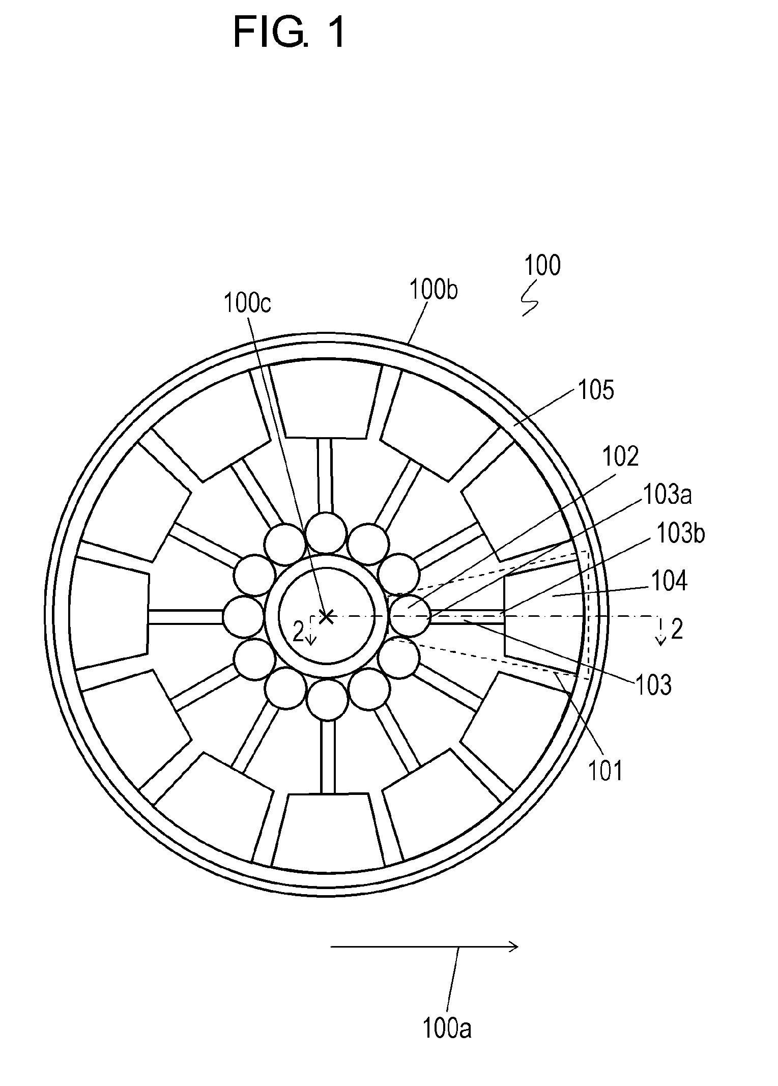
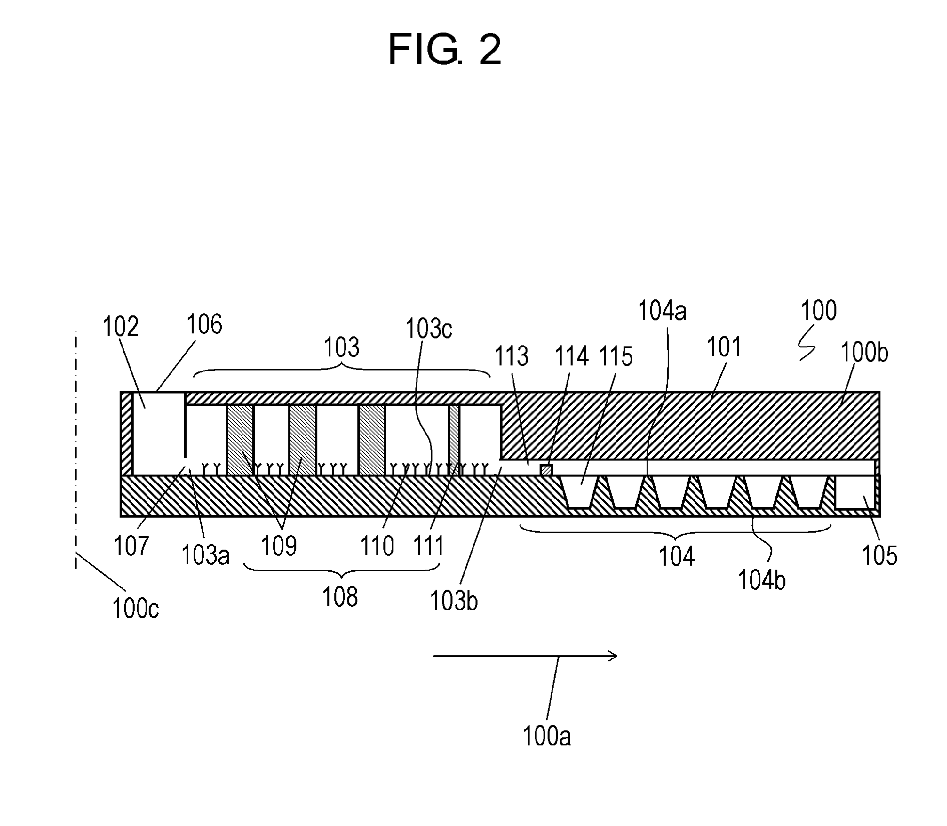
[0043]FIG. 1 is a top view of diagnosis kit 100 according to Exemplary Embodiment 1. Diagnosis kit 100 includes base plate 100b and diagnosis plates 101 disposed to base plate 100b. Base plate 100b has a circular shape about center axis 100c. Diagnosis plates 101 are arranged radially at equiangular intervals about center axis 100c to extend in predetermined directions 100a away from center axis 100c. Diagnosis plates 101 enable diagnosis kit 100 to measure (examine) plural biological specimens at once.

[0044]Diagnosis plates 101 can execute a process of diluting a biological specimen and staining nucleic acid thereof, a process of extracting red blood cells, i.e., separating the red blood cells from white blood cells and extract the red blood cells, and a process of disposing the extracted red blood cells. Diagnosis kit 100 can extract a target substance, the red blood cells, from the biological specimen containing the target substance, and examining the target substance.

[0045]The e...

exemplary embodiment 2

[0174]FIG. 9 is a sectional view of diagnosis kit 200 according to Exemplary Embodiment 2. In FIG. 9, components identical to those of diagnosis kit 100 according to Embodiment 1 shown from FIGS. 1 to 8 are denoted by the same reference numerals. Diagnosis kit 200 according to Embodiment 2 includes diagnosis plate 201 and test plate 204 instead of diagnosis plate 101 and test plate 104 of diagnosis kit 100 according to Embodiment 1. Test plate 204 has cavities 215 instead of cavities 115 of test plate 104 according to Embodiment 1. Cavities 215 have shapes different from cavities 115.

[0175]FIG. 10 is a top view of test plate 204. Cavities 215 are provided in surface 104a of test plate 204, similarly to test plate 104 according to Embodiment 1.

[0176]FIG. 11 is a top view of cavity 215. FIG. 12 is a sectional view of cavity 215 at line 12-12 shown in FIG. 11. Cavity 215 has opening 215a opened to surface 104a of test plate 104, bottom surface 215b, and inner wall 215e extending from b...

exemplary embodiment 3

[0187]FIG. 15 is a sectional view of diagnosis plate 301 of diagnosis kit 300 according to Exemplary Embodiment 3. In FIG. 15, components identical to those of diagnosis kit 100 according to Embodiment 1 shown from FIGS. 1 to 8 are denoted by the same reference numerals. Diagnosis kit 300 includes diagnosis plate 301 instead of diagnosis plate 101 according to Embodiment 1. Diagnosis kit 300 according to Embodiment 3 can extract white blood cells from blood and living tissue derived components, and analyze the white blood cells using test plate 104.

[0188]In diagnosis kit 300, diagnosis plate 301 has through-hole 330 formed therein between channel 103 and test plate 104 to provide a path to allow channel 103 to communicate with an outside of diagnosis kit 300. Through-hole 330 is located between channel 103 and test plate 104. In each of diagnosis plates 301 of diagnosis kit 300, through-hole 330 extends from liquid reservoir 113 in an upward direction perpendicular to direction 100a...


PUM

PropertyMeasurementUnit
lengthaaaaaaaaaa
heightaaaaaaaaaa
depthaaaaaaaaaa

Abstract

A diagnosis kit is configured to detect whether or not an extraneous organism exists in a red blood cell by using a biological specimen containing the red blood cell, and a stain solution capable of staining nucleic acid. The kit includes at least one diagnosis plate. The diagnosis plate includes a first chamber configured to store the stain solution and to have the biological specimen injected into the stain solution, a channel connected to the first chamber, and a test plate connected to the channel. A second chamber is connected with the test plate. The channel is configured to extract the red blood cell. The second chamber can collect a part of the biological specimen and a part of the stain solution. This diagnosis kit can detect extraneous organisms in the red blood cells easily with a small amount of the biological specimen.

Description

[0001]This application is a continuation of International Application PCT / JP2012 / 002383, filed on Apr. 5, 2012, claiming the foreign priority of Japanese Patent Application No. 2011-086040, filed on Apr. 8, 2011, and Japanese Patent Application No. 2011-149820, filed on Jul. 6, 2011, the contents of which are incorporated therein by reference.TECHNICAL FIELD[0002]The present invention relates to a diagnosis kit and a method of using the diagnosis kit used for an apparatus configured to extract or separate blood corpuscular components contained in whole blood of human and animal, or other tissue-derived fluids, and to perform diagnosis, for example, an apparatuses configured to extract red blood cells and diagnose a disease with which the blood cells are infected.BACKGROUND ART[0003]Separating only a specific substance from a sampled specimen containing plural kinds of substances is necessary in various fields. However, it is difficult to detect only a specific substance among a lot ...

Claims


Application Information

Patent Timeline
Patent Type & Authority Applications(United States)
IPC IPC(8): G01N33/50
CPCG01N33/5091B01L3/502753B01L2300/0681B01L2300/0803B01L2300/0851B01L2300/0861B01L2400/0409B01L2400/086G01N15/1484G01N21/07G01N21/253G01N21/31G01N33/569G01N33/56905G01N33/80G01N35/00069G01N2015/0073G01N2035/00158G01N2035/00524Y02A90/40
Inventor OKA, HIROAKINAKATANI, MASAYAHAYAMA, MASAAKITANEMURA, AKINA
Owner PANASONIC INTELLECTUAL PROPERTY MANAGEMENT CO LTD
Why Eureka
Try Eureka