Ultrasound imaging method and ultrasound imaging apparatus

an ultrasound imaging and ultrasonic technology, applied in the field of ultrasonic imaging, can solve the problems of bringing radiation complications, methods cannot show the exact and existing ultrasound imaging technologies are difficult to precisely determine the distance between the catheter tip and the target location. to achieve the effect of safe and reliable us

Inactive Publication Date: 2014-01-02
GENERAL ELECTRIC CO
View PDF4 Cites 28 Cited by
  • Summary
  • Abstract
  • Description
  • Claims
  • Application Information

AI Technical Summary

Benefits of technology

[0007]Embodiments of the present invention provide an ultrasound imaging method and an ultrasound imaging apparatus that can solve the above-mentioned problems.
[0032]Using the ultrasound imaging method and the ultrasound imaging apparatus of an embodiment of the present invention, it is possible to guide the real time placement of a catheter tip by marking distance scales along the target vessel centerline, and to guide the catheter insertion route through the real time varying vessel centerline when the target vessel is changed dynamically due to physiological events such as heart beats. Since the present invention is based on ultrasound scan instead of x-ray scan, and since ultrasound imaging does not involve harmful radiation, it is safely and reliably used for various patients including debilitated patients such as elderly patients and newborn babies. Furthermore, the present invention can support various catheters, such as ordinary catheters, and when a catheter tip is provided with a position sensor, the present invention may issue an alarm automatically when the catheter tip is proximate to or at the predetermined critical line of the heart entrance.

Problems solved by technology

This method cannot show the exact distance between catheter tip and target location because the catheter may be curved in the blood vessel.
X-ray imaging may achieve a resolution that is sufficient for a surgeon to differentiate micro vessels, but x-ray imaging may bring about complications related to radiation.
Although, to a certain degree, the ultrasound imaging technology can help surgeons in determining the insertion depth of the catheter, since blood vessels in a body are of space curves and blood vessels vary with physiological events such as heart beats, existing ultrasound imaging technologies are hard to exactly determine the distance between the catheter tip and the target location such as the heart entrance, and hence are hard to take a quantitative measurement of the distance.

Method used


Image

  • Ultrasound imaging method and ultrasound imaging apparatus
  • Ultrasound imaging method and ultrasound imaging apparatus
  • Ultrasound imaging method and ultrasound imaging apparatus

Examples


Embodiment Construction

[0046]In the following detailed description, some embodiments of the present invention will be described with reference to the accompanying drawings. Those skilled in the art will appreciate that the present invention should not be considered as limited to these embodiments.

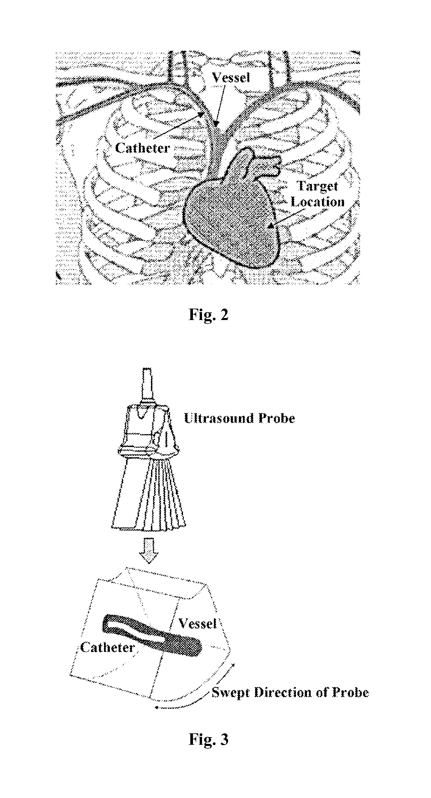
[0047]FIG. 2 is a schematic diagram illustrating a catheter inserting procedure performed by a surgeon along a blood vessel. As shown in FIG. 2, the catheter is inserted along a blood vessel until the catheter tip is positioned at the target location (e.g., heart entrance). As described above, during the catheter insertion, the distance between the catheter tip and the target location is important to place the catheter in the right position. With the use of the ultrasound imaging method and the ultrasound imaging apparatus of an embodiment of the present invention, the exact distance between the catheter tip and the target location can be measured in real time, and the exact position of the catheter tip in the bl...


PUM


Abstract

An ultrasound imaging apparatus is provided. The apparatus comprises an ultrasound probe configured to perform ultrasound sweeping of a region of interest including a target location and a target vessel in a real time volume ultrasound scan mode, an image acquiring unit configured to acquire three-dimension scan data of the target location and the target vessel, and a controlling unit configured to calculate the target vessel centerline length based on the acquired three-dimension scan data. The apparatus further comprises an image processing unit configured to automatically mark distance scales from the critical line located at the target location along the target vessel centerline at predetermined intervals based on the calculated centerline length, and a displaying unit configured to displaying the target location and the target vessel centerline marked with distance scales in real time.

Description

TECHNICAL FIELD[0001]Embodiments of the present invention relate to the field of ultrasound imaging, and in particular to an ultrasound imaging method and an ultrasound imaging apparatus for guiding the placement of a catheter in a vessel in real time by distance scales marked along the vessel centerline.BACKGROUND[0002]Some interventional surgical procedures need to insert medical instruments, such as a catheter, into a blood vessel of a patient. As shown in FIG. 2, the catheter is inserted along the blood vessel and finally positioned at a target location, such as the heart entrance of the patient, for subsequent medical processes. During the insertion of the catheter along the blood vessel, such as during a PICC (Peripherally Inserted Central Catheter) procedure, the distance between the catheter tip and the target location, such as the heart entrance, is very important to place the catheter in right position.[0003]Presently, a surgeon typically determines the position of the cat...

Claims


Application Information

Patent Timeline
Patent Type & Authority Applications(United States)
IPC IPC(8): A61B8/08A61B8/00A61M25/01A61B8/14
CPCA61B8/0891A61B8/14A61B8/0883A61B8/54A61B8/0841A61M25/0108A61B8/085A61B8/483A61M2025/0166G06T19/00G06T2207/10132G06T2207/30101G06T2207/30172G06T2210/41G06T2219/012G06T7/12
Inventor LIU, GANGZHAO, YIMINGXIA, WENTINGTEN EYCK, LAWRENCE GUY
Owner GENERAL ELECTRIC CO
Why Eureka
Try Eureka