
System and method for providing registration between breast shapes before and during surgery

Inactive Publication Date: 2014-02-13
TRUSTEES OF DARTMOUTH COLLEGE THE
View PDF7 Cites 41 Cited by
  • Summary
  • Abstract
  • Description
  • Claims
  • Application Information

AI Technical Summary

Benefits of technology

The present invention provides a system and method for registering breast shapes before and during surgery using a volumetric image and an optically scanned surface image. The method involves identifying an air / tissue boundary in the volumetric image and processing it with an image filter to emphasize the boundary. The surface optically scanned image is then registered with the filtered volumetric image. The technical effect of this invention is to improve the accuracy and precision of breast surgery procedures.

Problems solved by technology

For example, the wire localization method adds a separate procedure to the surgical resection, thereby complicating and lengthening the process.
Additional precision can be gained by placing additional wires, but this may increase the length of time for the localization procedure.
Therefore, wire localization can be an inefficient and imprecise technique.
For example, the soft tissue of an internal organ may be displaced or deformed during surgery, making it difficult to correlate the location of a feature in an intraoperative optical image to the location of the feature in a preoperative magnetic resonance image.
Therefore, the location of an internal point of interest from a preoperative volumetric image may be difficult to discern due to the distortion of the internal tissue relative to the breast surface.
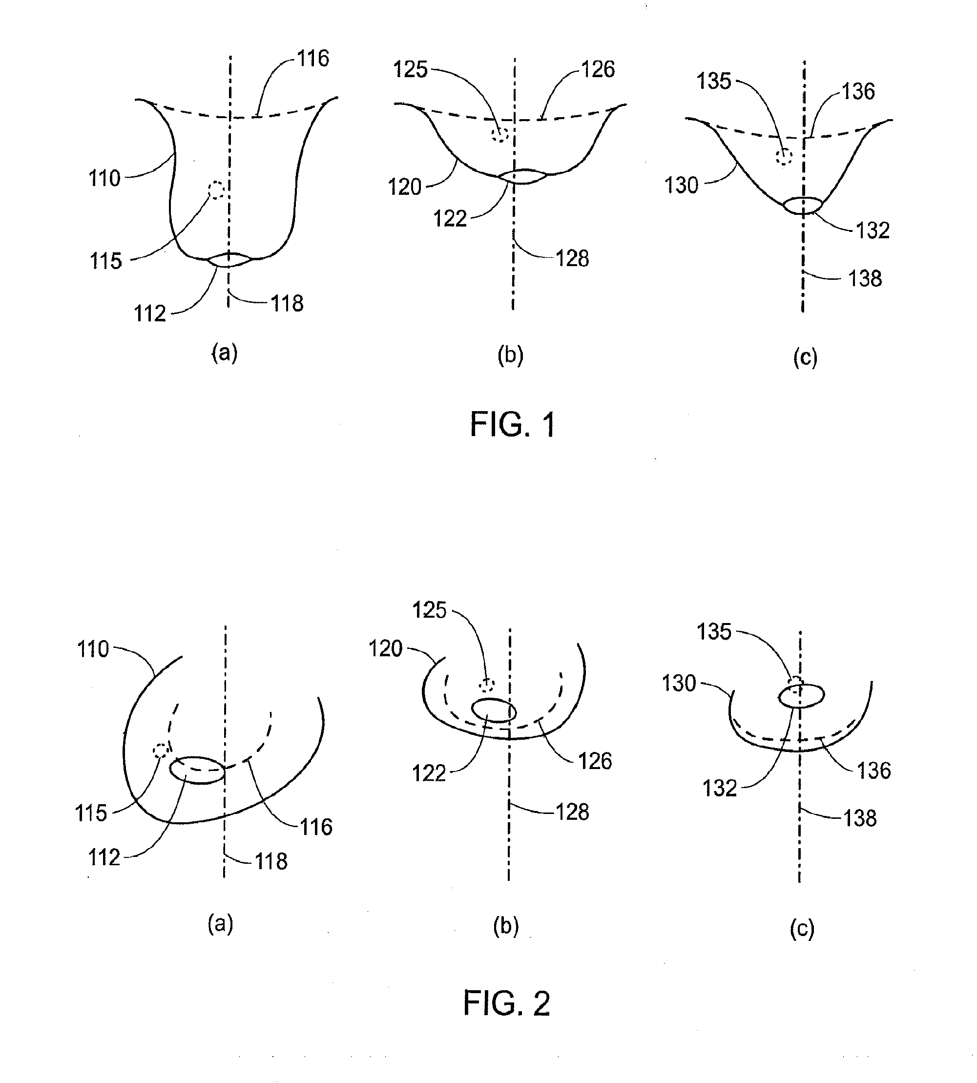
That is, the relation of surface features, such as the nipple, to internal tissue, such as a cancerous growth, may not be consistent between the preoperative prone volumetric image and the intraoperative optical scan image.
Such variations illustrate the potential difficulties of locating an internal region of interest in relation to external features when images are taken with the subject in a different position from the position of the subject in the operating room.
However, even when a large number of fiducials are used, difficulty may arise when registering a first image with a second image where there has been significant tissue displacement between the first and second image.

Method used


Image

  • System and method for providing registration between breast shapes before and during surgery
  • System and method for providing registration between breast shapes before and during surgery
  • System and method for providing registration between breast shapes before and during surgery

Examples


first embodiment

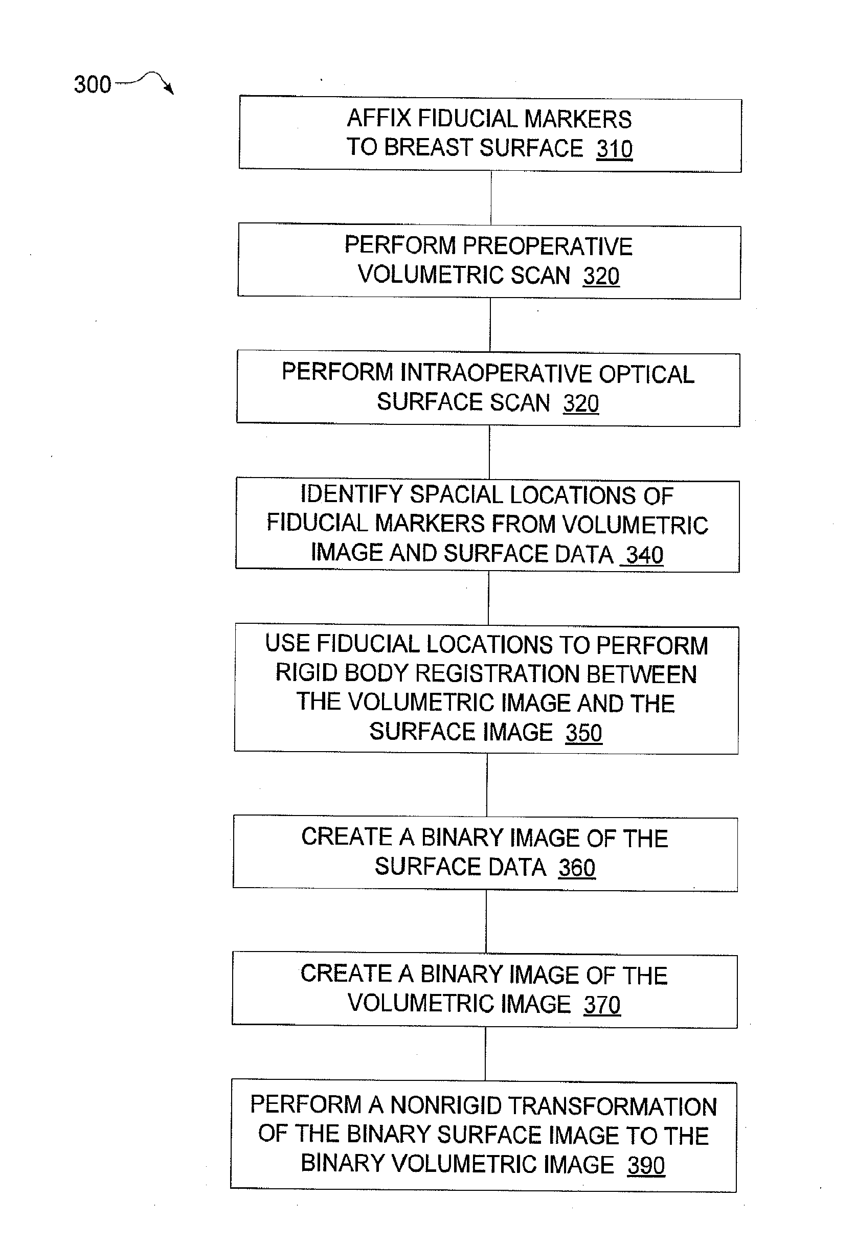
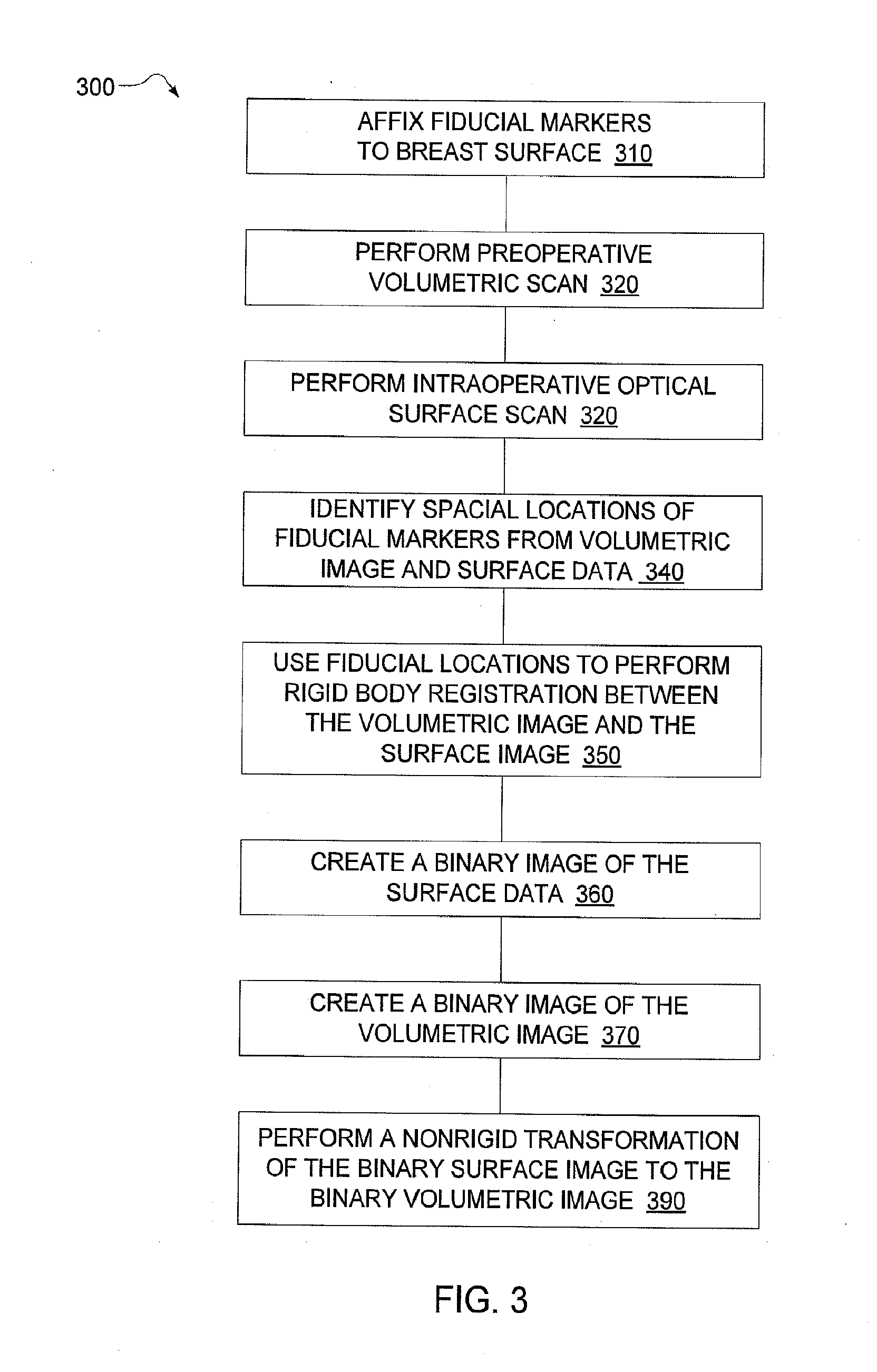
[0034]FIG. 3 is a flow chart 300 showing a method for registering intraoperative optical scan images with a preoperative MR image. It should be noted that any process descriptions or blocks in flow charts should be understood as representing modules, segments, portions of code, or steps that include one or more instructions for implementing specific logical functions in the process, and alternative implementations are included within the scope of the present invention in which functions may be executed out of order from that shown or discussed, including substantially concurrently or in reverse order, depending on the functionality involved, as would be understood by those reasonably skilled in the art of the present invention.

[0035]As shown by block 310, fiducial markers are affixed to the breast surface. As discussed above, the fiducial markers provide a common reference for images taken under different circumstances. For a first example, the fiducials may be used to provide a com...

second embodiment

[0053]There may be scenarios where the surface of the optical scanner image and the surface of the MR image do not perfectly align following a rigid transformation. Under a method for registering intra-operative optical scan images with preoperative volumetric images, Finite Element Modeling (FEM) may be used in situations where rigid transformations may yield errors. Such modeling may take into account the physical properties of the tissue in the images, rather than only attempting to linearly correlate the distance between image features and constants, such as fiducials. By modeling the physical properties of the tissue, FEM may more accurately register prone volumetric images with supine surface images.

[0054]In some situations, FEM may leverage additional information about the tissue in the volumetric and surface images to more accurately model the transformation. For example, breast tissue may exhibit different elastic properties in a first region of the breast than the elastic ...


PUM


Abstract

A registration framework is presented that registers volumetric breast images captured before surgery with intraoperative surface images. The framework may be implemented using either image-based or model-based registration techniques. The method contains the steps of: identifying an air / tissue boundary from a volumetric image created at a first time; processing the volumetric image with an image filter to emphasize the air / tissue boundary; and registering a surface optically scanned image, with the filtered volumetric image, where the surface optically scanned image is created at a second time.

Description

FIELD OF THE INVENTION[0001]The present invention relates to medical imaging, and more particularly, is related to registration of preoperative volumetric images to intraoperative surface image data.BACKGROUND[0002]Many women with breast cancer have their tumors detected by screening mammography or breast magnetic resonance imaging (MRI), before the tumors become clinically palpable. Most of these women with small breast cancers will typically choose breast conserving surgery. The goal of breast conserving surgery is to completely resect the tumor with negative margins and simultaneously preserve the shape of the breast. The standard technique for breast conserving surgery for patients with non-palpable breast cancer is to place a wire into the cancer preoperatively (in radiology under, for example, mammographic, ultrasound or MRI guidance) and then, in the operating room (OR), to excise the tissue around the wire. This technique, initially developed in the 1970's, has several limit...

Claims


Application Information

Patent Timeline
IPC IPC(8): G06T7/00
CPCG06T7/0012G06T2207/30068G06T7/33
Inventor BARTH, JR., RICHARD J.JI, SONGBAIPAULSEN, KEITH D.PALLONE, MATTHEW J.
Owner TRUSTEES OF DARTMOUTH COLLEGE THE
Learn More