Cell Model and Method for Screening c-Fms Kinase Inhibitors
a cell model and kinase inhibitor technology, applied in the field of cytobiology and pharmacy, can solve the problems of high expression, difficult to detect accurately, and abnormal proliferation of mononuclear macrophages or high expression, and achieve the effects of high content, high sensitivity, and high efficacy
- Summary
- Abstract
- Description
- Claims
- Application Information
AI Technical Summary
Benefits of technology
Problems solved by technology
Method used
Image

Examples
example 1
Establishment of a Cell Line Stably Expressing Human c-Fms and STAT1
[0057](1) A eukaryotic expression vector pCORON / puro-cfms comprising full-length cDNA of human c-Fms (donated by Dr. Charles Sherr and Dr. Martine Roussel from St. Jude Children's Research Hospital, USA) was constructed, and a human osteosarcoma cell line wherein GFP and STAT1 were fused and expressed, was transfected with the vector by lipofectin.
[0058]A new cell line stably expressing human c-Fms was obtained by means of resistance screening and limiting dilution. DMEM medium that contains 4500 mg / L glucose, 10% fetal bovine serum, 4 mM L-glutamine, 500 m / mL G418 and 2 μg / mL puromycin was used, and the cell line was cultured in an incubator at 37° C., 5% CO2, 80% humidity.
[0059]Compared with the cells prior to transfection, the established cell had no significant change in cellular morphology, and had a stable growth state.
[0060]The cell line prepared in this example (i.e. the human osteosarcoma cell line of the p...
example 2
Characterization of the Newly Established Cell Line
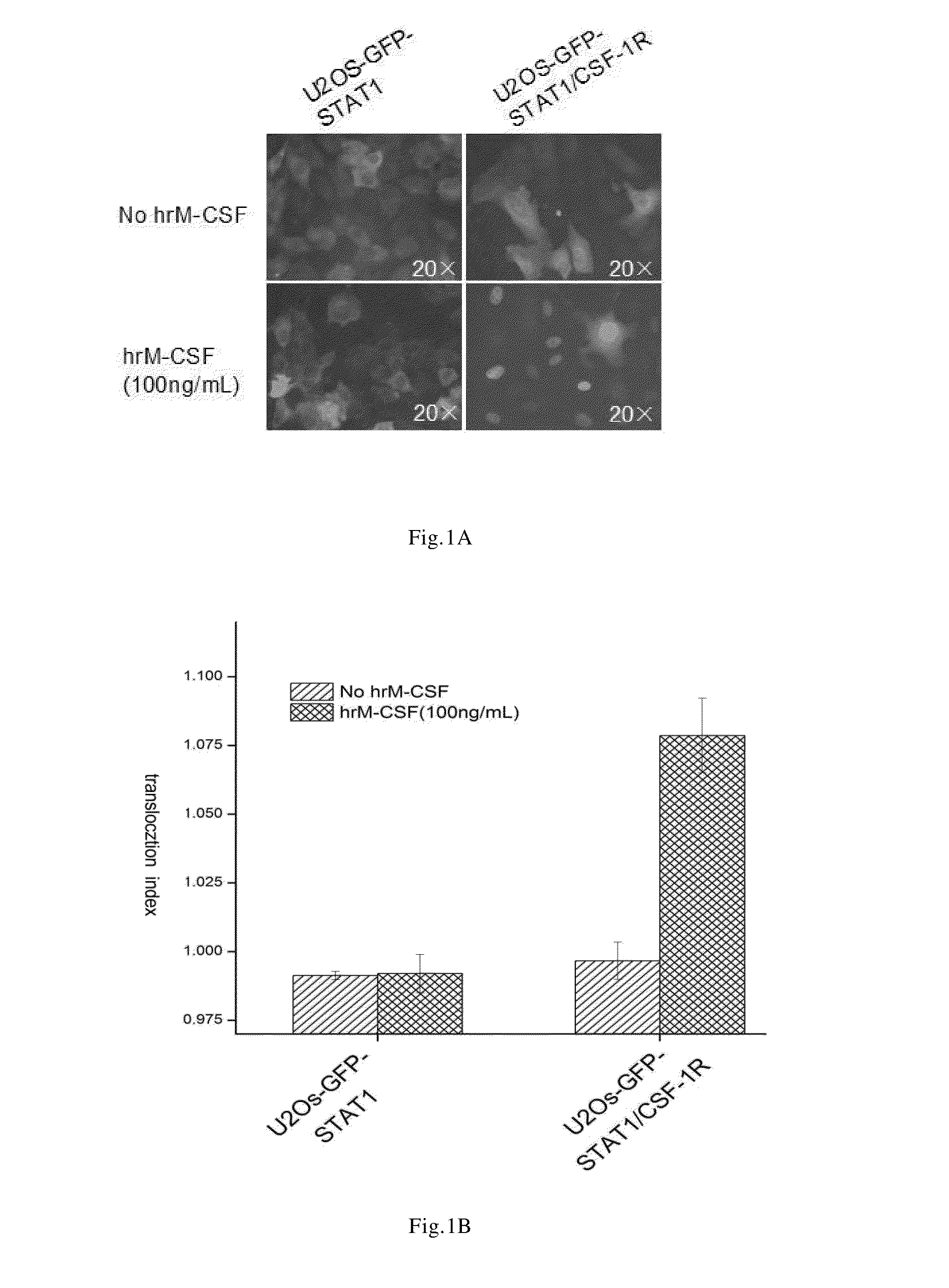
[0061]The cell line prepared in the Example 1 was treated with recombinantly expressed human M-CSF(hrM-CSF), so as to activate c-Fms and induce GFP-STAT1 nuclear translocation. In the established cell line, human macrophage colony stimulating factor receptors were dimerized upon binding to the specific ligand M-CSF, and meanwhile the steric conformation was changed and the kinase domain was activated. The downstream signal pathway of c-Fms was activated by transphosphorylation. The phosphorylated GFP-STAT1 were also dimerized and entered nucleus, namely, nuclear translocation occurred.
[0062]In order to quantitatively detect the extent of nuclear translocation of GFP-STAT1 induced by hrM-CSF, in this Example, In Cell Analyzer 1000 or In Cell Analyzer 2000 was used to obtain the cell image of GFP-STAT1 nuclear translocation. The cell image obtained was analyzed by using Nuclear Trafficking Analysis Module, wherein the translocation in...
example 3
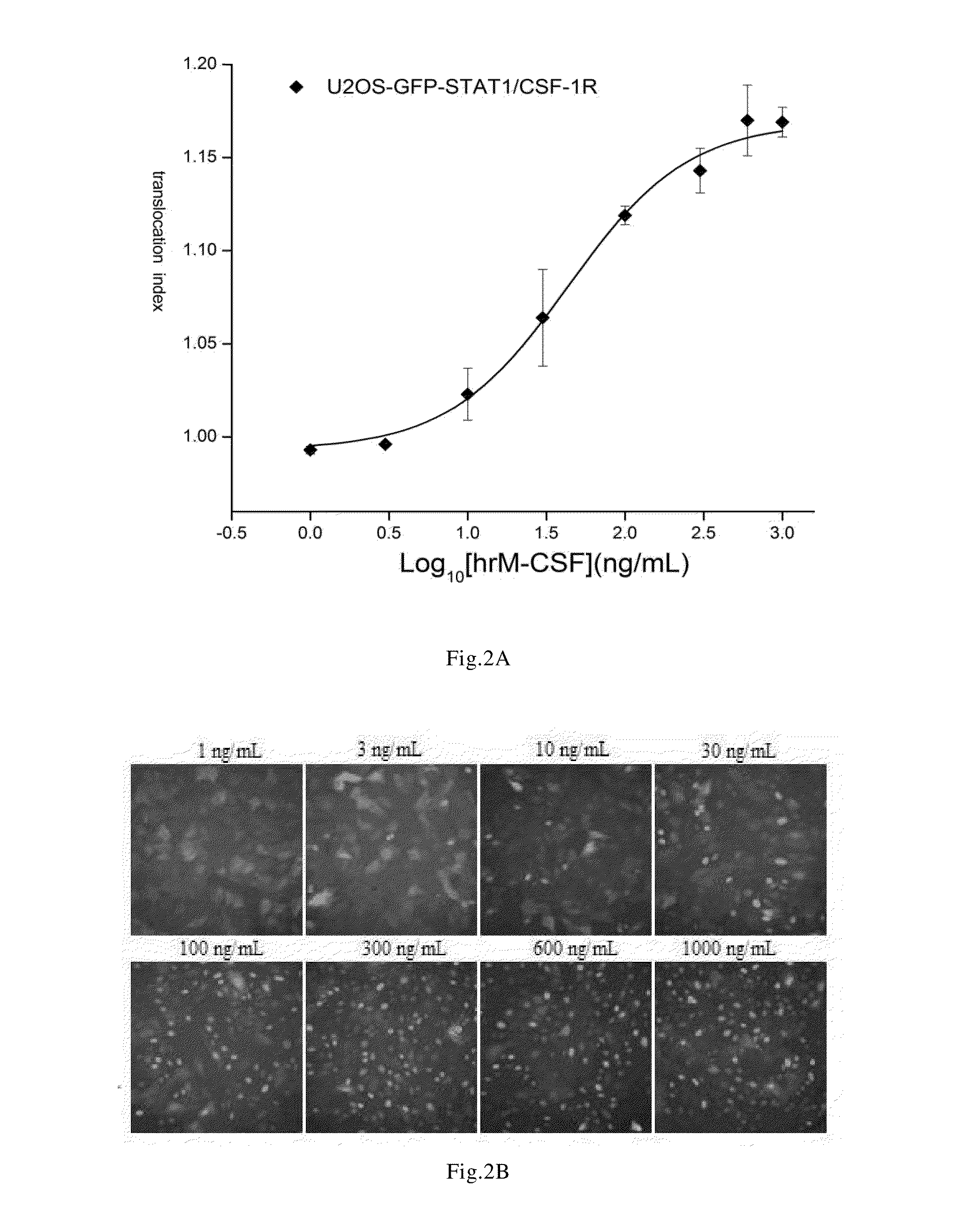
Determination of the Median Effective Concentration of hrM-CSF For Inducing GFP-STAT1 Nuclear Translocation
[0070]The steps were as follows:[0071]1) using the cells prepared in the Example 1 to prepare a cell suspension of 1×10⁵ cells / mL, seeding the cells to a 96-well culture plate (which was black and transparent at the bottom) at 100 μL / well;[0072]2) culturing the cells in an incubator at 37° C., 5% CO2, 80% humidity for 24 hours;[0073]3) washing the cells twice with the cell analytic liquid at 100 μL / well for each time, discarding the solution, and adding the cell analytic liquid at 50 μL / well;[0074]4) adding hrM-CSF diluted with the cell analytic liquid, to obtain a final concentration of 1, 3, 10, 30, 100, 300, 600, 1000 ng / mL respectively;[0075]5) after incubating the cells in an incubator at 37° C., 5% CO2, 80% humidity for 30 minutes, adding the cell fixation solution preheated at 37° C. at 100 μL / well, slightly shaking the culture plate to mix the mixture uniformly, and pla...
PUM
| Property | Measurement | Unit |
|---|---|---|
| Temperature | aaaaa | aaaaa |
| Fraction | aaaaa | aaaaa |
| Fraction | aaaaa | aaaaa |
Abstract
Description
Claims
Application Information
 - R&D Engineer
- R&D Manager
- IP Professional
- Industry Leading Data Capabilities
- Powerful AI technology
- Patent DNA Extraction
Browse by: Latest US Patents, China's latest patents, Technical Efficacy Thesaurus, Application Domain, Technology Topic.
© 2024 PatSnap. All rights reserved.Legal|Privacy policy|Modern Slavery Act Transparency Statement|Sitemap