
Imaging with Multiple Aperture Medical Ultrasound and Synchronization of Add-On Systems

a technology of medical ultrasound and add-on systems, applied in the field of medical imaging techniques, can solve the problems of poor lateral resolution, small aperture, and use of ultrasonic imaging for medical purposes

Inactive Publication Date: 2014-03-13
MAUI IMAGING
View PDF2 Cites 28 Cited by
  • Summary
  • Abstract
  • Description
  • Claims
  • Application Information

AI Technical Summary

Benefits of technology

The present invention relates to a method and apparatus for synchronizing an add-on ultrasound system with a host ultrasound probe. The technical effect of this invention is that it allows for the efficient and accurate synchronization of the add-on system with the host probe, resulting in improved image quality and reduced latency. This is achieved through an algorithm that calculates the start of frame of the transmitted ultrasound pulses, based on the identification of an interval between amplitude peaks or a large change in peak amplitudes during successive scan lines. The synchronization can be achieved using a tap that couples the host probe to the add-on system, or by processing data from the tap. The invention also allows for the display of ultrasound images from the add-on system, providing a more convenient and efficient means of viewing ultrasound images.

Problems solved by technology

This design configuration lies at the heart of one of the most significant limitations in the use of ultrasonic imaging for medical purposes; namely, poor lateral resolution.
Theoretically the lateral resolution could be improved by increasing the aperture of the ultrasonic probe, but the practical problems involved with aperture size increase have kept apertures small and lateral resolution poor.
For scanners intended for abdominal and other use, the limitation on aperture size is not so obvious, but it is a serious limitation nevertheless.
The problem is that it is difficult to keep the elements of a large aperture array in phase because the speed of ultrasound transmission varies with the type of tissue between the probe and the area of interest.
When the size of the aperture is increased to improve the lateral resolution, the additional elements of a phased array may be out of phase and may actually degrade the image rather than improving it.
However, adding resolution to existing system would face additional challenges, such as synchronizing an add-on system to the existing host ultrasound machine.

Method used


Image

  • Imaging with Multiple Aperture Medical Ultrasound and Synchronization of Add-On Systems
  • Imaging with Multiple Aperture Medical Ultrasound and Synchronization of Add-On Systems
  • Imaging with Multiple Aperture Medical Ultrasound and Synchronization of Add-On Systems

Examples


Embodiment Construction

[0028]Various embodiments of an ultrasound imaging system are described.

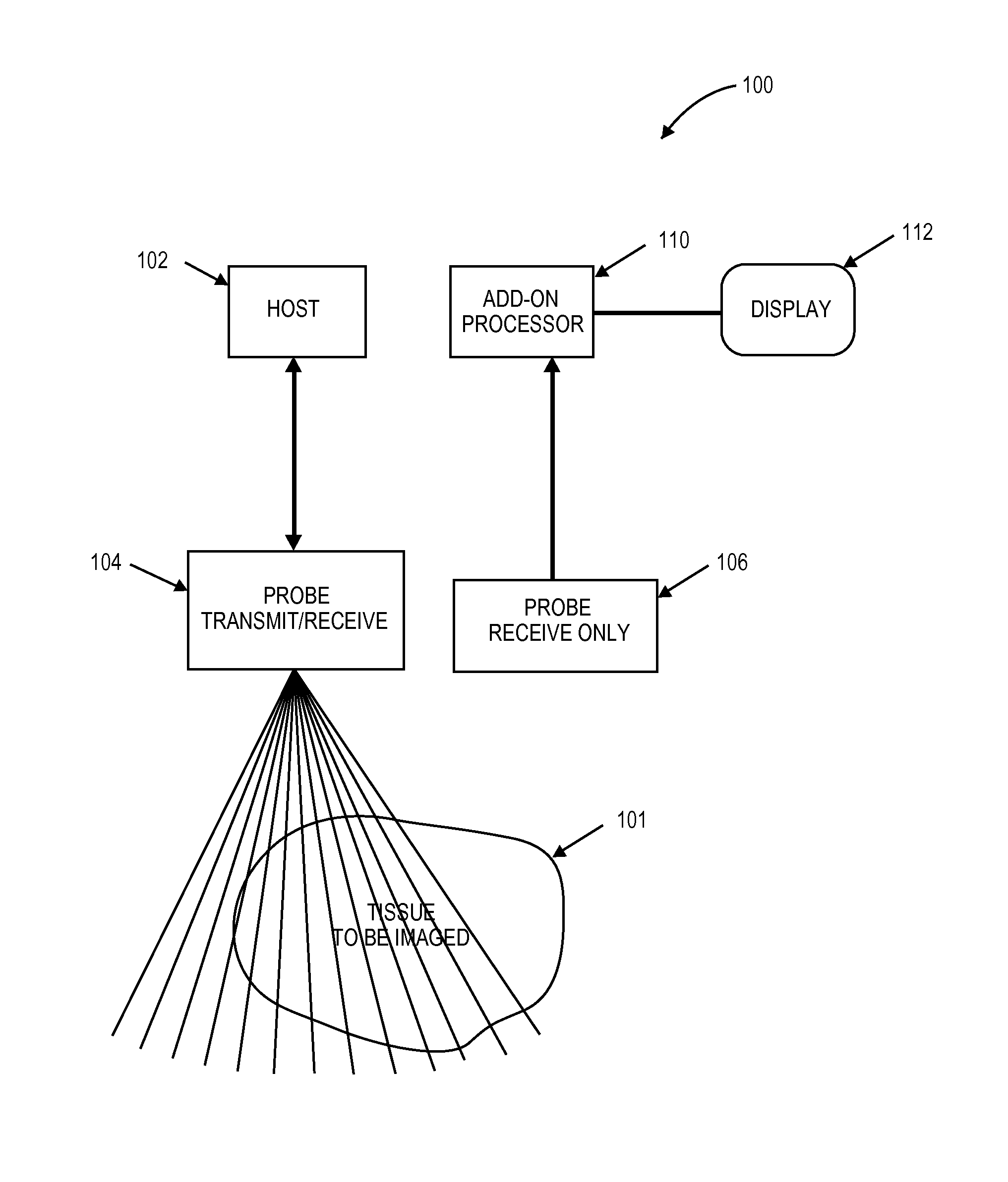
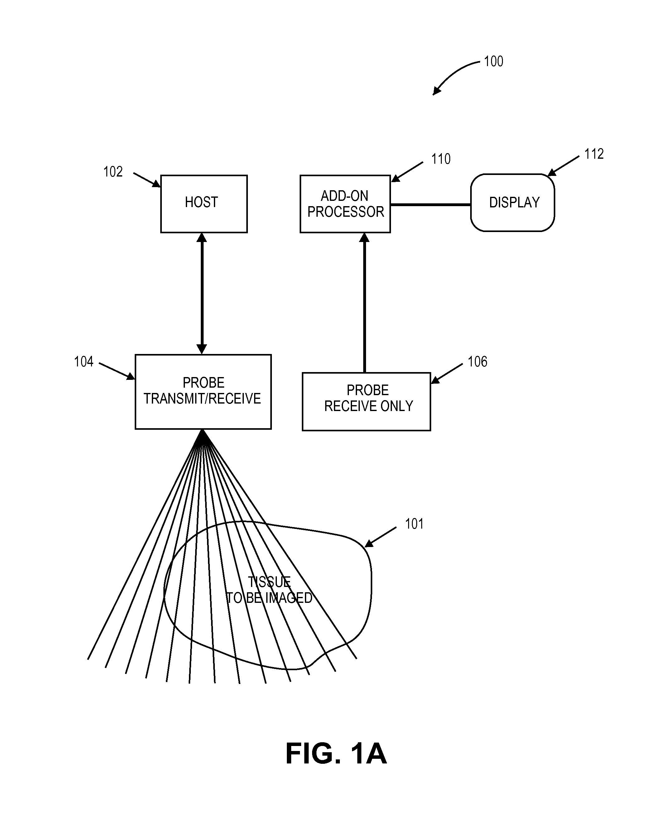
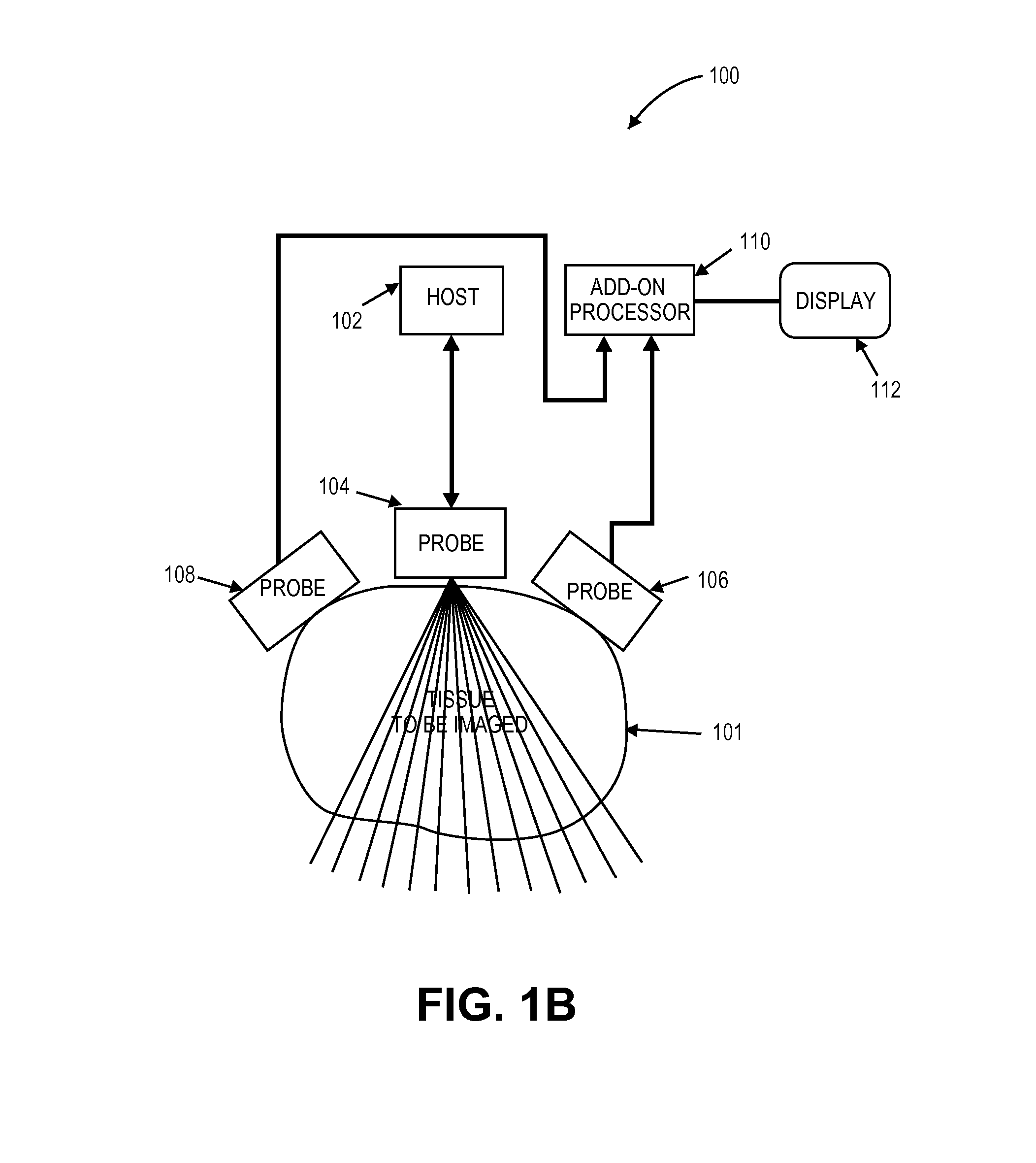
[0029]Returned echoes in ultrasonography can be detected by a separate relatively non-directional receiving probe located away from the insonifying probe (e.g., the transmitting probe), and the non-directional receive transducer can be placed in a different acoustic window from the insonifying probe. This non-directional receiving probe can be called an omni-directional or receiving probe because it can be designed to be sensitive to a wide field of view.

[0030]If the echoes detected at the receiving probe are stored separately for every pulse from the insonifying transducer, the entire two-dimensional image can be formed from the information received by a single receiving probe. Additional copies of the image can be formed by additional omni-directional probes collecting data from the same set of insonifying pulses.

[0031]In one embodiment, an add-on device can be designed as a receive-only device while using an ...


PUM


Abstract

The benefits of a multi-aperture ultrasound probe can be achieved with add-on devices. Synchronization and correlation of echoes from multiple transducer elements located in different arrays is essential to the successful processing of multiple aperture imaging. The algorithms disclosed here teach methods to successfully process these signals when the transmission source is coming from another ultrasound system and synchronize the add-on system to the other ultrasound system. Two-dimensional images with different noise components can be constructed from the echoes received by individual transducer elements. The disclosed techniques have broad application in medical imaging and are ideally suited to multi-aperture cardiac imaging using two or more intercostal spaces.

Description

CROSS REFERENCE TO RELATED APPLICATIONS[0001]This application is a continuation of U.S. application Ser. No. 13 / 002,778, filed Apr. 6, 2011, which is the national stage of International Application No. PCT / US2009 / 053096, filed Aug. 7, 2009. Said PCT / US2009 / 053096 application claims the benefit of U.S. Provisional Application No. 61 / 087,571, filed Aug. 8, 2008, and U.S. Provisional Application No. 61 / 169,264, filed Apr. 14, 2009, all which are herein incorporated by reference.[0002]This application is related to U.S. application Ser. No. 11 / 532,013, filed Oct. 11, 2007, and U.S. patent application Ser. No. 11 / 865,501, filed May 1, 2008, which are herein incorporated by reference.INCORPORATION BY REFERENCE[0003]All publications and patent applications mentioned in this specification are herein incorporated by reference to the same extent as if each individual publication or patent application was specifically and individually indicated to be incorporated by reference.FIELD OF THE INVE...

Claims


Application Information

Patent Timeline
Patent Type & Authority Applications(United States)
IPC IPC(8): A61B8/08
CPCA61B8/5207A61B8/54G01S15/8997G01S15/8913G01S7/52087G01S7/52046G01S15/8927G01S15/8929A61B8/4477A61B8/00A61B8/4281
Inventor SPECHT, DONALD F.BREWER, KENNETH D.
Owner MAUI IMAGING
Learn More