
Surgical probe incorporating a dilator

a technology of dilator and probe, which is applied in the field of surgical procedures, can solve the problems of unfavorable trauma and an appreciable amount of time, and achieve the effect of avoiding damage to critical structures in the vicinity

Active Publication Date: 2014-09-18
GLOBUS MEDICAL INC
View PDF4 Cites 9 Cited by
  • Summary
  • Abstract
  • Description
  • Claims
  • Application Information

AI Technical Summary

Benefits of technology

The device described in this patent allows for the placement of a probe without damaging critical structures in the area. It includes an electrode for supplying an electrical signal during insertion. Additionally, it has a dilator with an inflatable member that expands to displace surrounding soft tissue and create space for surgical procedures. This allows for visualization of the structures to be operated on and improves the accuracy of surgical interventions.

Problems solved by technology

This, however, requires an appreciable amount of time and results in undesirable amount of trauma to the tissue between the incision where the probe has been inserted and the required dilated opening through the psoas muscle and other tissues surrounding the spinal column.

Method used


Image

  • Surgical probe incorporating a dilator
  • Surgical probe incorporating a dilator
  • Surgical probe incorporating a dilator

Examples


Embodiment Construction

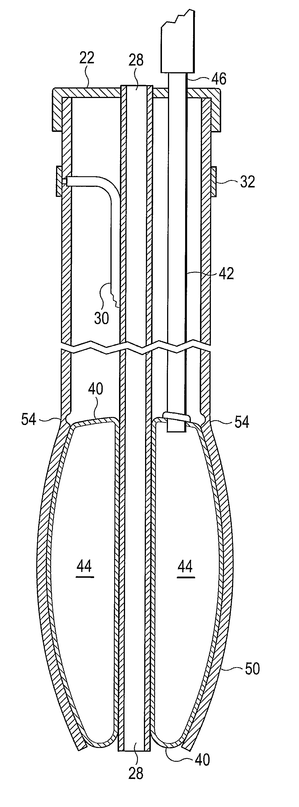
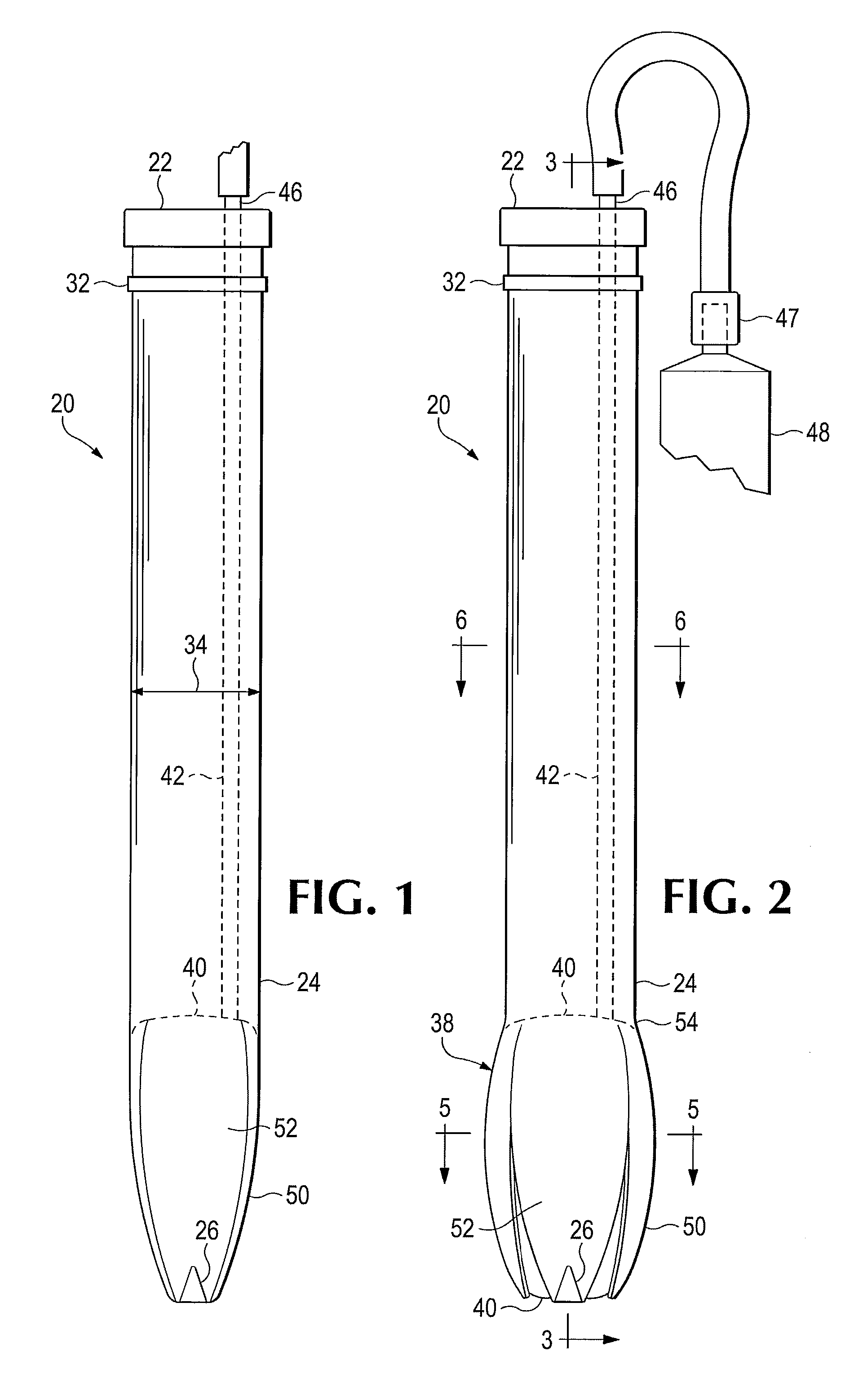
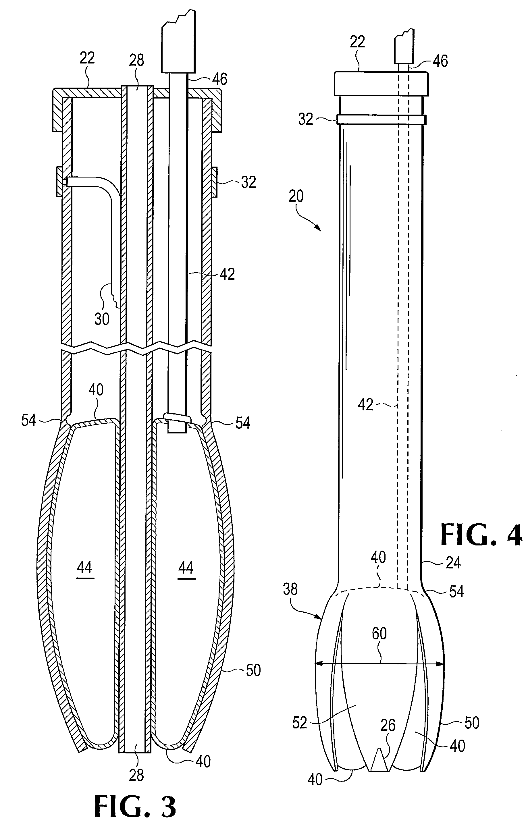
[0030]Referring to FIGS. 1-6 of the drawings, a probe 20 which is a first exemplary embodiment of the device disclosed herein includes a main body having a proximal end portion 22 and a distal end portion 24 that may be tapered to a relatively sharp end. The probe 20 is shown with its transverse, or lateral, dimensions considerably exaggerated, for the sake of clarity. In one embodiment the main body may be of a molded polymeric plastic resin material. Exposed at the extreme distal tip and extending a short distance along the distal end portion 24 there is an electrode 26. A centrally located cannula 28 extends longitudinally through the probe 20, with a central bore extending from the proximal end 22 to and through the distal end portion 24, as may be seen best in FIG. 3.

[0031]An insulated electrical conductor 30 is connected electrically with the electrode 26, extending within the body of the probe 20, and is electrically connected with a terminal 32 such as a ring of electrically...


PUM


Abstract

A surgical probe and a method for forming and enlarging an access opening through a psoas muscle to provide for minimally invasive lateral approach for surgical access to a lumber intervertebral disc. A distal end portion of the probe is equipped with an electrode useful for confirming proper location of the probe and includes an inflatable dilator body for enlarging an access opening through tissue adjacent to a spinal column. The probe includes a cannula through which a K wire can be extended to anchor the probe to a patient.

Description

BACKGROUND[0001]The present invention relates to surgical procedures, and in particular to a probe incorporating a dilator and a method for its use in connection with minimally invasive surgery.[0002]Minimally invasive surgery techniques have been used in the past for spinal disc replacement procedures.[0003]There are known devices for dilatation of soft tissue structures of the human body, including blood vessels, other ducts, bowels, and other lumens. Certain dilators are known to incorporate inflatable members utilized to separate soft tissue, including muscular tissue, in order to provide visibility and access to portions of the anatomy that are of interest.[0004]In some surgical procedures it is important to determine where certain structures are located in order not to damage them. For example, it is desired not to sever nerves or some blood vessels or other ducts or conduits for fluid. While fluoroscopy can be used to guide surgeons in the placement of probes used to guide ce...

Claims


Application Information

Patent Timeline
IPC IPC(8): A61B17/88
CPCA61B17/8855A61B17/1626A61B17/3439A61N1/0551A61B2017/00261A61B2017/320048A61B5/4893A61B17/3421
Inventor TATSUMI, ROBERT LOUIS
Owner GLOBUS MEDICAL INC
Learn More