
Removal of tumor cells from intraoperative autologous blood salvage

a tumor cell and autologous technology, applied in the field of tumor cell removal from intraoperative autologous blood salvage, can solve the problems of preoperative anemia, contaminated blood transfusion, and increased economic questions about blood donation prior to surgery, and achieve excellent viral diagnostics, reduce the reluctance of surgeons to use autotransfusion in cancer surgery, and reduce the effect of reluctan

Active Publication Date: 2014-12-18
LINDIS BLOOD CARE GMBH
View PDF2 Cites 2 Cited by
  • Summary
  • Abstract
  • Description
  • Claims
  • Application Information

AI Technical Summary

Benefits of technology

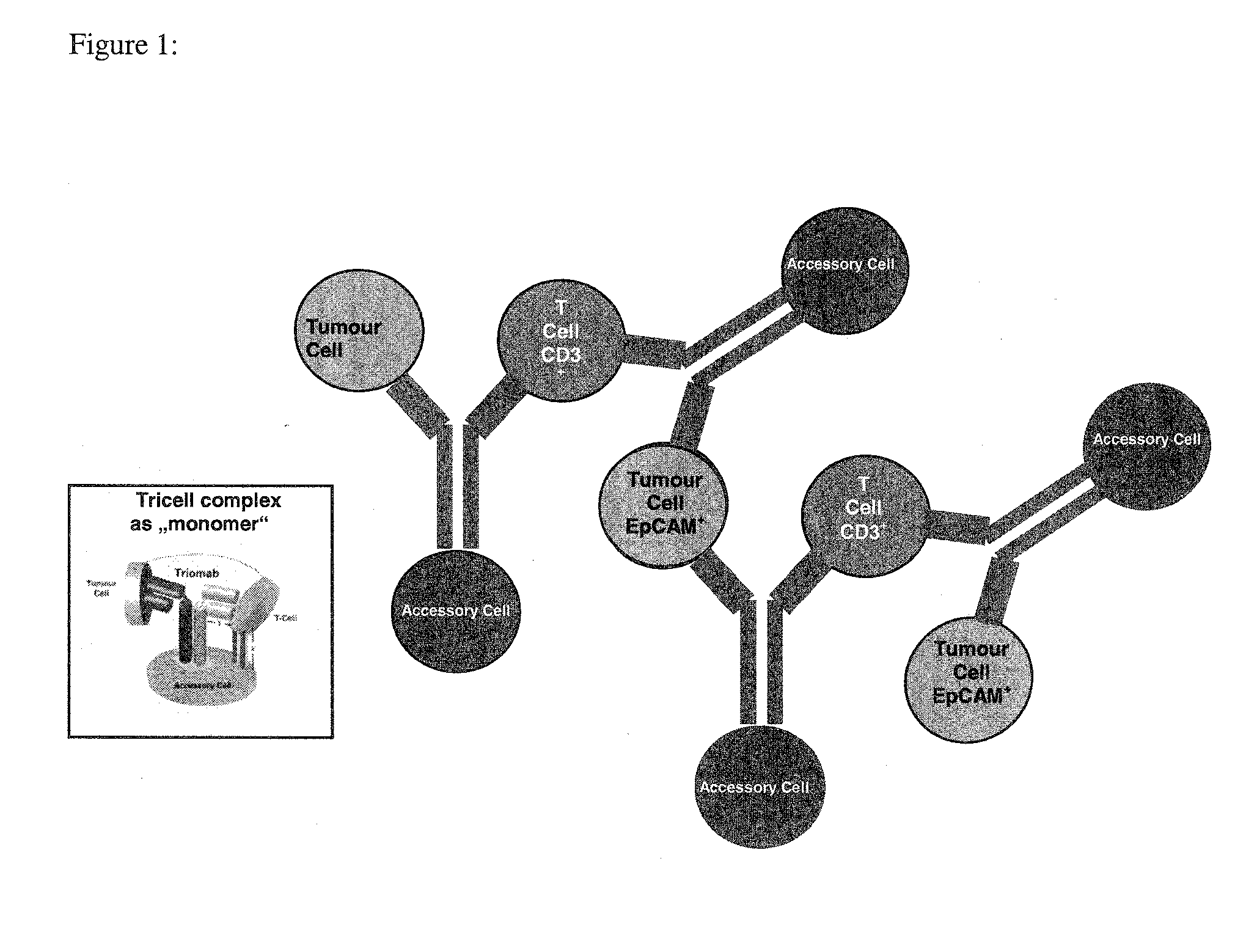
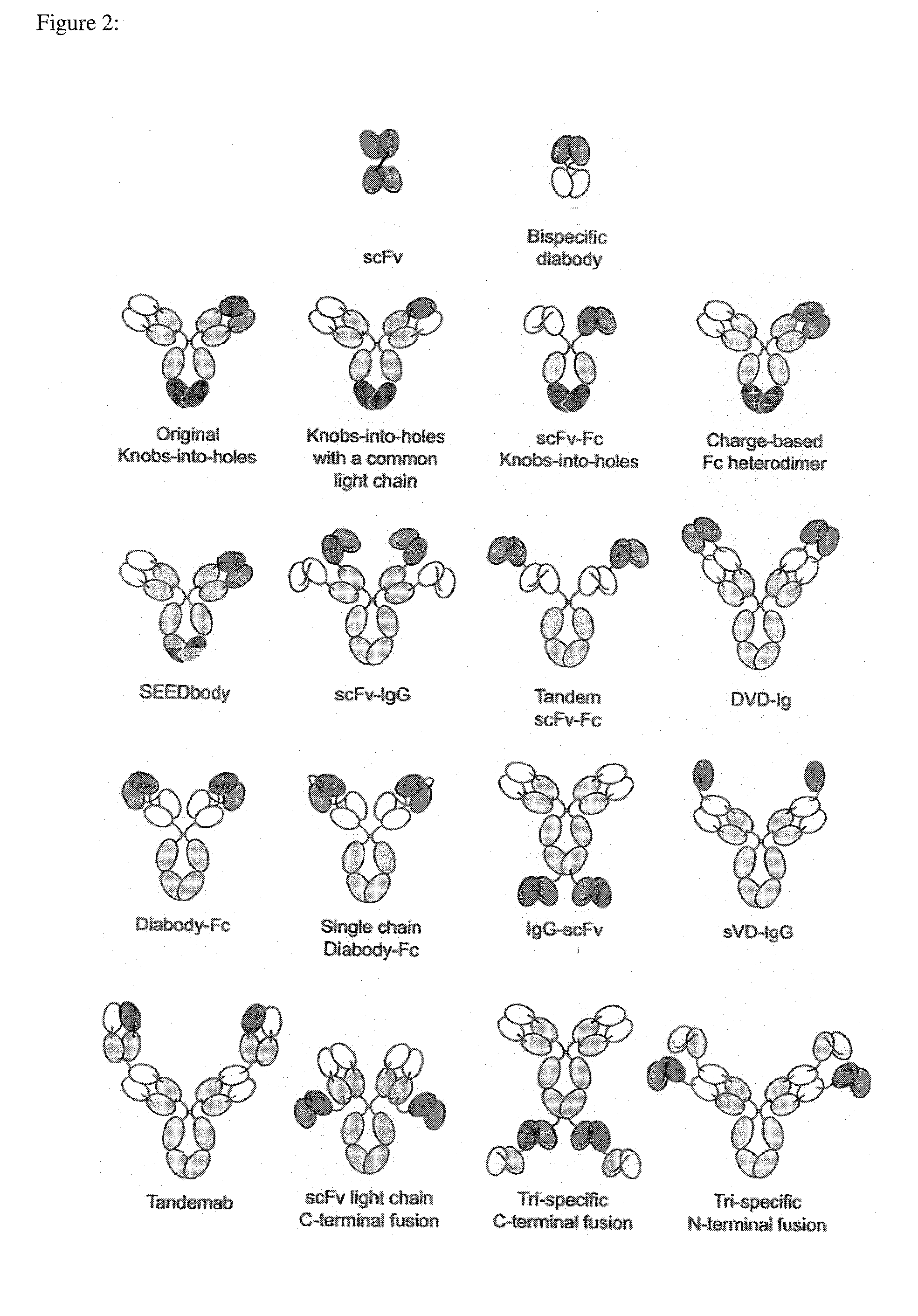
The present invention is based on a new method that uses antibodies to recognize and bind to tumor cells, and then remove them from the body. This method can also involve the recognition of immune cells like T cells and Fc-receptor positive accessory cells. The invention can also use protein scaffolds that mimic antibodies. Overall, this invention provides a way to potentially target and remove tumor cells from the body.

Problems solved by technology

Besides increased incidences of preoperative anemia, blood donation prior surgery raises serious additional economical questions due to the high overall transfusion rates (Carless et al., Transfus. Med. 14: 123, 2004).
Strikingly, the transmission of infectious diseases by means of contaminated blood transfusions appears to be a minor risk compared to the enormous risks of ABO-incompatible blood due to administrative or human failure along the blood donation and transfusion process (Sazama, Transfusion 30: 583, 1990; Dzik et al., Transfusion 43: 1190, 2003).
Pre- and intraoperative autotransfusion of red blood cells bypasses per se these inherent risks of allogeneic blood transfusion.
Due to the complex logistic demand of this IBS / irradiation approach including additional staff requirement, dosimetry issues, suitable as well as certified irradiation equipment in the clinical department, this latter procedure is not in favor of broad application.

Method used


Image

  • Removal of tumor cells from intraoperative autologous blood salvage
  • Removal of tumor cells from intraoperative autologous blood salvage
  • Removal of tumor cells from intraoperative autologous blood salvage

Examples


examples

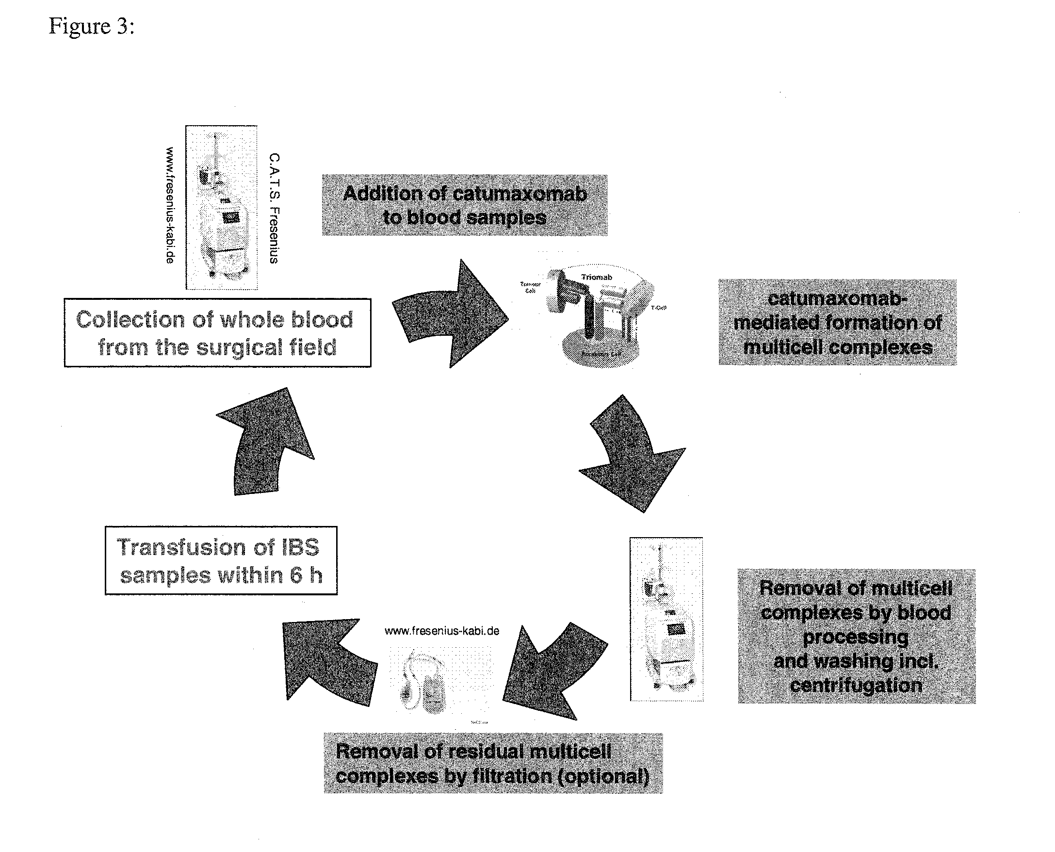
Anti-EpCAM Mediated Removal of Tumor Cells Through Multicell Complex Depletion by Centrifugation and / or Filtration During IBS

[0152]Expression profiles of the transmembrane glycoprotein EpCAM classified by tissue microarray staining occurs in carcinomas of various tumors (Table. 3). This broad expression EpCAM pattern represents an interesting hallmark of carcinomas which may be not only considerable for therapeutical application but also for improved IBS protocols during cancer surgeries. Nevertheless, it should be noted that most soft-tissue tumors and all lymphomas were completely EpCAM negative.

TABLE 3Tissue microrarray: EpCAM expressionprofile on various tumor entitiesNegativeStrongTumorNumber ofExpressionWeak / ModerateExpression*Entitysamples (n)(%)Expression# (%)(%)Prostate14141.910.987.2Colon111860.31.997.7Lung1128713.522.563.9Gastric14732.56.890.7Ovarian22722.612.185.31Data taken from Went et al., Brit. J. Cancer 94: 128, 2006.2Data taken from Spizzo et al., Gyneological Onco...


PUM

PropertyMeasurementUnit
temperatureaaaaaaaaaa

Abstract

The invention relates to a method performed ex vivo for removal of tumor cells from intraoperatively collected blood salvage, to antibodies and scaffold proteins which mimic antibodies for use in said ex vivo method, to the use of said ex vivo method for removal of tumor cells from intraoperatively collected blood salvage followed by reintroducing the so obtained purified blood salvage or of concentrates of erythrocytes purified by said method to a patient from whom said intra-operatively collected blood was obtained, as well as to blood salvage or a concentrate of erythrocytes, both obtainable by said method for reinfusion to said patient.

Description

FIELD OF THE INVENTION[0001]The invention relates to a method performed ex vivo for removal of tumor cells from intraoperatively collected blood salvage, to antibodies and scaffold proteins which mimic antibodies for use in said ex vivo method, to the use of said ex vivo method for removal of tumor cells from intraoperatively collected blood salvage followed by reintroducing the so obtained purified blood salvage or of concentrates of erythrocytes purified by said method to a patient from whom said intraoperatively collected blood was obtained, as well as to blood salvage or a concentrate of erythrocytes, both obtainable by said method for reinfusion to said patient.BACKGROUND OF THE INVENTION[0002]Since the AIDS epidemic of the early 1980s the interest in alternatives to allogeneic blood transfusion has grown, particularly for elective surgery. One alternative that currently accounts for over 5% of the blood donated in the United States and some countries in Europe is autologous tr...

Claims


Application Information

Patent Timeline
Patent Type & Authority Applications(United States)
IPC IPC(8): C12N5/00C07K16/30C12N5/078C07K16/46A61K35/18
CPCC12N5/0093C07K16/468C07K2317/31C12N5/0641C07K16/30A61K35/18C07K16/2809C07K2317/24C07K2317/626
Inventor LINDHOFER, HORSTSTROEHLEIN, MICHAELHEISS, MARKUS
Owner LINDIS BLOOD CARE GMBH
Learn More