Systems and methods for displaying medical images

a medical image and system technology, applied in the field of systems and methods for displaying medical images, can solve problems such as delay in treatment and/or diagnosis, and achieve the effect of facilitating facilitating the inserting of medical implements

Inactive Publication Date: 2015-09-17
EVENA MEDICAL
View PDF4 Cites 72 Cited by
  • Summary
  • Abstract
  • Description
  • Claims
  • Application Information

AI Technical Summary

Benefits of technology

[0093]The method can include illuminating the target area with near infrared light to produce a near infrared image on the display after confirming the vein using the ultrasound image; and using the near infrared image to facilitate the inserting of the medical implement into the vein.

Problems solved by technology

However, many methods and systems for displaying medical images require consulting stationary screens adapted only for displaying a single type of image.
Such limitations can cause delay in treatment and / or diagnosis.

Method used


Image

  • Systems and methods for displaying medical images
  • Systems and methods for displaying medical images
  • Systems and methods for displaying medical images

Examples


Embodiment Construction

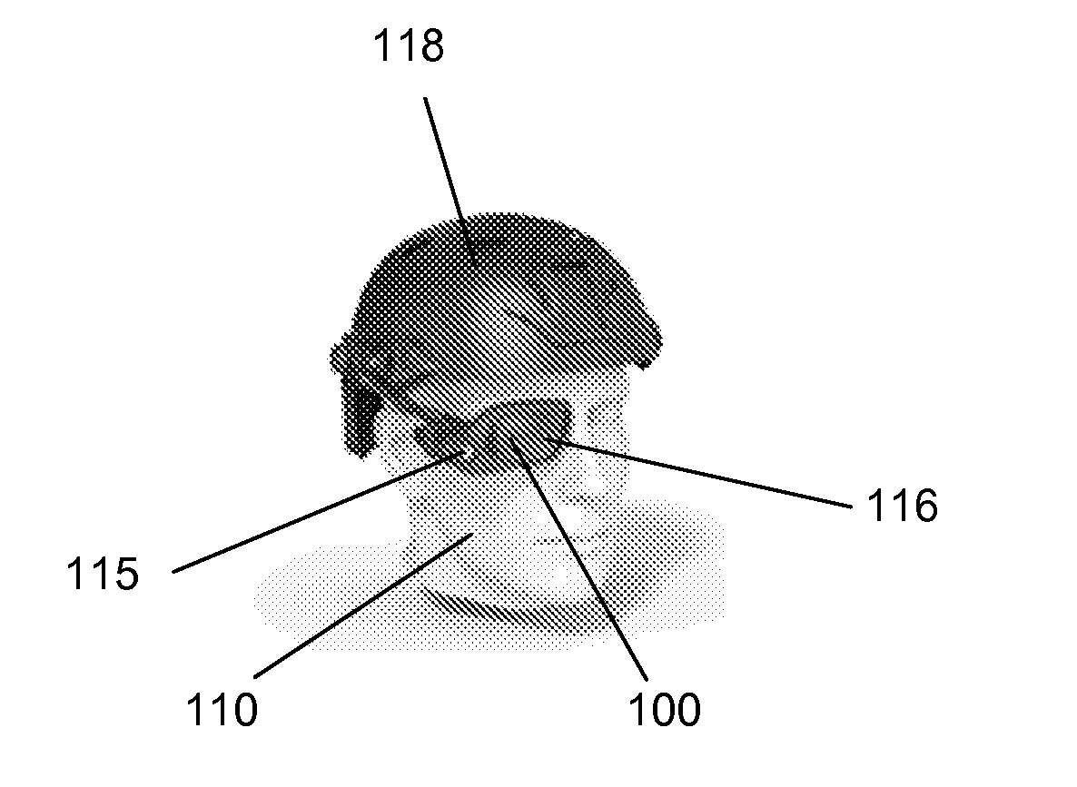
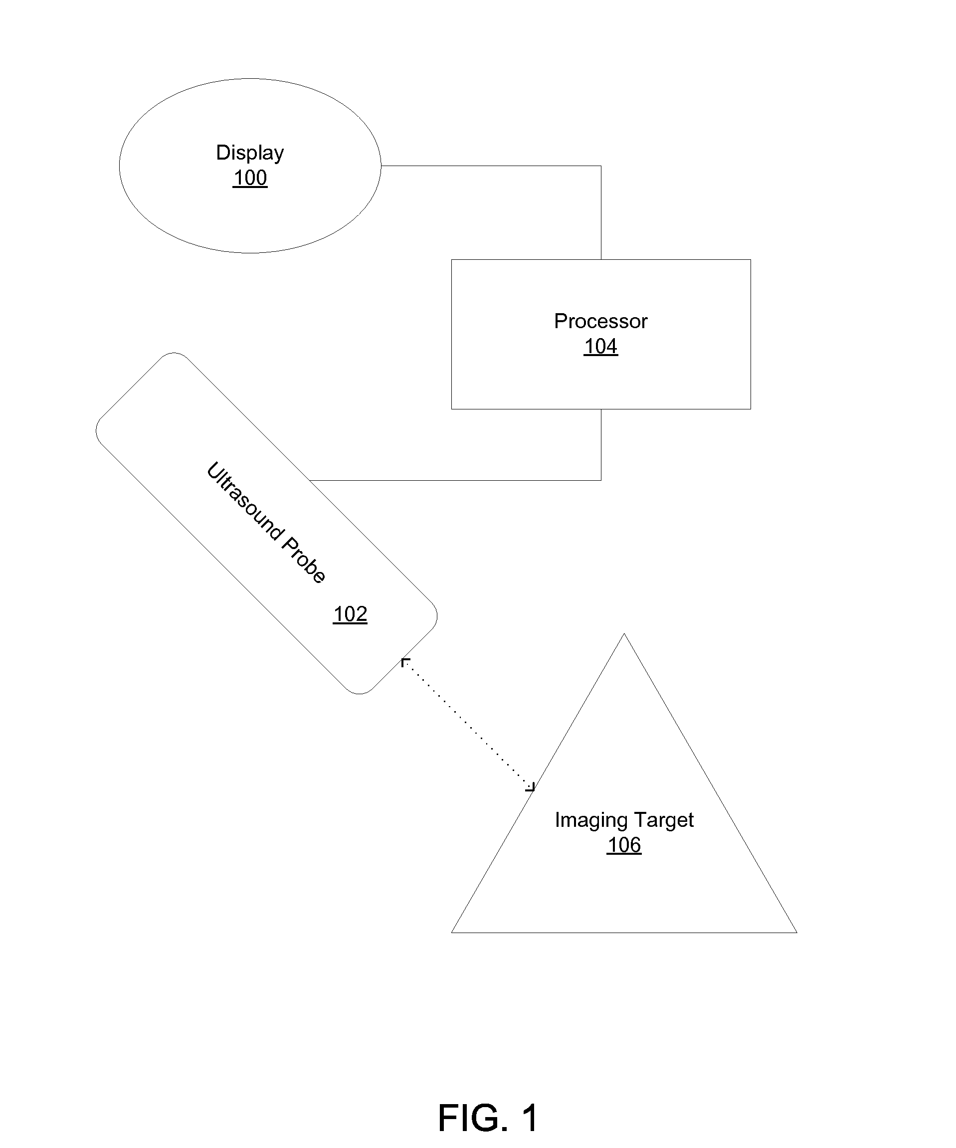
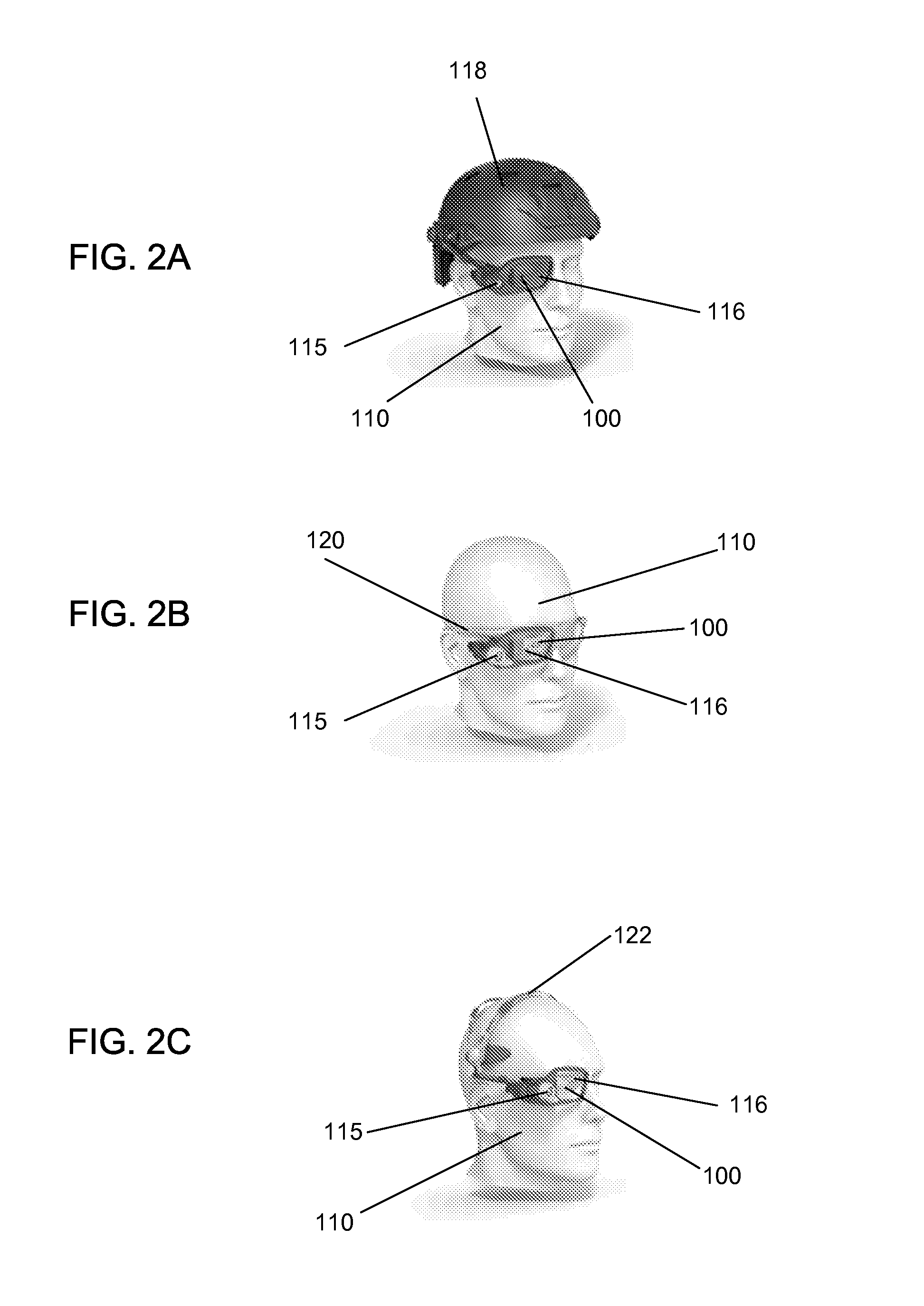
[0007]A method for displaying an ultrasound image can include identifying a target body portion of a patient to be imaged; emitting ultrasound signals from an ultrasound probe, wherein the ultrasound signals can cause echo signals to be reflected from tissue within the target body portion; receiving the echo signals; generating an ultrasound image of the target body portion based at least in part on the echo signals; and displaying the ultrasound image on a head-mounted display.

[0008]The method can include transmitting the ultrasound image to a remote display accessible to a remote healthcare professional; receiving treatment instructions from the remote healthcare professional; and administering treatment based at least in part on the instructions from the remote healthcare professional.

[0009]The method can include generating echo signal data based at least in part on the echo signals; and transmitting the echo signal data wirelessly to an image processor that generates the ultraso...


PUM


Abstract

Various embodiments relate to methods and systems for displaying an ultrasound image of a patient's target body portion using a wearable display module. A wearable display module can be mounted on a user's head. A wearable display module can display ultrasound images as a heads up display so that a user can view an ultrasound image while maintaining lines of sight with other viewpoints. Various embodiments relate to methods and systems for displaying both an ultrasound image and a light image (e.g., a near infrared image). A single display can be configured to display the light image and the ultrasound image on a single surface, in close proximity, and at the same time or in sequence. This disclosure provides in some embodiments for the use of wireless communication between various imaging probes, sensors, image processors, and display modules in order to facilitate more effective viewing of images.

Description

RELATED APPLICATIONS[0001]This application claims the benefit under 35 U.S.C. §119(e) of U.S. Provisional Patent Application No. 61 / 895,302, filed Oct. 24, 2013, and titled SYSTEMS AND METHODS FOR DISPLAYING MEDICAL IMAGES, and U.S. Provisional Patent Application No. 61 / 988,098, filed May 2, 2014, and titled SYSTEMS AND METHODS FOR DISPLAYING MEDICAL IMAGES. The entire contents of each of the above-identified applications are hereby incorporated by reference herein and made a part of this specification.INCORPORATION BY REFERENCE[0002]U.S. patent application Ser. No. 13 / 802,604 (the '604 Application), titled VEIN IMAGING SYSTEMS AND METHODS, filed on Mar. 13, 2013, and published as U.S. Patent Publication No. US 2014 / 0039309 on Feb. 6, 2014, is hereby incorporated by reference in its entirety.BACKGROUND[0003]1. Field of the Disclosure[0004]Some embodiments of this disclosure relate to systems and methods for displaying medical images, such as subcutaneous images taken of a patient wi...

Claims


Application Information

Patent Timeline
Patent Type & Authority Applications(United States)
IPC IPC(8): A61B8/00A61B8/08A61B8/14
CPCA61B8/462A61B8/14A61B8/565A61B8/5207A61B8/464A61B5/0035A61B5/0077A61B5/7445A61B8/4416A61B8/5261
Inventor BALL, FRANK J.CESPEDES, IGNACIO E.HARRIS, MELVYN L.GRUEBELE, DAVID J.HOSEIT, PAUL M.
Owner EVENA MEDICAL
Why Eureka
Try Eureka