Low emmission full mouth xray apparatus

a xray apparatus and low emmission technology, applied in the field of low emmission full mouth xray apparatus, can solve the problems of multiple x-ray bursts, equipment focus in the blind, operator exiting the room while the x-ray burst is occurring, etc., and achieves the effect of reducing the amount of escaped x-ray energy

Active Publication Date: 2015-10-01
GANZ JONATHAN
View PDF0 Cites 0 Cited by
  • Summary
  • Abstract
  • Description
  • Claims
  • Application Information

AI Technical Summary

Benefits of technology

[0009]The apparatus the present invention provides the ability to take an x-ray image of an entire mouth in one exposure while significantly reducing the amount of escaped x-ray energy. This is accomplished by using a full mouth insert that has slots for two strips of film, one for the upper set of teeth and one for the lower set of teeth, or an embedded imaging sensor.
[0012]Additionally, the full mouth insert has a waveguide means that directs the beam of x-ray energy from the horn of the transmitter to the inside of the mouth. Advantageously, the waveguide of the present invention is configured such that conventional x-ray machines may be used to accomplish the x-ray process. Misalignment of the x-ray machine is minimized since the waveguide of the present invention interfaces in an accurate and repeatable manner with the horn of the contemporary x-ray machine. Once the x-ray energy is present in the inside of the inner u-shaped segment, it is directed outward, through the teeth and then into a barrier that significantly reduces any excess radiation from exiting the apparatus. In this way the apparatus allows an accurate image of the entire mouth with only one shot with very low possibility of misalignment.

Problems solved by technology

While functional, this method has several serious drawbacks.
First, the patient is exposed to multiple x-ray bursts.
A third drawback of contemporary methods is that the operator of the apparatus must exit the room while the x-ray burst is applied.
A fourth drawback of current methods is the need for the operator to focus the equipment in the blind.
While this method improves the time required to take and analyze x-ray images, it still requires the four separate exposures to the x-rays and has the same excess radiation problem.

Method used


Image

  • Low emmission full mouth xray apparatus
  • Low emmission full mouth xray apparatus
  • Low emmission full mouth xray apparatus

Examples


first embodiment

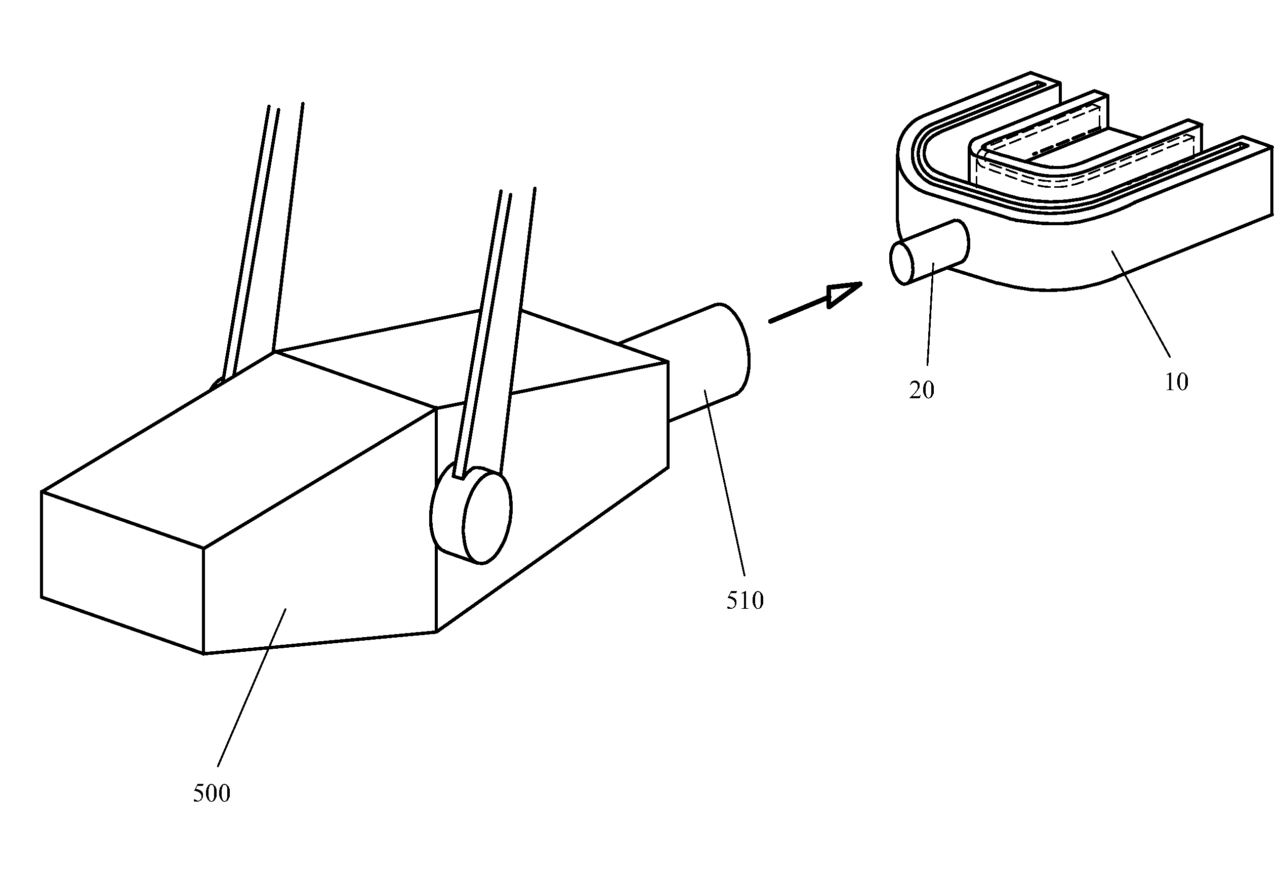
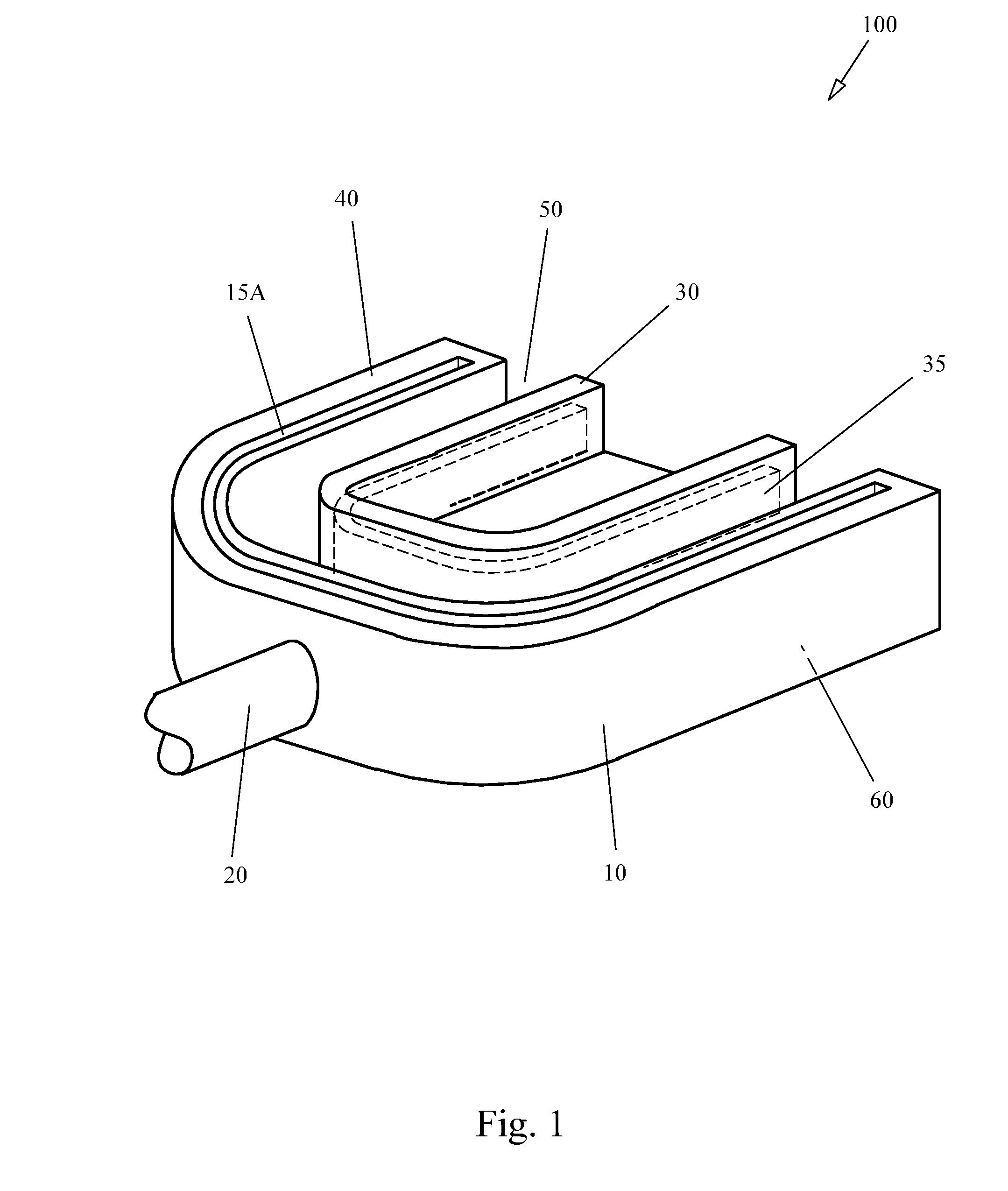
[0023]Full mouth insert 10 is made from some pliable material, for example plastic, allowing the waveguide means 35 to be encased and the upper and lower narrow film slots 15A and 15B to be formed on a mass produced basis. In this first embodiment the full mouth insert 10 is made from plastic, but as will be recognized by those of skill in the art, other materials, such as hard rubber, could be used without departing from the spirit of the invention, thus the scope of the invention is limited only by the claims. Not shown in FIG. 1 but described in detail below, the full mouth insert 10 also has a set of x-ray impeding barriers which prevent x-ray energy from escaping the apparatus either to the outside of the patient's mouth or to the inside of the patient's mouth.

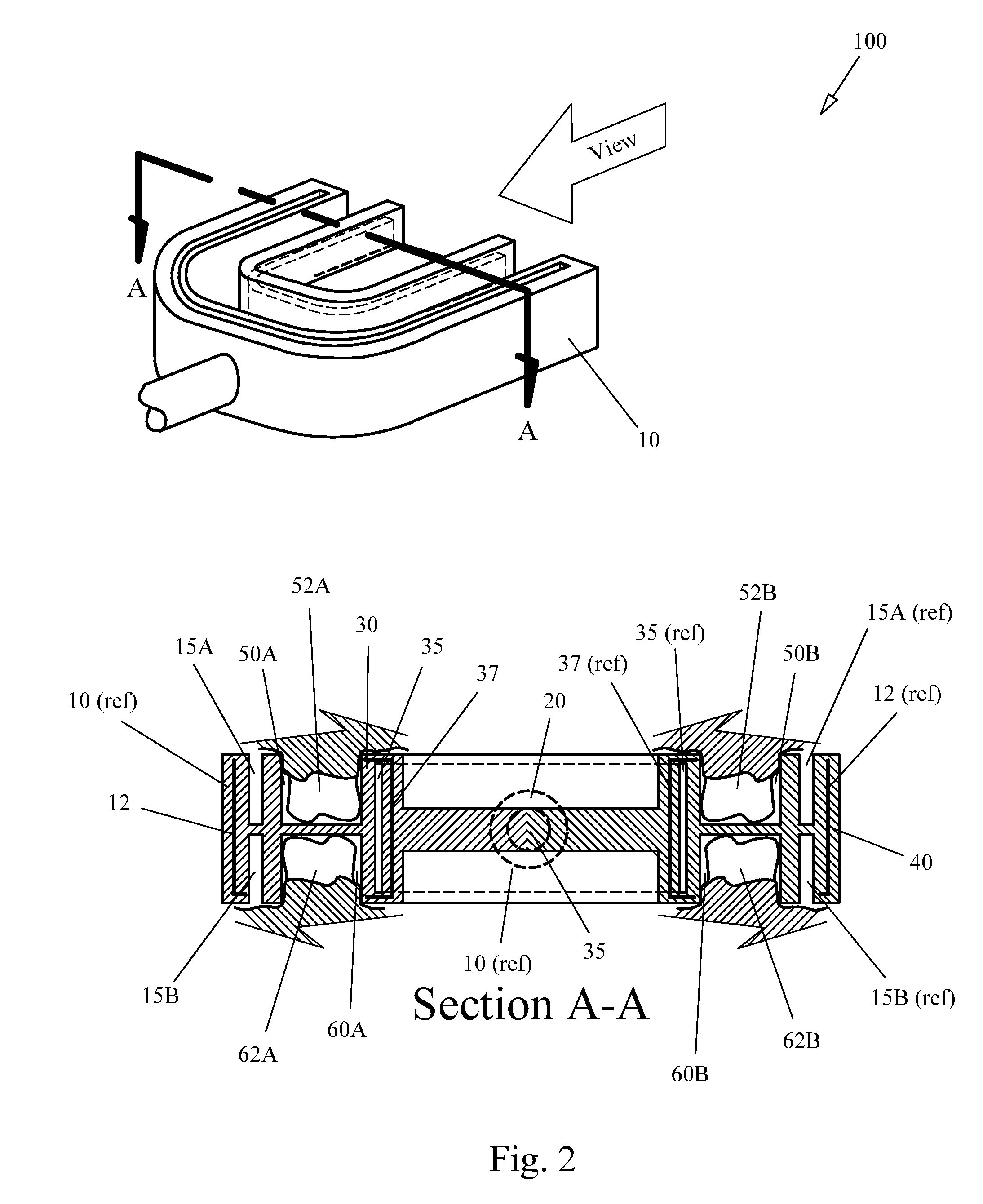
[0024]Looking now at FIG. 2, the apparatus of the present invention 100 shows the full mouth insert 10 in latitudinal cross section A-A. Note that the viewing perspective is from the rear of the full mouth insert 10 towar...

second embodiment

[0033]FIG. 5 provides an overview 200 of a second, preferred embodiment of the apparatus of the present invention. This second embodiment is for use with contemporary computer based imaging systems. A full mouth insert 210 has a pair of cavities 250 and 260 to receive the teeth of a patient to be imaged. The upper tooth cavity 250 accommodates the upper set of teeth while the lower tooth cavity 260 (not shown in detail) accommodates the lower teeth. The full mouth insert 210 can be made in several sizes to allow use on any size mouth, from early childhood to adult.

[0034]As was the case for the first embodiment, a waveguide 235 is embedded in the inner u-shaped cavity 230 and is connected to the focusing tube 220. Also as was the case for the first embodiment, the x-ray barriers in both the outer and inner u-shaped cavities 230 and 240 are present. The fact that they are not shown is for the sake of clarity, but those of skill in the art will recognize that their presence and functio...


PUM


Abstract

The apparatus the present invention provides the ability to take an x-ray image of an entire mouth in one exposure while significantly reducing the amount of escaped x-ray energy. This is accomplished by using a full mouth insert that has slots for two strips of film, one for the upper set of teeth and one for the lower set of teeth, or an embedded imaging sensor.

Description

[0001]This application for patent claims the benefit of U.S. provisional application 61 / 808,453 filed Apr. 4, 2013.BRIEF DESCRIPTION[0002]The subject of this invention relates to x-ray imaging, specifically, to a full mouth x-ray apparatus. The disclosed invention presents an apparatus that allows the entire mouth to be imaged in one exposure, with very low radiation escape providing superior protection for the patient. The disclosed invention may be used with contemporary medical / dental x-ray devices.BACKGROUND OF THE INVENTION[0003]The need for the medical / dental practitioners to take x-ray images of the human mouth remains as necessary modernly as it has historiocally. Over time the technology has remained the same, but the equipment used to implement the technology has continued to advance.[0004]Contemporarily, if an x-ray image of patient's full mouth is needed, it is done in segments. That is, a film card or image sensor is placed in one quadrant or another of the patient's mo...

Claims


Application Information

Patent Timeline
Patent Type & Authority Applications(United States)
IPC IPC(8): A61B6/14
CPCA61B6/145A61B6/04A61B6/4208
Inventor GANZ, JONATHAN
Owner GANZ JONATHAN
Why Eureka
Try Eureka