Angiopoietin-related protein 4 (cangptl4) as a diagnostic biomarker for acute lung damage
an angiopoietin-related protein and diagnostic biomarker technology, applied in the field of cterminal fragment of angiopoietin-related protein 4 cangptl4, can solve the problems of multiple antibiotic resistance strains, chest pain, fever and difficulty breathing, and huge financial burden on the health system worldwid
- Summary
- Abstract
- Description
- Claims
- Application Information
AI Technical Summary
Benefits of technology
Problems solved by technology
Method used
Image
Examples
example 2
cAngptl4 Expression Coincides and Co Localizes with Inflammation-Induced Lung Damage in Influenza Infection on Mice (FIG. 2)
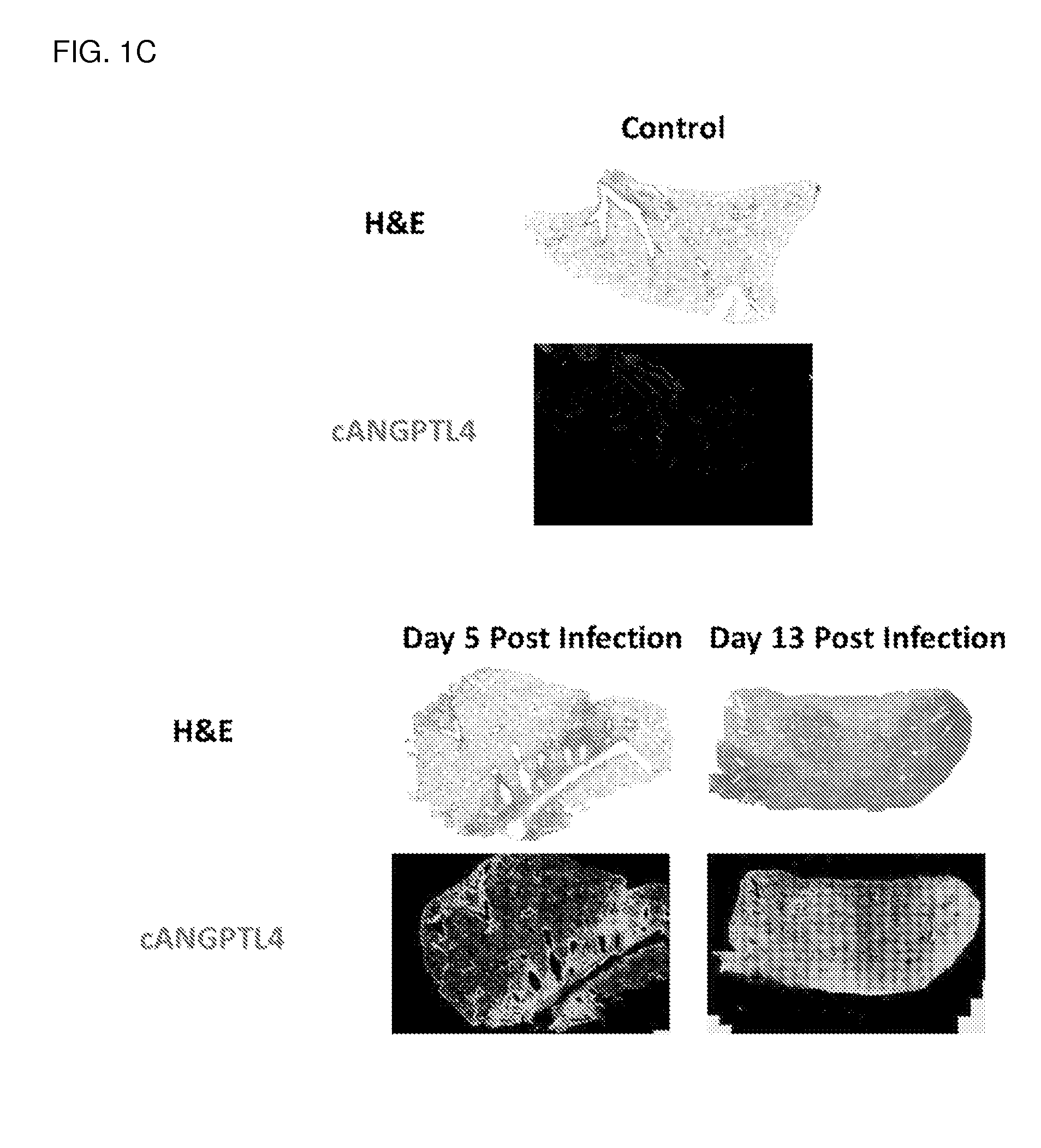
[0171]Mice were infected with PR8 virus and lungs were harvested at day 0 (used as control), day 5 and day 13 post infection. In control mice, no inflamed region and little cAngptl4 expression can be detected in alveolar spaces. The expression of cAngptl4 was restricted to the well-defined blood vessel structure. For infected mice, dense staining in H&E marked lung regions that were heavily infiltrated by immune cells at day 5 and 13 post infection, which is a typical event in inflammation and caused local lung damage. In contrast to control mice, the expression of cAngptl4 is not limited to blood vessels, but was extensively detected in the lung regions of massive infiltration of immune cells. Bronchioles consistently express cANGPTL4 throughout the entire disease progression.
example 3
Identification of the Source for cANGPTL4 Production During Influenza Infection (FIG. 3)
[0172]The lung tissue sections were further analyzed to identify the main sources for cANGPTL4. (a) Dual immunohistochemistry staining was performed using major cell markers and cANGPTL4. Clara cells (CC10 positive) were shown to be consistently stained with anti-cANGPTL4 antibody throughout the disease progression. (b) Tunica media of the blood vessels was also shown to be stained with anti-cANGPTL4 antibody for the entire disease progression, while day 13 inflammation makes the staining of cANGPTL4 on tunica media less distinguishable from the surrounding tissues that are also stained with cANGPTL4. Tunica media at day 5 showed much higher staining of cANGPTL4 than the other time points, indicating active production of ANGPTL4. (c) Infected type II alveolar epithelial cells produces cANGPTL4 while non-infected type II alveolar epithelial cells do not.
example 4
cANGPTL4 Detection in Various Forms of Samples. (FIG. 4)
[0173](a) cANGPTL4 is a secreted protein and is expected to be detected from samples in various forms including serum, sputum and BALF, allowing convenient diagnosis using methods such as western blot or size-filtered protein ELISA for cANGPTL4; (b) cANGPTL4 was detected in BALF protein by western blot using anti-cANGPTL4 antibody. The BALF was collected from PR8 influenza-infected mice at different time point post infection. Day 11 and Day 13 samples have significantly more cANGPTL4 than Day 0 protein, showing cANGPTL4 can be conveniently detected from airway secretions. (c) Secondary infection following flu infection increases cANGPTL4 expression in mouse model. For mice mock infected with heat-inactivate influenza virus and S. Pneumoniae, cANGPTL4 signal was only detected at bronchioles but not in other regions such as alveolar space. For mice infected with 1000 CFU S. Pneumoniae only, similar cANGPTL4 signal was observed as...
PUM
| Property | Measurement | Unit |
|---|---|---|
| Composition | aaaaa | aaaaa |
Abstract
Description
Claims
Application Information
 - R&D
- Intellectual Property
- Life Sciences
- Materials
- Tech Scout
- Unparalleled Data Quality
- Higher Quality Content
- 60% Fewer Hallucinations
Browse by: Latest US Patents, China's latest patents, Technical Efficacy Thesaurus, Application Domain, Technology Topic, Popular Technical Reports.
© 2025 PatSnap. All rights reserved.Legal|Privacy policy|Modern Slavery Act Transparency Statement|Sitemap|About US| Contact US: help@patsnap.com


