
Extraoral dental scanner

a dental scanner and scanner body technology, applied in the field of extraoral dental scanners, can solve problems such as almost gapless data sets

Active Publication Date: 2016-01-14
DENTSPLY SIRONA INC
View PDF5 Cites 14 Cited by
  • Summary
  • Abstract
  • Description
  • Claims
  • Application Information

AI Technical Summary

Benefits of technology

This patent describes a dental scanner that allows for precise measurements and modeling of dental parts. The scanner has a small measurement volume, which leads to higher measurement precision, and the camera and shaping part can move independently. This allows for easy positioning of the dental part and model rotation without interfering with the measurement process. The scanner can automatically scan most relevant model geometries from multiple angles using a special combination of optics and axis movement. Overall, this patent presents a solution for improving dental procedures by providing precise and efficient measurement tools.

Problems solved by technology

Here the high freedom of movement of the axial system is fully exploited, which results in almost gapless data sets.

Method used


Image

  • Extraoral dental scanner
  • Extraoral dental scanner
  • Extraoral dental scanner

Examples


Embodiment Construction

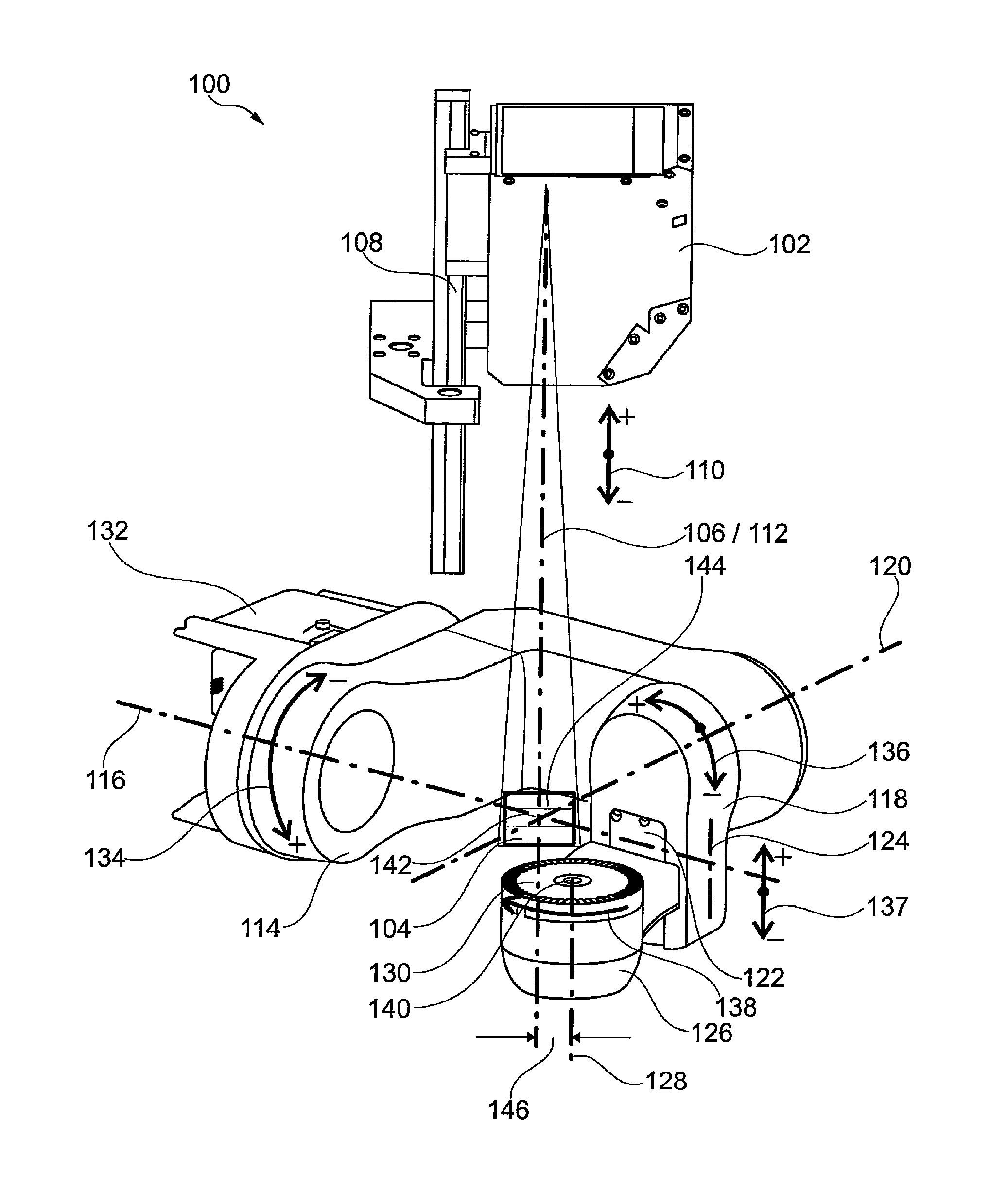
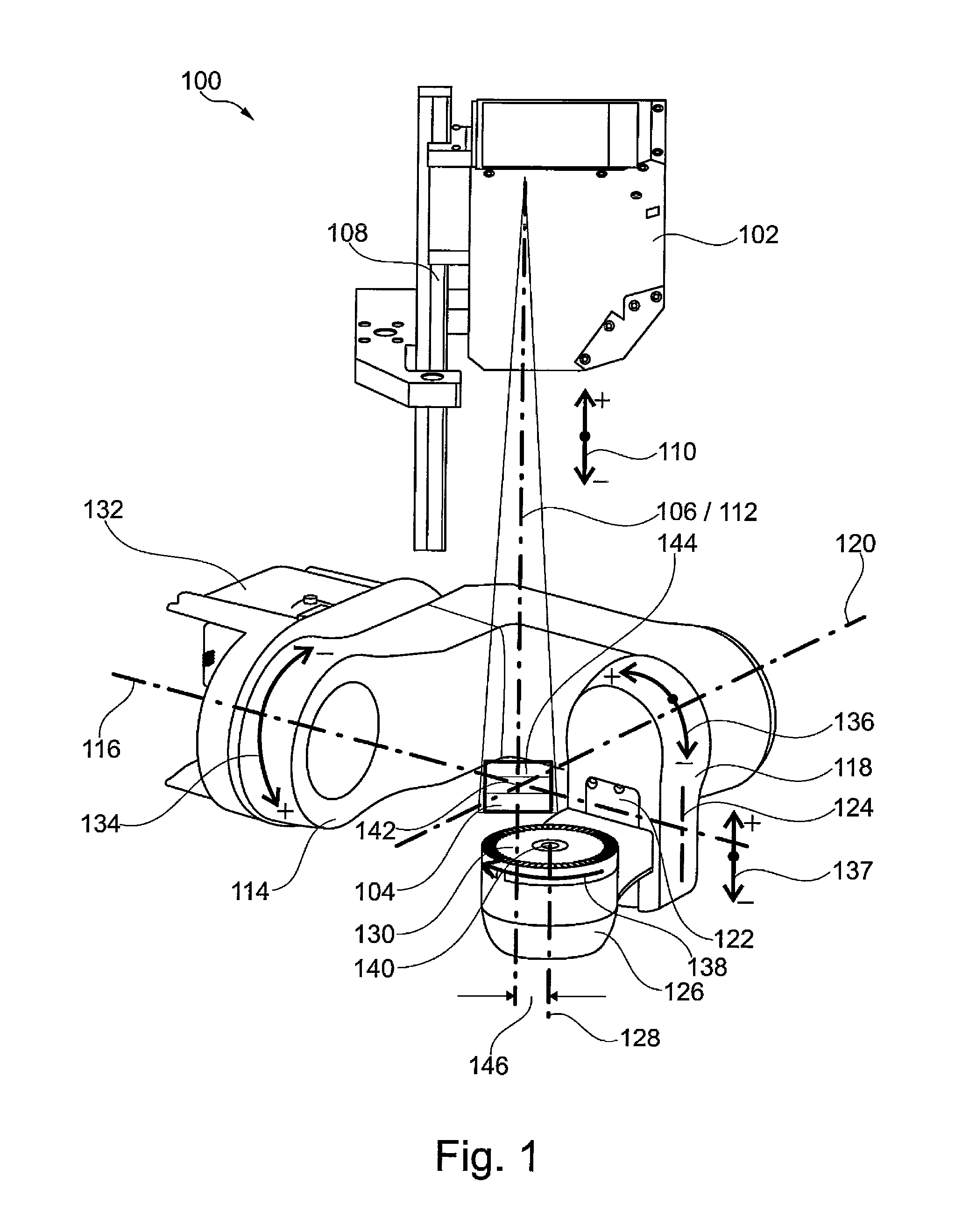
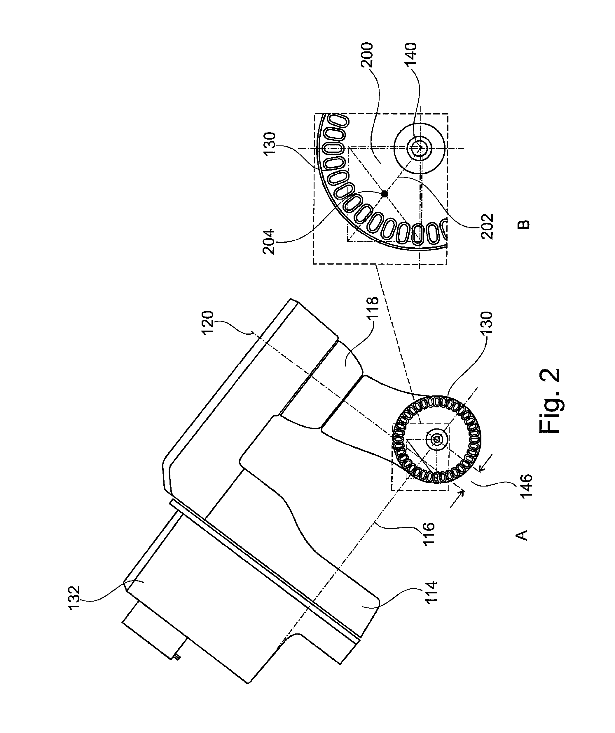
[0075]The extraoral dental scanner 100 shown schematically in its neutral position in the partial view in FIG. 1 and used for the three-dimensional capture of the surface of a dental shaped part has the following principal elements or assemblies which are arranged inside a housing (not shown):[0076]a 3D measuring camera 102 for the three-dimensional capture of the surface of the dental shaped part in a measurement volume 104 of the 3D measurement camera 102, the 3D measurement camera 102 having an optical axis 106 and a measurement volume 104;[0077]means for the relative positioning of the 3D measurement camera 102 and of the dental shaped part, namely:[0078]a camera elevation module (linear drive module) 108 for the vertical movement of the 3D measurement camera 102 in either direction 110 along a linear axis 112 (first axis), whose position is identical to that of the optical axis 106 of the 3D measurement camera 102;[0079]a tilting module 114 with an axis of rotation 116 (second ...


PUM


Abstract

An extraoral dental scanner for three-dimensional capture of the surface of a dental shaped part (300) with a 3D measuring camera (102) having an optical axis (106), wherein the means for the machine-controlled relative positioning of the 3D measuring camera (102) and the dental shaped part (300) are embodied in such a way that the means for taking up and positioning the dental shaped part (300) can be moved into a parking position outside a region that can be captured optically by the 3D measuring camera (102), with a work plate (708) for manually positioning the dental shaped part (300) in the measurement volume (144) of the 3D measuring camera (102), wherein the work plate (708) is aligned perpendicularly to the optical axis (106) and wherein the work plate (708), as viewed from the 3D measuring camera (102), is arranged behind the means for taking up and positioning the dental shaped part (300), makes it possible to record uninterrupted 3D image data with very short recording times both by automatic and by manual positioning of dental shaped parts of different sizes and embodiment variants.

Description

FIELD OF THE INVENTION[0001]The invention concerns an extraoral dental scanner. With extraoral dental scanners the dental shaped parts to be measured must be positioned during recording in a suitable position in a measurement volume of the scanner. By a dental shaped part is meant dental models such as, for example, maxillary / mandibular models articulated in the articulator, individual full-jaw models, individual single-tooth models, partial jaw models, wax-up models, full-jaw impression trays, counter-bite impression trays. The model is recorded in different views so as to obtain the most complete uninterrupted data possible. Here the surfaces to be scanned should be aligned as perpendicularly as possible to the optical axis of the measurement camera in order to obtain a high quality of data. The measurement volume is a virtual demarcated space which, for example, can have the geometrical shape of an approximate cuboid, within which the relevant areas on the surface of the dental s...

Claims


Application Information

Patent Timeline
IPC IPC(8): A61C13/34A61C9/00G01B11/24H04N13/02
CPCA61C13/34G01B11/24H04N13/0203A61C9/0093A61C9/006G01B5/0004G01B11/25G01B11/2522H04N13/204
Inventor JUMPERTZ, RAINER
Owner DENTSPLY SIRONA INC
Learn More