Method for Inhibiting Proliferation of High Lix1I-Expressing Tumor Cell, and Tumor Cell Proliferation-Inhibiting Peptide
- Summary
- Abstract
- Description
- Claims
- Application Information
AI Technical Summary
Benefits of technology
Problems solved by technology
Method used
Image
Examples
reference example 1
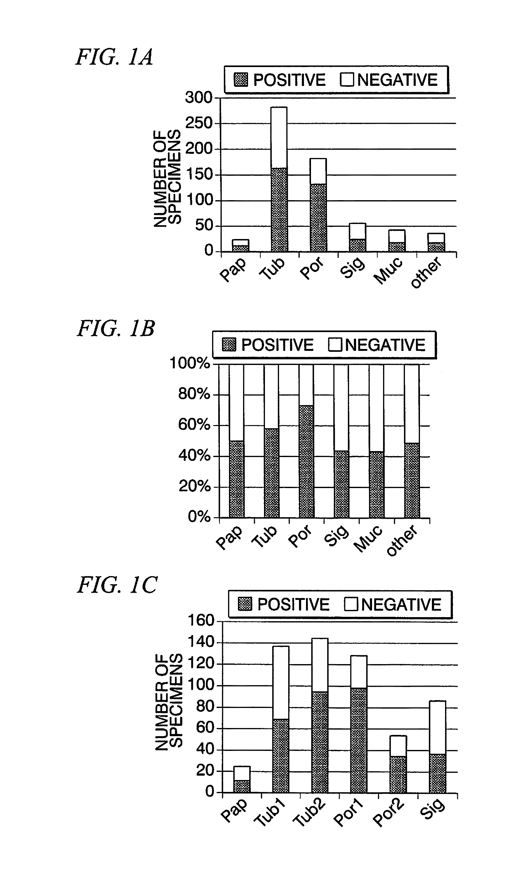
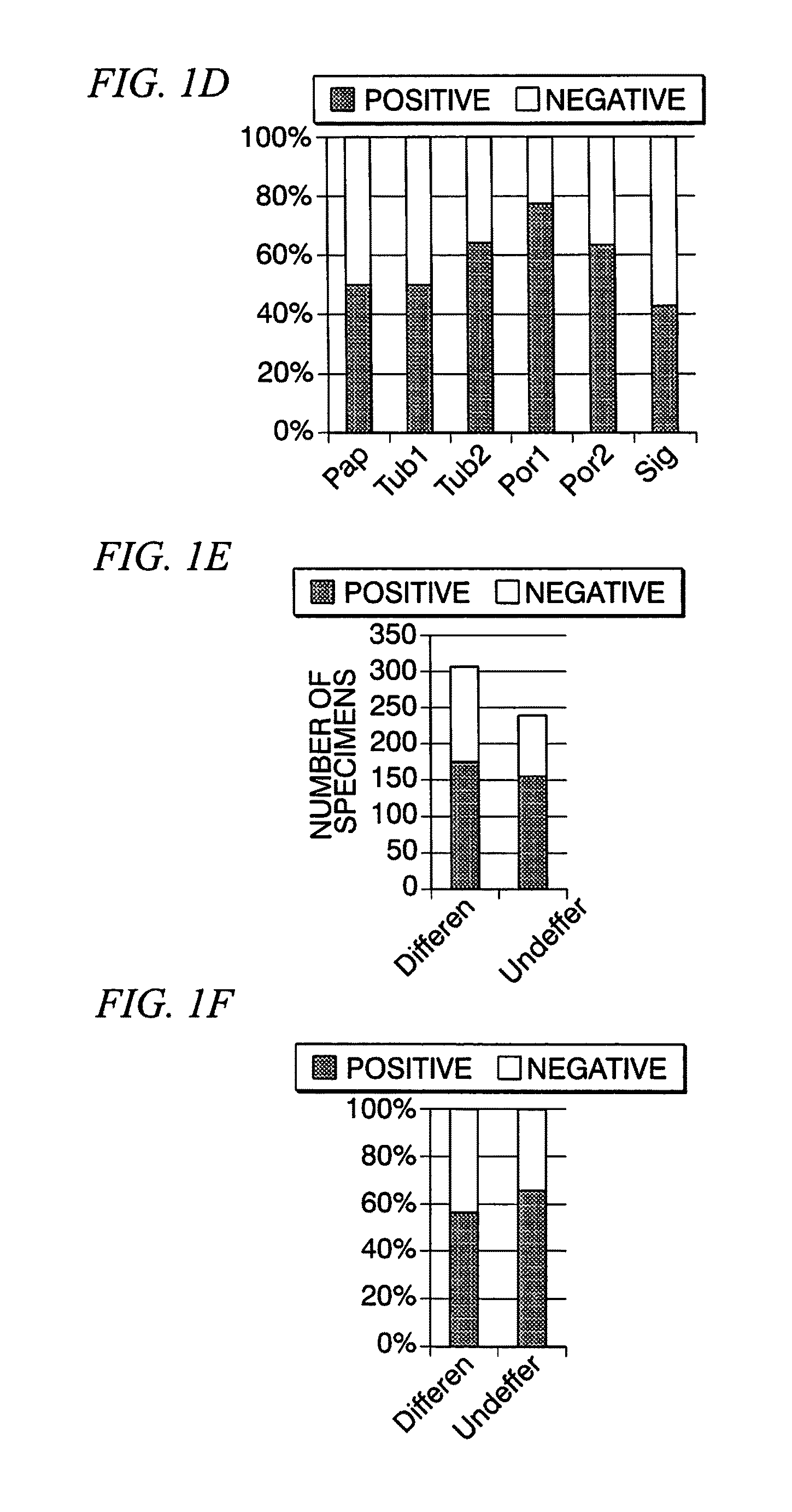
[0234]The expression levels of LIX1L genes of gastric cancer clinical specimens collected from patients with gastric cancer were quantitatively detected through immunohistochemical staining for comparison. These gastric cancer clinical specimens were classified into papillary adenocarcinoma (Pap), tubular adenocarcinoma (Tub), poorly differentiated adenocarcinoma (Por), signet ring cell carcinoma (Sig), mucinous carcinoma (Muc), and others. The tubular adenocarcinoma was further classified into a well-differentiated type (Tub 1) and a moderately-differentiated type (Tub2), and the poorly differentiated adenocarcinoma was further classified into a solid type (Por1) and a nonsolid type (Por2). Among these, the papillary adenocarcinoma and the tubular adenocarcinoma were differentiated tumor cells and the others were undifferentiated tumor cells.
[0235]In general, a normal cell and a tumor cell coexist in one tumor tissue. Therefore, a specimen of which the proportion of a high LIX1L-ex...
reference example 2
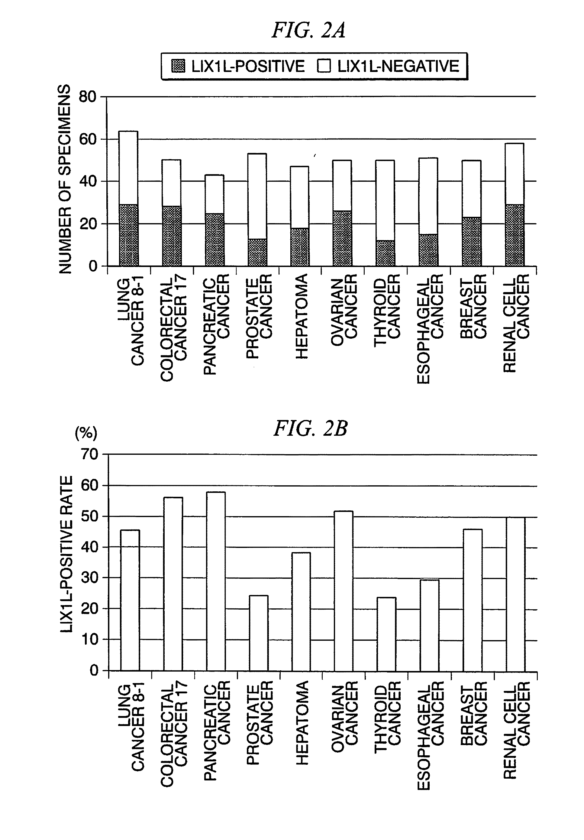
[0240]Similarly to Reference Example 1, immunohistochemical staining was performed on clinical specimens collected from patients with solid tumors (lung cancer, colorectal cancer, pancreatic cancer, prostate cancer, hepatoma, ovarian cancer, thyroid cancer, esophageal cancer, breast cancer, and renal cell cancer) other than gastric cancer, and the number of LIX1L-positive specimens and LIX1L-negative specimens in various kinds of cancer and the proportion thereof were obtained.
[0241]The number of LIX1L-positive specimens and LIX1L-negative specimens in various kinds of cancer obtained from the results of the immunohistochemical staining is shown in FIG. 2A, and results in which the frequency of LIX1L-positive tumors in various kinds of cancer is calculated from the results thereof is shown in FIG. 2B. In all kinds of cancer, about 20% to 60% of the tumors were LIX1L-positive tumors.
reference example 3
[0242]The expression levels of LIX1L genes in gastric cancer cell strains were quantitatively measured from both a protein level and an mRNA level through a Western blotting method and an RT-PCR method for comparison.
[0243]Two kinds of strains including KATO-III strains and OCUM-1 strains were used as gastric cancer cell strains. Both cell strains are undifferentiated tumors. Each of the KATO-III strains was analyzed after being cultured for 10 days to 14 days in a culture solution in which 10 vol. % of fetal calf serum (FCS) with respect to an RPMI 1640 culture medium was added to the RPMI 1640 culture medium. Each of the OCUM-1 strains was analyzed after being cultured for 10 days to 14 days in a culture solution in which 10 vol. % of FCS with respect to a DMEM culture medium was added to the DMEM culture medium. Specifically, regarding each analysis, first, the cultured cells were collected, and Western blotting was used for some of the cultured cells and RT-PCR was used for the ...
PUM
| Property | Measurement | Unit |
|---|---|---|
| Length | aaaaa | aaaaa |
| Permeability | aaaaa | aaaaa |
| Cell proliferation rate | aaaaa | aaaaa |
Abstract
Description
Claims
Application Information
 - R&D
- Intellectual Property
- Life Sciences
- Materials
- Tech Scout
- Unparalleled Data Quality
- Higher Quality Content
- 60% Fewer Hallucinations
Browse by: Latest US Patents, China's latest patents, Technical Efficacy Thesaurus, Application Domain, Technology Topic, Popular Technical Reports.
© 2025 PatSnap. All rights reserved.Legal|Privacy policy|Modern Slavery Act Transparency Statement|Sitemap|About US| Contact US: help@patsnap.com


