Dual Needle Core Biopsy Instrument

a biopsy instrument and needle core technology, applied in the field of dual needle core biopsy instruments, can solve the problems of wasting medical resources and dollars, destined to fail, and affecting the patient's overall health, and causing the patient to be more compromised and still needing an alternate treatment protocol, and ineffective treatmen

Active Publication Date: 2016-03-17
ACKROYD ROBERT K
View PDF9 Cites 4 Cited by
  • Summary
  • Abstract
  • Description
  • Claims
  • Application Information

AI Technical Summary

Benefits of technology

[0010]A dual needle core biopsy instrument that can take two samples directly adjacent to each other at exactly the same time. This instrument would ensure that two samples of the tissue taken for biopsy would be representative of each other. One would be sent for microscopic diagnostic evaluation and the second would be submitted for genetic studies. The instrument will also ensure that only diagnostic tissue would be sent for the more expensive molecular testing. If the tumor tissue was not represented by microscopy, the molecular test would not be processed.
[0011]The novel design concept is based on two (instead of just one) core sample needles incorporated into a single triggering instrument. These core needles would be separated no more than 0.1 mm to 3 mm apart. This would ensure that two adjacent representative samples of tissue are taken simultaneously.

Problems solved by technology

Treatments destined to fail impart increased morbidity, unnecessary side effects, and leave the patient more compromised and still in need of an alternate treatment protocol.
Ineffective treatment also wastes both medical resources and dollars.
If additional core biopsies are available for molecular studies, there is no guarantee that each individual core will have diagnostic tissue present.
This presents the possibility of false negative molecular results.
Needle core biopsies are suitable for these tests, however, without microscopic confirmation, it is impossible to guarantee that the sample taken for genetic testing adequately represents the tumor being biopsied.

Method used


Image

  • Dual Needle Core Biopsy Instrument
  • Dual Needle Core Biopsy Instrument
  • Dual Needle Core Biopsy Instrument

Examples


first embodiment

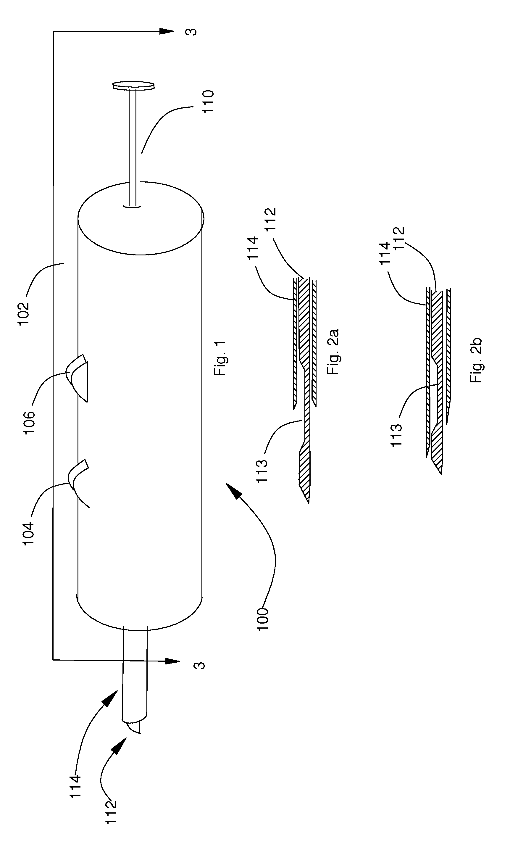
[0021]FIG. 1 is a front perspective view of a single needle core biopsy instrument 100. Illustrated in FIG. 1 is a case 102, a first sliding button 104, a second sliding button 106, a cannula cylinder 114, a plunger 110, and the needle 112. In the following, the term forward means towards the needle 112 end and the term backward means towards the plunger 110 end. Similarly, the terms clockwise and counterclockwise refer to the figure under discussion.

[0022]FIGS. 2a and 2b illustrate sectional views of a portion of the needle 112 and the cannula cylinder 114 with the needle 112 in an extended and retracted position respectively. The needle 112 has a tissue receiving recess 113 that collects the tissue material during the biopsy collection process.

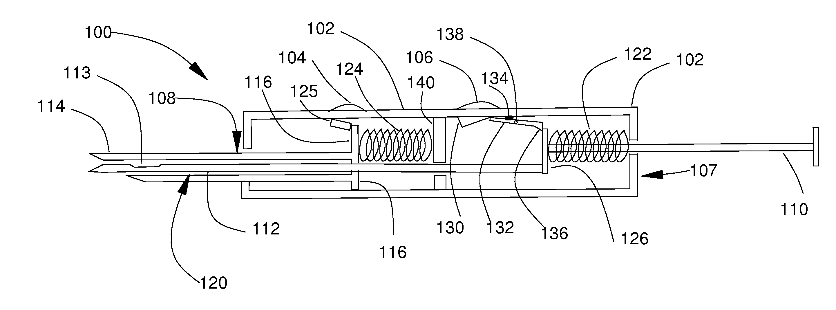
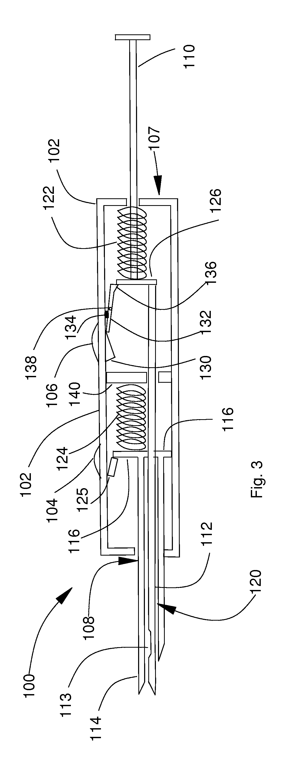
[0023]FIG. 3 is a sectional view of the needle core biopsy instrument, the section indicated in FIG. 1. It has a case component 107, a cannula component 108, a needle component 120, and a trigger component 128. The case component 107 has a c...

second embodiment

[0039]Referring to FIG. 9, the dual needle core biopsy instrument 200 is comprised of a dual needle case 202, a first sliding button 104, a second sliding button 106, dual cannula cylinders 214, a plunger 110, and dual needles 212. Similar to the description for the single needle core biopsy instrument 100 the term forward means towards the dual needles 212 end and the term backward means towards the plunger end.

[0040]FIGS. 9a and 9b illustrate sectional views of a portion of the dual needle 212 and the dual cannula cylinders 214 with the dual needles 212 in an extended and retracted position respectively. Note that the dual needles 212 each has a tissue receiving recess 113 that collects the tissue material during the biopsy collection process.

[0041]FIG. 10 is a sectional view of the dual needle core biopsy instrument 200, the section indicated in FIG. 8. It has a dual needle case component 207, a dual cannula component 208, a dual needle component 220, and a trigger component 128....


PUM


Abstract

The embodiments presented herein present a single needle core biopsy instrument and modification required that convert the single needle core biopsy instrument to a dual needle core biopsy instrument. Depending on the design, the dual needle core biopsy instrument permits the simultaneous extraction of tissue or bone specimens taken at between 0.1 mm and 3 mm apart.

Description

[0001]This application claims the benefit of the U.S. Provisional Patent Application No 62 / 049783 filed Sep. 12, 2014 by the present inventor. This provisional patent application is incorporated herein by reference.STATEMENT REGARDING FEDERALLY SPONSORED RESEARCH OR DEVELOPMENT[0002]Not ApplicableREFERENCE TO SEQUENCE LISTING, TABLE, OR A COMPUTER PROGRAM LISTING[0003]Not ApplicableCOMPACT DISK APPENDIX[0004]Not ApplicableBACKGROUND OF THE DISCLOSURE[0005]The era of Molecular diagnostics and personalized medicine is now upon us and it will continue to rapidly grow into the next decade. Novel techniques for the diagnosis and treatment of Cancer and other diseases are evolving in an exponential basis as the genetic code of disease is dissected from tissue and blood samples.[0006]As more and more molecular markers are discovered, we are able to subdivide all cancer types into different genetic subtypes. We are discovering that many morphologically identical tumors responded differently...

Claims


Application Information

Patent Timeline
Patent Type & Authority Applications(United States)
IPC IPC(8): A61B10/02
CPCA61B10/0233A61B2010/0225A61B2010/0208A61B10/025
Inventor ACKROYD, ROBERT, K.
Owner ACKROYD ROBERT K
Why Eureka
Try Eureka