A kind of preparation method and specimen of intraosseous blood vessel anatomy display display specimen

A production method and blood vessel technology, applied in the field of biological specimens, to achieve the effect of reducing postoperative recurrence

Active Publication Date: 2022-03-29
青岛山大齐鲁医院(山东大学齐鲁医院(青岛))
View PDF0 Cites 0 Cited by
  • Summary
  • Abstract
  • Description
  • Claims
  • Application Information

AI Technical Summary

Problems solved by technology

[0012] However, in the tibiofemoral LC channel in children that we found in this study, the tunnel passes from the joint cavity into the intercondyle, and the small blood vessels of the middle knee vessel contained in it enter the epiphysis of the tibia or femur, and run longitudinally through the entire epiphysis and epiphysis. Plate, and into the metaphysis, became a penetrating vessel not seen in previous reports

Method used


Image

  • A kind of preparation method and specimen of intraosseous blood vessel anatomy display display specimen
  • A kind of preparation method and specimen of intraosseous blood vessel anatomy display display specimen
  • A kind of preparation method and specimen of intraosseous blood vessel anatomy display display specimen

Examples


Embodiment 1

[0094] This embodiment provides a method for making intraosseous blood vessel anatomy display specimens. The cases, image data and human specimens in this embodiment are all from Qilu Hospital of Shandong University and Qingdao Campus of Qilu Hospital of Shandong University.

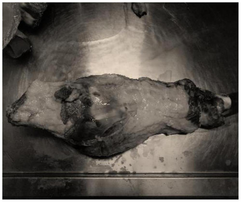
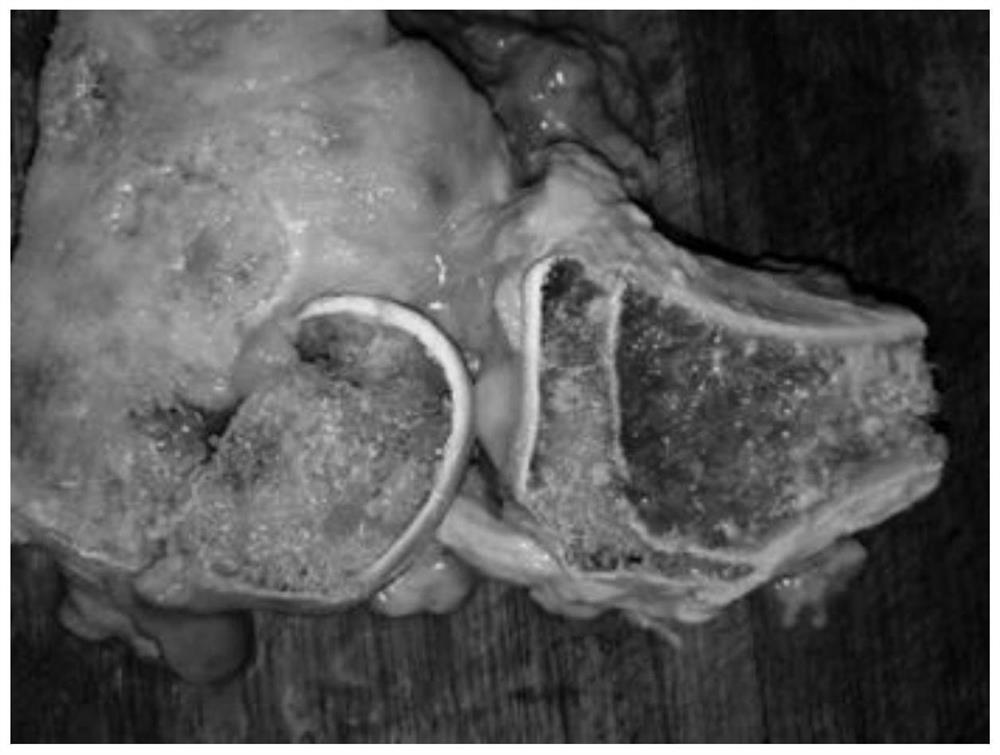
[0095] Tumor knee anatomy specimens:

[0096] A total of 22 human complete tumor knee joint specimens were collected from May 2017 to March 2020. The sources of materials included fresh specimens after amputation (12 cases) and fresh specimens after tumor resection (10 cases).

[0097] Inclusion criteria: 1. Distal femoral and tibial invasive tumors resected with clear pathological diagnosis; 2. Primary malignant bone tumors of the tibia and femur, with tumor segment resection or amputation and clear pathological diagnosis; 3. Knee joint cavity Intracondylar primary invasive tumors, resection of tumor segments, postoperative pathologically clear; 4. Those who meet the inclusion criteria 1.2.3, the preop...

Embodiment 2

[0115] This example provides a method for making an adult intraosseous LC pathway display specimen. In this example, the cases, image data, and human specimens are all from Qilu Hospital of Shandong University and Qingdao Campus of Qilu Hospital of Shandong University.

[0116] From May 2017 to March 2020, a total of 200 cases (200 knees) of the knee joint CT1mm thin-slice plain scan + 3D reconstruction were used in the image center database, and 100 cases (100 knees) of the knee joint were MRI plain scan or enhanced images. Including knee joint trauma, knee degenerative disease, meniscus injury, anterior cruciate ligament, posterior cruciate ligament, lateral collateral ligament injury, primary invasive tumor of the knee joint, knee joint malignant tumor patients and general physical examination patients. CT data are mainly used to demonstrate the ubiquity and location distribution of the tibial intercondylar foramen and femoral intercondylar foramen, and MRI data are mainly u...

Embodiment 3

[0127] This example provides a method for making a display specimen of intraosseous LC pathway in children. The cases, imaging data and human specimens in this example are all from Qilu Hospital of Shandong University and Qingdao Campus of Qilu Hospital of Shandong University.

[0128] From May 2017 to March 2020, there were 50 cases (50 knees) of children in the imaging center database with CT 1mm thin-slice plain scan + 3D reconstruction, and 30 cases (30 knees) with knee MRI 1mm thin-slice plain scan or enhanced images . Including patients with knee joint injury, discoid meniscus, osteomyelitis, lower extremity osteochondroma, knee joint malignant tumor and general physical examination. CT data are mainly used to demonstrate the prevalence of tibial intercondylar foramen and femoral intercondylar foramen in children, and the channel properties of the LC pathway in the epiphysis and metaphysis, and MRI data are mainly used to demonstrate the content of the LC pathway and wa...


PUM


Abstract

The preparation method and specimen of intraosseous blood vessel anatomical display display specimen of the present invention, wherein the method comprises: obtaining fresh femur or tibial specimen; removing cortical bone, immersing and fixing in formalin; using EDTA decalcifying agent to soak and decalcify; using CT thin Layer scanning to find the femoral intercondylar foramen or tibial intercondylar foramen; enter the anatomy through the above-mentioned holes to reveal the blood vessels and the intraosseous vascular channels containing the blood vessels; put the dissected bone specimens into the container box and add resin After the bone specimen is submerged, the resin gradually enters into the bone specimen; put the container box into the incubator, remove the container box after the resin hardens, and take out the specimen; cut off the excess resin, and polish to complete the production. By using the manufacturing method of the present invention, 100% of intraosseous blood vessel channels and intraosseous blood vessels can be exposed, and the existence and distribution of intraosseous blood vessels in the bone can be visually displayed; if it is a pediatric bone specimen, it can also be revealed that intraosseous blood vessels pass through the epiphyseal plate situation.

Description

technical field [0001] The invention relates to the technical field of biological specimens, in particular to a method for preparing a specimen for anatomical display of intraosseous blood vessels and the specimen. Background technique [0002] Because there are many classic anatomical structures in and around the knee joint, and it is a predilection site for many common sports injuries, degeneration, tumors, and traumas, it has been a hot topic of research by orthopedics and anatomy scholars for nearly a century. There are classic anatomical structures such as tibial plateau, femoral condyle and intercondylar fossa, anterior and posterior cruciate ligaments, medial and lateral menisci, etc., and its macroscopic structure and related clinical significance have been more maturely studied. In recent years, the knee joint has been newly named Anatomical structures are rare. [0003] There is also a relatively mature theoretical system about the source of blood supply and vascu...

Claims


Application Information

Patent Timeline
Patent Type & Authority Patents(China)
IPC IPC(8): A01N1/00G09B23/30G09B23/28
CPCA01N1/00G09B23/306G09B23/28
Inventor 程坤李建民李玉椿李秋尧侯海青金利新高晓斐冯琨杨志成杨强邵显昊蒙思瑜
Owner 青岛山大齐鲁医院(山东大学齐鲁医院(青岛))
Why Eureka
Try Eureka