
Systems and methods for adaptive histopathology image unmixing

an image and histopathology technology, applied in image enhancement, image analysis, instruments, etc., can solve the problems of erroneous estimation of the nuclear component, difficult extraction of endmember spectra from the image and solving for abundance, and difficult accurate estimation of the abundance of quantum dots (i.e., unmixed images)

Active Publication Date: 2016-09-01
VENTANA MEDICAL SYST INC
View PDF9 Cites 11 Cited by
  • Summary
  • Abstract
  • Description
  • Claims
  • Application Information

AI Technical Summary

Benefits of technology

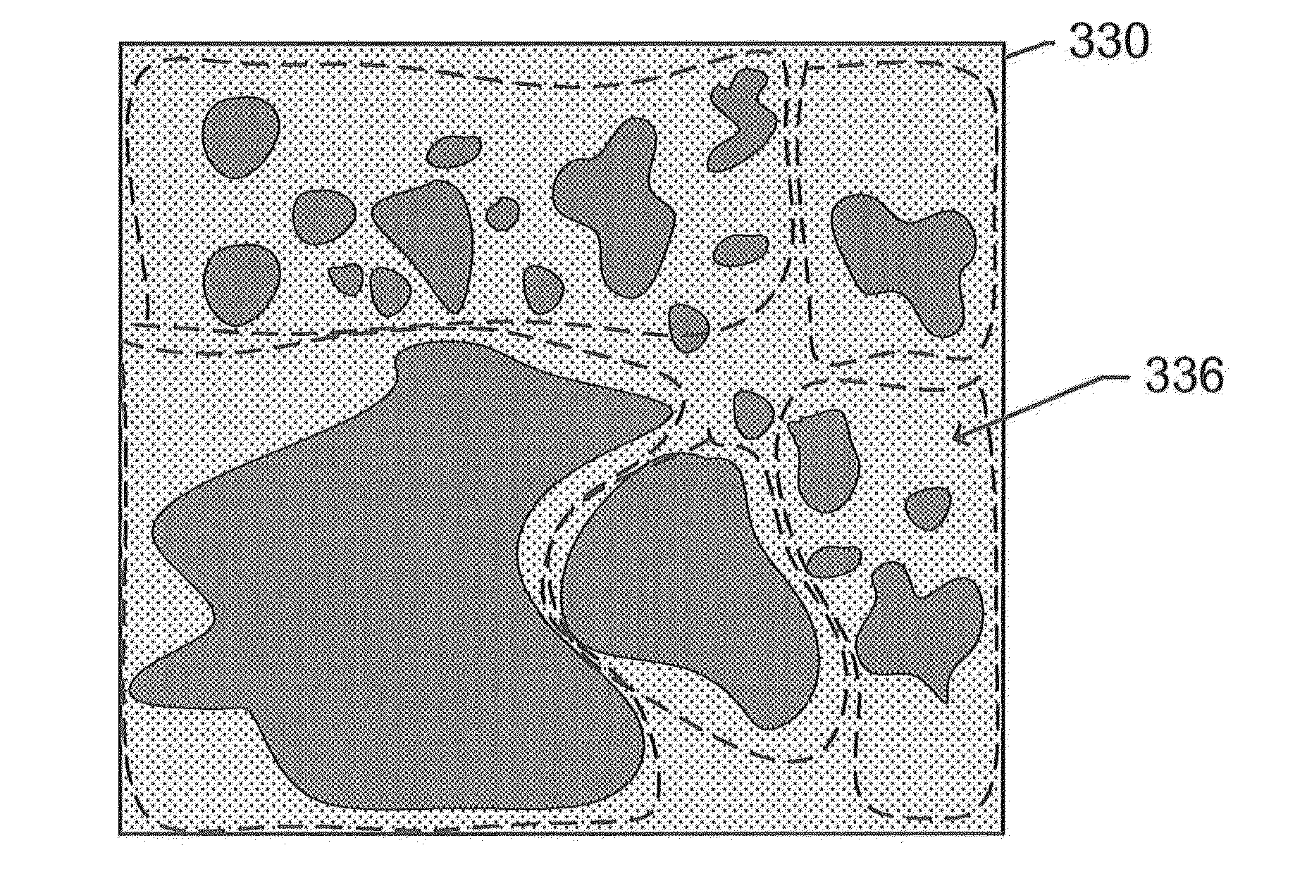
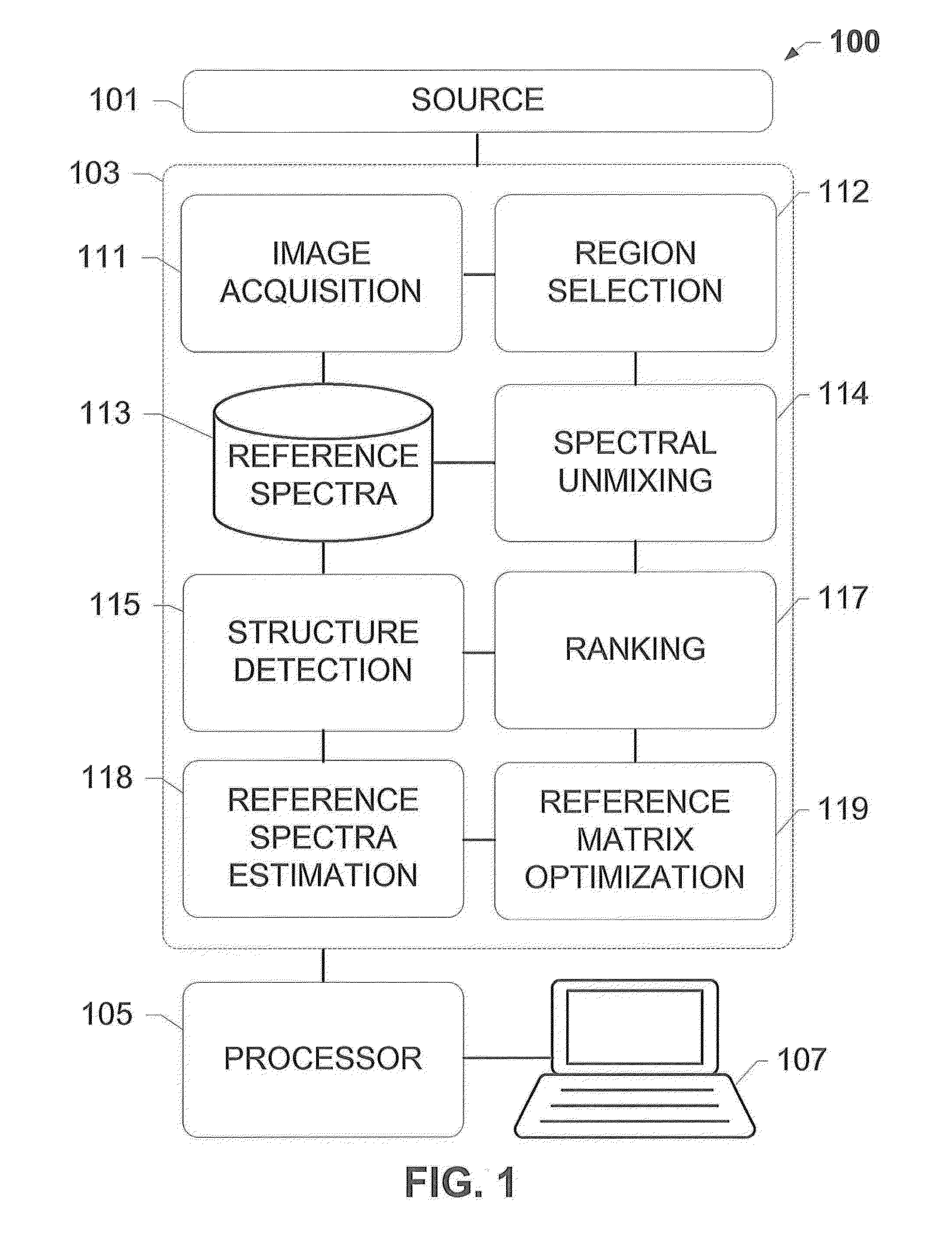
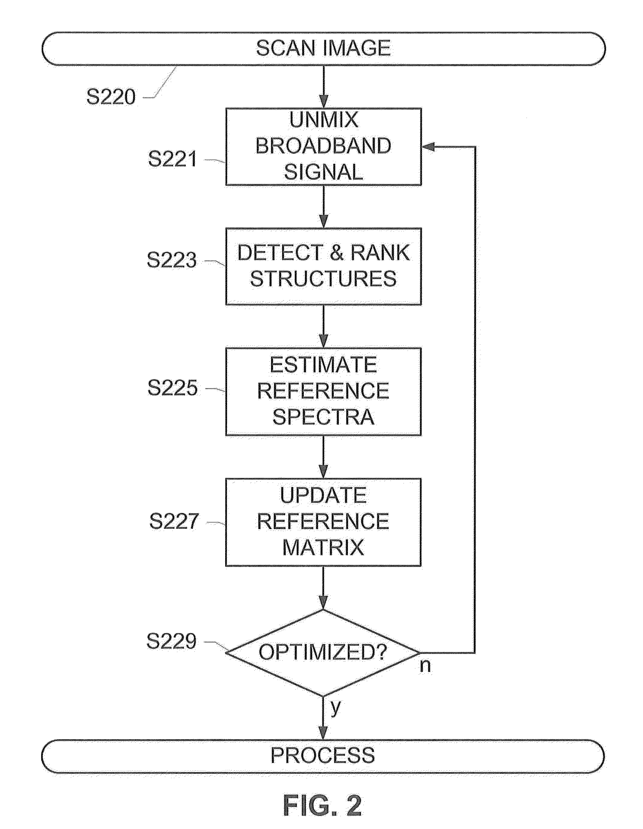
The patent describes a system and method for unmixing images that contain a mixture of signals. The system detects structures in the image and ranks them based on their confidence level. It then creates a reference matrix with the initial reference spectra or colors and uses it to unmix the image. The process is iterated until an optimal reference matrix is achieved. The optimized reference matrix results in improved unmixing of the image compared to existing methods. The system can be used with different types of images and can be executed in parallel on multiple regions of the image.

Problems solved by technology

Unlike the applications in the domain of remote sensing where different objects such as grass and rocks can be easily identified from the scene, the biomarkers are often co-localized in histopathology fluorescent images, therefore making it difficult to extract the endmember spectra from the image and solve for the abundances.
The broad-band spectra overlap with the narrow-band spectra, making the accurate estimation of the quantum dot abundances (i.e., the unmixed images) even more difficult.
In addition, part of the DAPI spectrum may be wrongly estimated as AF because the spectral signatures of DAPI and AF are similar to each other, which may lead to an erroneous estimation of the nuclear component.
However, due to the non-linearity of the problem, this method is not guaranteed to converge to a physically meaningful and relevant solution.
NMF automatically solves for the reference spectral matrix, however the algorithm is not able to identify which reference spectrum corresponds to DAPI and which one corresponds to AF.
Additional frameworks proposed based on the orthogonality assumption (i.e., the assumption that the reference spectrum of DAPI is orthogonal to that of quantum dots) of the endmember do not yield meaningful results for real data.

Method used


Image

  • Systems and methods for adaptive histopathology image unmixing
  • Systems and methods for adaptive histopathology image unmixing
  • Systems and methods for adaptive histopathology image unmixing

Examples


Embodiment Construction

[0022]The subject disclosure presents systems and methods for adaptively optimizing the broadband reference spectra for an image comprising a plurality of fluorescent channels or updating the reference color for a bright-field image. For each region of an image, we update the reference vector, i.e. a reference spectrum in the case of a fluorescence image and a reference color in the case of a bright field image spectra and unmix the region till convergence. As an example application, this disclosure presents the details of the reference spectral refinement method for multi-spectral images, but the same technique can be applied to the bright-field images.

[0023]Taking the fluorescent image as an example, this adaptive optimization is based on structures detected in an unmixed broadband channel of the image. For instance, a slide holding a sample material may be scanned using a scanner coupled to a fluorescence or brightfield microscope system to generate a scanned image. The image is ...


PUM


Abstract

The present invention relates to systems and methods for adaptively optimizing broadband reference spectra for a multi-spectral image or adaptively optimizing reference colors for a bright-field image. The methods and systems of the present invention involve optimization techniques that are based on structures detected in an unmixed channel of the image, and involves detecting and segmenting structures from a channel, updating a reference matrix with signals estimated from the structures, subsequently unmixing the image using the updated reference matrix, and iteratively repeating the process until an optimized reference matrix is achieved.

Description

CROSS-REFERENCE TO RELATED APPLICATIONS[0001]This is a continuation of International Patent Application No. PCT / EP2014 / 070793 filed Sep. 29, 2014, which claims priority to and the benefit of U.S. Provisional Patent Application No. 61 / 884,974 filed Sep. 30, 2013, both of which prior applications are incorporated herein by reference as if set forth in their entirety.FIELD OF INVENTION[0002]The present invention relates to the unmixing of images, for example, histopathology images, in fluorescence microscopy or bright-field microscopy. More particularly, the present invention relates to dynamically and adaptively refining reference spectra for a multi-spectral image or reference colors for the bright-field image.BACKGROUND[0003]In a multiplex fluorescent slide of a tissue specimen, different nuclei and tissue structures are simultaneously stained with specific fluorescent dyes, organic fluorescent dyes, or fluorescent counter stains, each of which fluoresces in a different spectral ban...

Claims


Application Information

Patent Timeline
Patent Type & Authority Applications(United States)
IPC IPC(8): G06T7/00G06K9/00
CPCG06T7/0081G06T7/0097G06K9/0014G06T2207/30024G06T2207/10064G06T2207/20148G06T2207/20144G06T2207/10056G06T2207/10024G06T7/11G06V20/695
Inventor CHEN, TINGCHUKKA, SRINIVASSARKAR, ANINDYA
Owner VENTANA MEDICAL SYST INC
Learn More