Laparoscopic retractor devices

a retractor device and tissue technology, applied in the field of tissue retraction devices, can solve the problems of difficult minimally invasive approach, inconvenient operation, and difficulty in retraction of tissue that is inflamed or requires gentle handling, so as to minimize the risk of pinching or impinging tissue, improve minimally invasive intra-operative visualization, and facilitate operation.

Active Publication Date: 2016-09-29
MAYO FOUND FOR MEDICAL EDUCATION & RES
View PDF2 Cites 19 Cited by
  • Summary
  • Abstract
  • Description
  • Claims
  • Application Information

AI Technical Summary

Benefits of technology

[0016]Particular embodiments of the subject matter described in this document can be implemented to realize one or more of the following advantages. First, in some embodiments the laparoscopic retractor devices provided herein are readily adjustable in size from a low profile delivery configuration to a substantially wider expanded configuration for performing atraumatic retraction of tissue and organs.
[0017]Second, the laparoscopic retractor devices provided herein have intuitive and easy to operate designs. Further, the laparoscopic retractor devices provided herein do not include separable pieces or multiple movable parts. In some embodiments, the laparoscopic retractor devices provided herein are designed to minimize the risk of pinching or impinging tissue during the adjustment of the retractors between the contracted and expanded configurations. Advantageously, clinicians can operate the retractor devices without a risk of pieces becoming detached within a body cavity.
[0018]Third, the laparoscopic retractor devices provided herein provide atraumatic laparoscopic retraction of tissue by wrapping a mesh material around the tissue in a gentle cradle-like fashion. In that manner, the potential of causing tissue damage such as ischemia, tears, ruptures, and the like, is at least partially mitigated.
[0019]Fourth, various surgeries can be performed in a minimally invasive fashion using the devices and methods provided herein. Minimally invasive intra-operative visualization is improved using the devices and methods provided herein. Such minimally invasive techniques can reduce recovery times, patient discomfort, and treatment costs.

Problems solved by technology

However, there are areas of the abdominal cavity in which the minimally invasive approach is still challenging.
For example, small bowel loops frequently obscure the paraaortic surgical field, especially in obese patients.
Retraction of tissue that is inflamed or requires gentle handling is not well-facilitated using current laparoscopic, endoscopic, and robotic surgery retraction instrumentation.
The crushing can cause ischemia, as well as rupture of both hollow and solid organs.
Further, in some scenarios tumors that are being retracted and dissected may rupture, spilling succus as well as tumor cells.
Further, the laparoscopic retractor devices provided herein do not include separable pieces or multiple movable parts.

Method used


Image

  • Laparoscopic retractor devices
  • Laparoscopic retractor devices
  • Laparoscopic retractor devices

Examples


Embodiment Construction

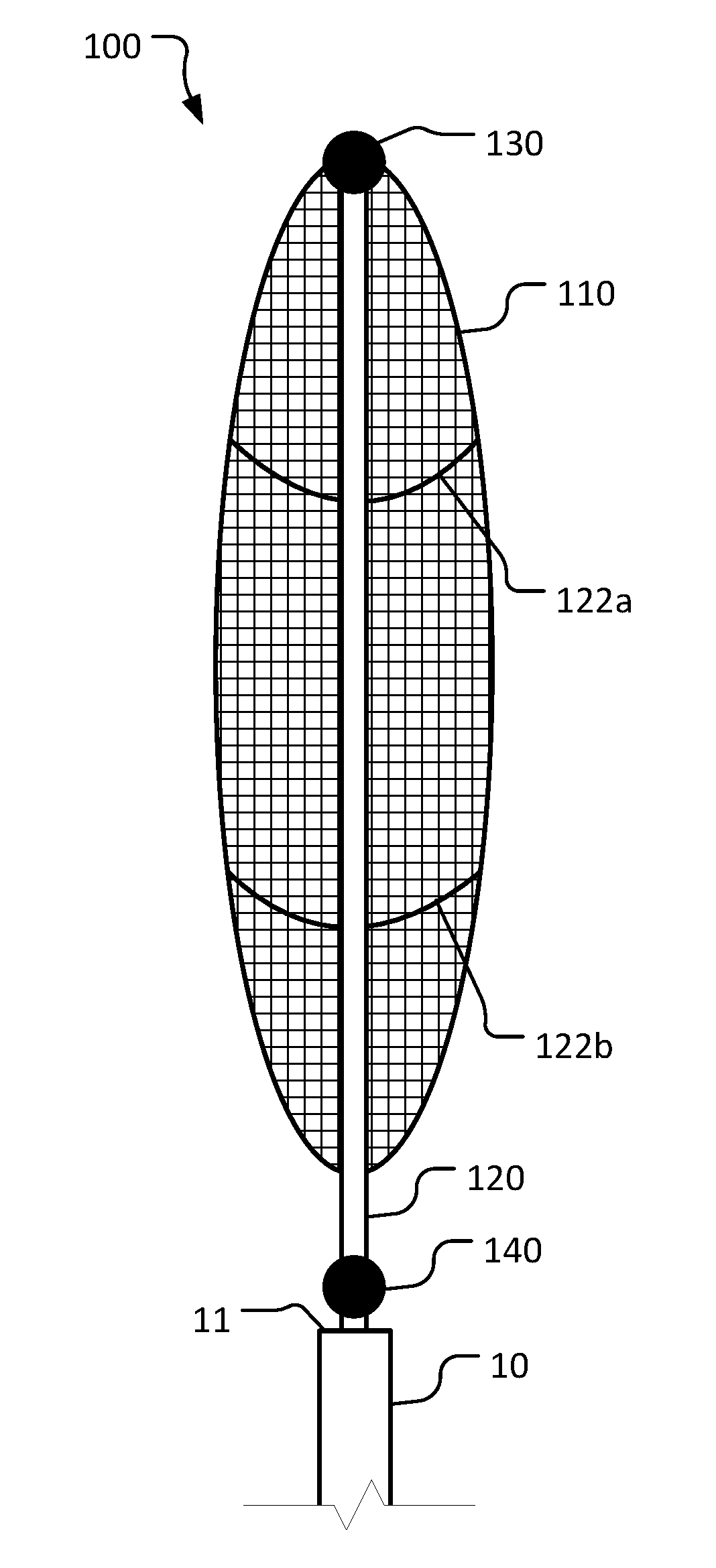
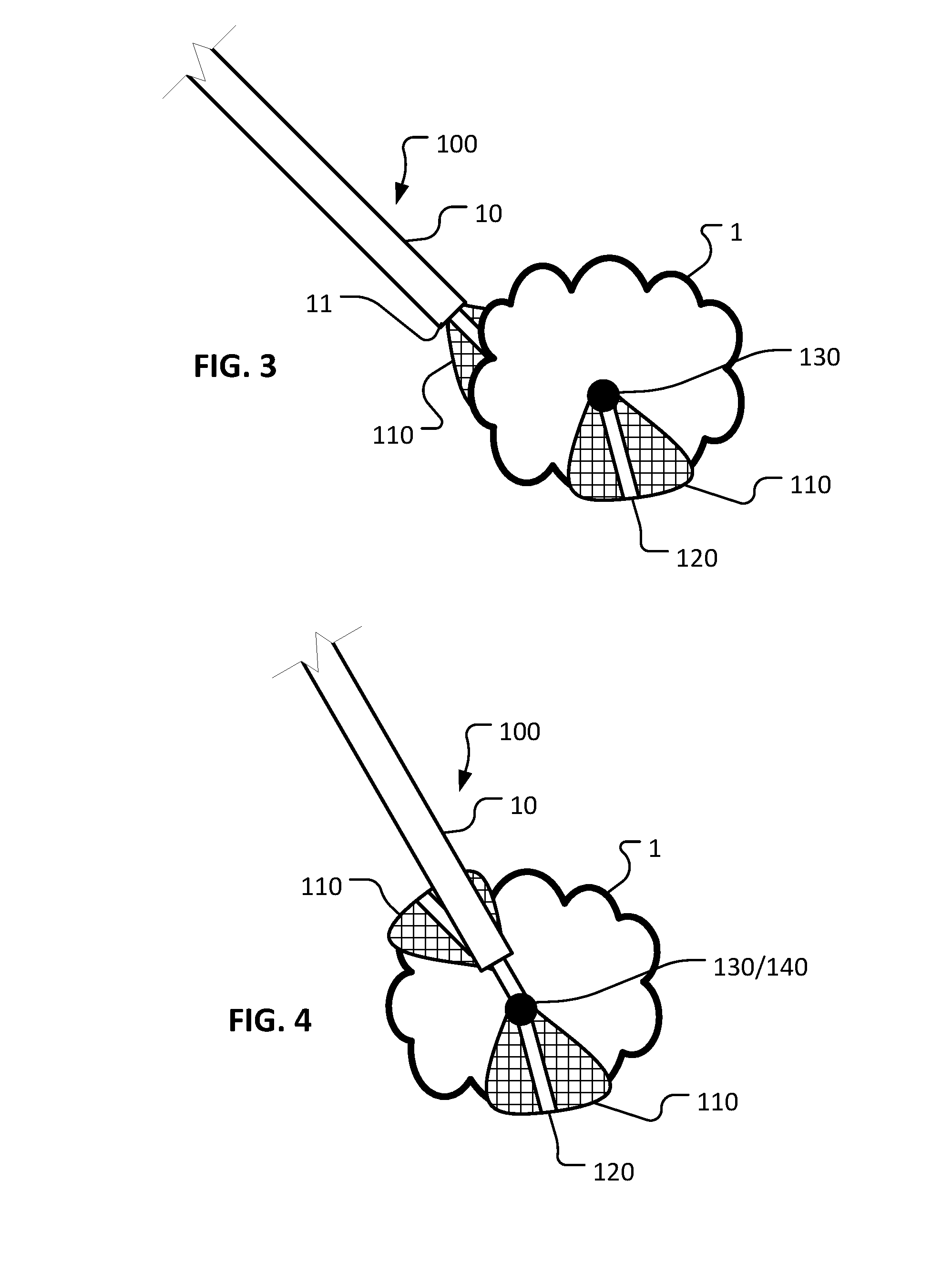
[0027]This document provides devices and methods for laparoscopic retraction of tissue. For example, this document provides devices and methods for atraumatic laparoscopic retraction of tissue by wrapping a mesh material around the tissue in a cradle-like fashion.

[0028]Many currently available laparoscopic retractors utilize a grasping or crushing modality to move tissues and mobilize tissue structures. One alternative to laparoscopic retraction using a traumatic crushing process are apparatuses that use a material to “hug” or “wrap” around the structure being manipulated. An analog of such atraumatic methods of retraction and tissue manipulation is used in some open procedures, as there is significantly more access to the body cavity being operated upon. The difficulty in using this method for laparoscopic surgery is in the limited access granted by laparoscopic port sites to allow either a hand or an instrument that is large enough to “wrap” the object.

[0029]This disclosure descri...


PUM


Abstract

Some devices for atraumatic retraction of tissue include a transcatheter deployable mesh hammock that is well-suited for laparoscopic, endoscopic, and robotic surgeries. Methods for atraumatic retraction of tissue include wrapping a mesh material around the tissue in a cradle-like fashion. In some embodiments, magnetic coupling of the proximal and distal ends of the mesh material can provide tissue containment and retractor securement to facilitate tissue retraction.

Description

CROSS-REFERENCE TO RELATED APPLICATIONS[0001]This application claims the benefit of U.S. Provisional Application Ser. No. 62 / 138,791, filed Mar. 26, 2015. The disclosure of the prior application is considered part of (and is incorporated by reference in) the disclosure of this application.BACKGROUND[0002]1. Technical Field[0003]This document relates to devices and methods for retraction of tissue. For example, this document relates to laparoscopic, endoscopic, and robotic surgery devices and methods for atraumatic retraction of tissue by wrapping a mesh material around the tissue in a cradle-like fashion.[0004]2. Background Information[0005]Optimal visualization of the operative field is a fundamental principle in surgery. Any technique that improves intra-operative visualization has the potential to make surgery safer and more cost efficient.[0006]With the advent of minimally invasive surgery, the utilization of laparoscopic, endoscopic, and robotic surgery is becoming the standard...

Claims


Application Information

Patent Timeline
Patent Type & Authority Applications(United States)
IPC IPC(8): A61B17/02
CPCA61B17/0218A61B2017/00477A61B2034/301A61B2017/00867A61B2017/0225A61B2017/00876
Inventor AHO, JOHNATHON M.THIELS, CORNELIUS A.HOLST, KIMBERLY A.
Owner MAYO FOUND FOR MEDICAL EDUCATION & RES
Why Eureka
Try Eureka