Agents for use in the treatment of retinal inflammation
- Summary
- Abstract
- Description
- Claims
- Application Information
AI Technical Summary
Benefits of technology
Problems solved by technology
Method used
Image

Examples
example 1
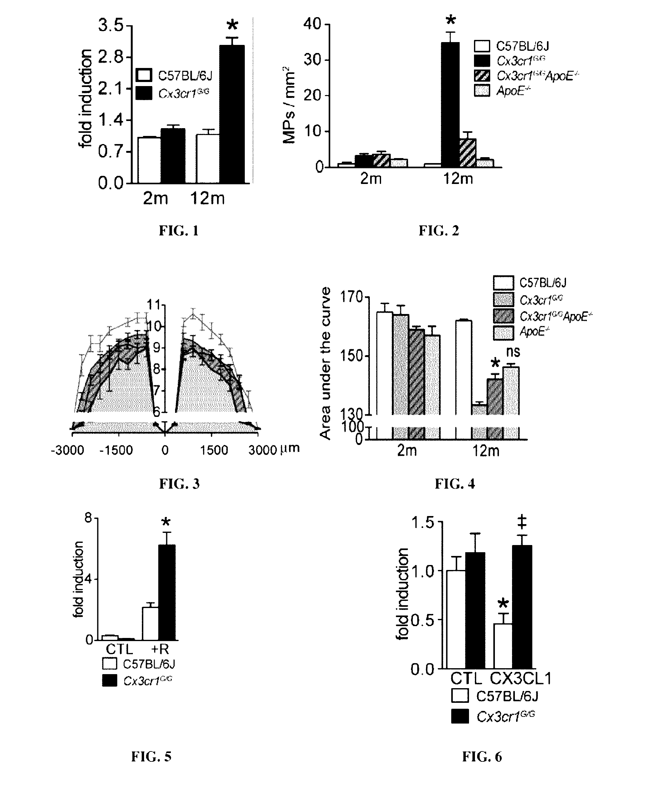
Subretinal Mononuclear Phagocytes (MPs) Cluster in and Around Soft Drusen in Early AMD and Express ApoE
[0126]Physiologically, the subretinal space does not contain significant numbers of MPs, possibly due in part to immunosuppressive RPE signals (Streilein et al. 2002).
[0127]Mononuclear phagocytes are nevertheless known to be present in the subretinal space and on the apical side of Retinal pigment epithelium cells adjacent to the lesions of atrophic AMD.
[0128]Experiments conducted on Retinal pigment epithelium / choroidal flatmounts of soft drusen from donors with intermediate AMD demonstrate that numerous CD18+ and IBA-1+ cells are contained within soft drusen (partially covered by the Retinal pigment epithelium), but also adjacent to the soft drusen on the surrounding autofluorescent Retinal pigment epithelium (data not shown). Higher magnification with lateral Z stack projections through a subretinal IBA-1+mononuclear phagocyte further demonstrates the close physical contact of th...
example 2
Subretinal Mononuclear Phagocytes (MPs) Accumulate on the RPE in the Vicinity of Atrophic Lesions and Large Drusen
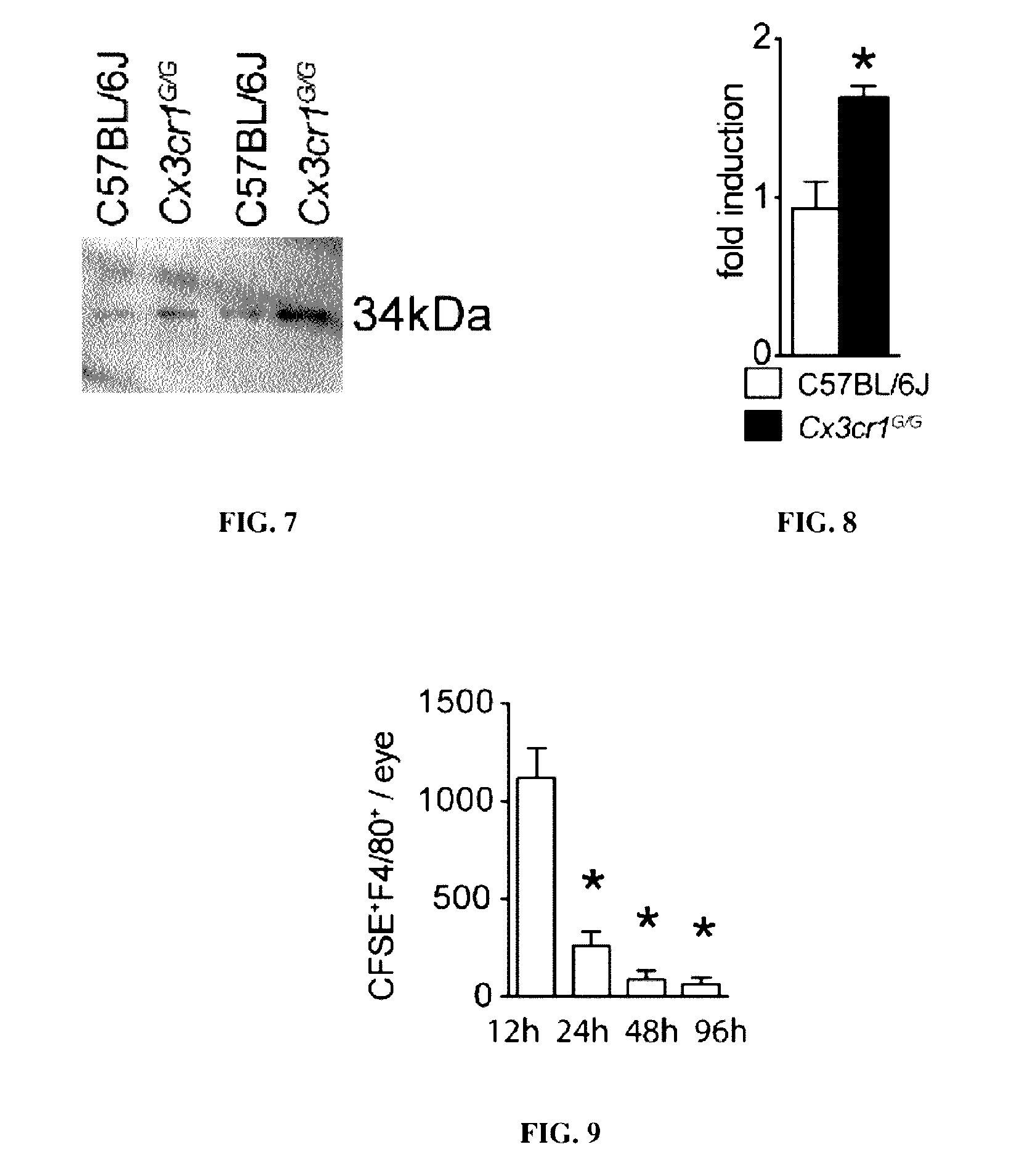
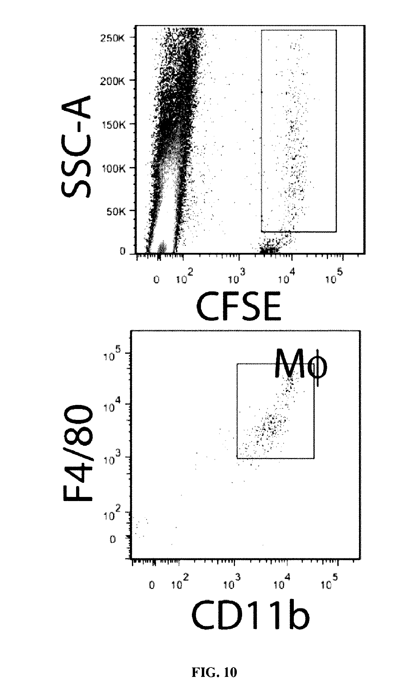
[0130]In late AMD, immunohistochemical studies on sections have revealed the presence of subretinal MPs on RPE cells adjacent to the lesions of atrophic AMD (Gupta et al, 2003; Sennlaub et al, 2013) and MPs were found in subretinal neovascular membranes (Oh et al. 1999). Because the small, dispersed MPs are difficult to detect on sections, MP-marker-IBA-1 immunohistochemistry was thus performed on healthy and diseased macular RPE / choroidal flatmounts (IBA-1 green fluorescence, RPE autofluorescence visible as orange due to its autofluorescence in the red and green channel). Confocal microscopy confirmed that subretinal IBA-1+MPs are only very occasionally observed in healthy age-matched donor central RPE (data not shown). Within the atrophic lesions of GA patients where the RPE has disappeared, MPs were numerous, but were also invariably observed on the apical side of the...
example 3
Subretinal MPs Accumulated on the RPE in the Vicinity of Atrophic Lesions and Large Drusen Express APOE
[0132]MPs have been reported to express APOE at high levels (Basu et al, 1982; Nakai et al, 1996; Peri & Nusslein-Volhard, 2008; Rosenfeld et al, 1993). Immunohistochemistry of APOE and IBA-1 on paraffin sections of human tonsils, which were used as a positive control, confirmed that IBA-1+MPs can strongly express APOE (data not shown). Similarly, on retinal flatmounts of donor eyes with large drusen. APOE staining was observed in and around subretinal IBA-1+MPs (data not shown). The double labeling was performed on the subretinal side of retinas to avoid masking by the RPE autofluorescence. APOE staining was performed on paraffin sections of controls and donor eyes with geographic atrophy lesions. A substrate revealing method (alkaline phosphatase / Fast Red) that is visible in bright field was used to circumvent confusion with RPE autofluorescence. In sections from control eyes the...
PUM
| Property | Measurement | Unit |
|---|---|---|
| Concentration | aaaaa | aaaaa |
Abstract
Description
Claims
Application Information