Structured finding objects for integration of third party applications in the image interpretation workflow

Active Publication Date: 2017-06-01
KONINKLJIJKE PHILIPS NV
View PDF7 Cites 10 Cited by
  • Summary
  • Abstract
  • Description
  • Claims
  • Application Information

AI Technical Summary

Benefits of technology

[0009]One advantage resides in organizing the radiology reading process around structured finding objects in order to facilitate collection and recordation of appropriate support for radiology fi

Problems solved by technology

Under these time constraints, the radiologist may find it difficult to recognize and make use of available tools such as the EMR, past radiology examinations stored in the PACS, and various CADx or electronic treatment guideline resources.
Opera

Method used


Image

  • Structured finding objects for integration of third party applications in the image interpretation workflow
  • Structured finding objects for integration of third party applications in the image interpretation workflow
  • Structured finding objects for integration of third party applications in the image interpretation workflow

Examples


Embodiment Construction

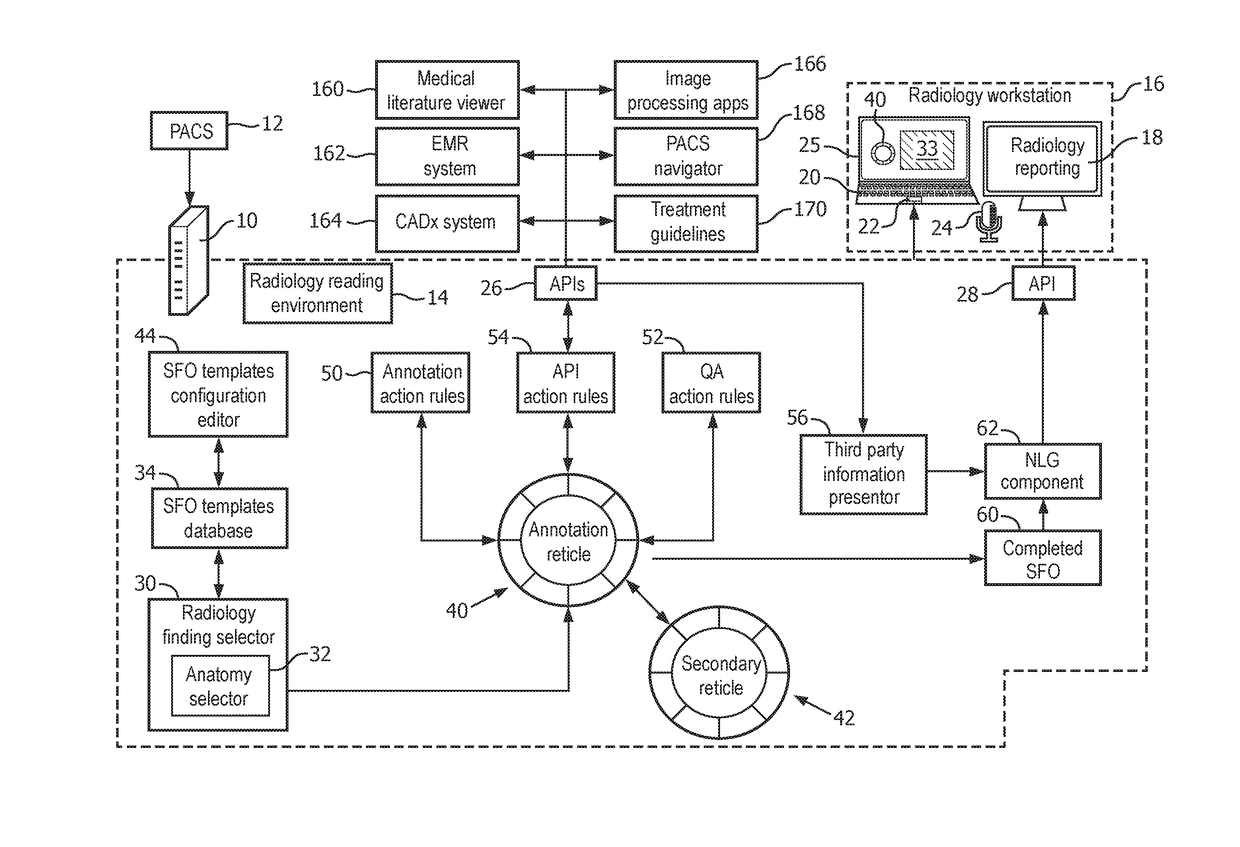
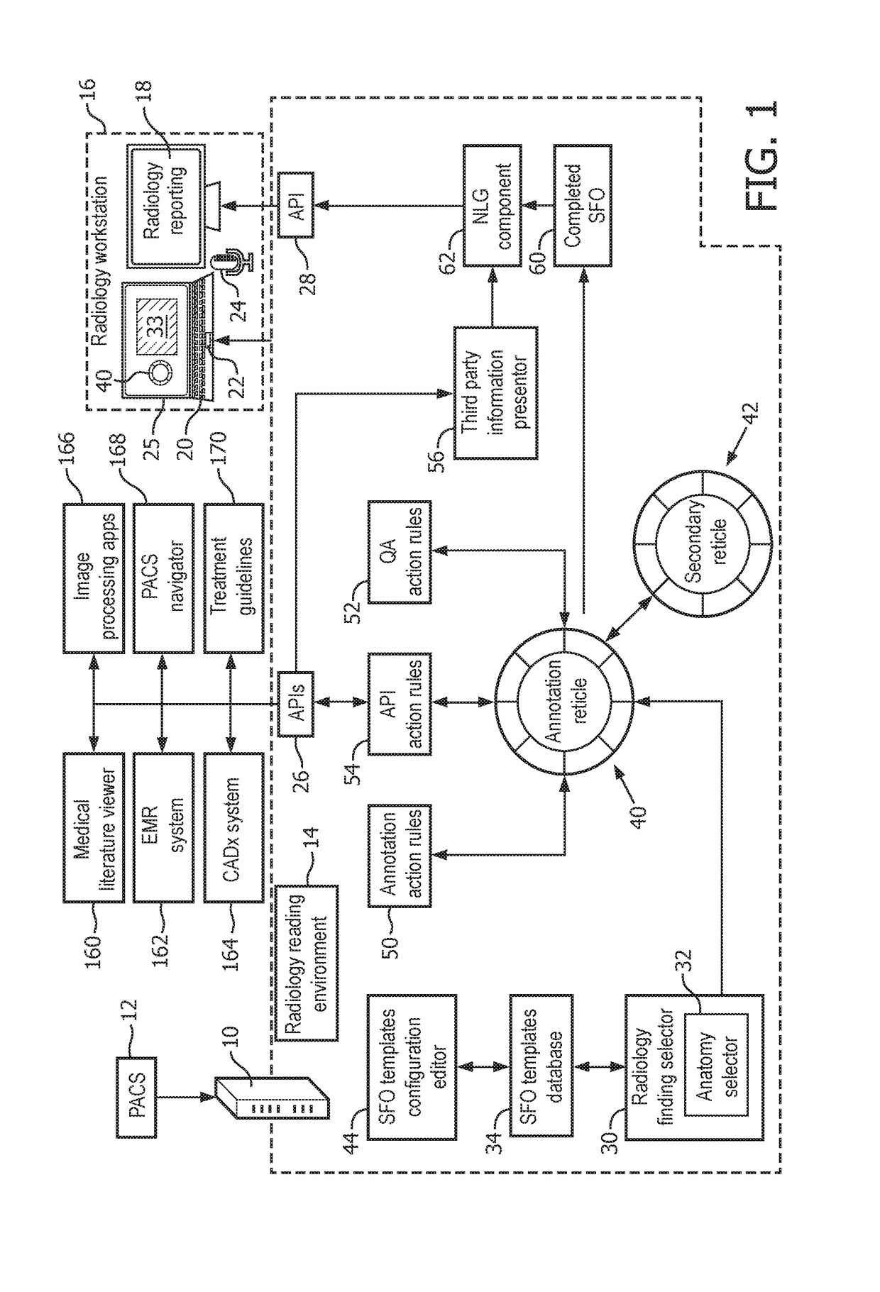
[0015]Disclosed radiology reading devices and methods utilize the concept of a structured finding object (SFO), which is a digital entity, preferably codified in a radiological ontology such as RadLex (promulgated by the Radiological Society of North America, RSNA) or Systematized Nomenclature of Medicine—Clinical Terms (SnoMed CT, promulgated by the College of American Pathologists, CAP). The SFO at least partially characterizes an image finding in a structured format using syntax such as Annotation Imaging Mark-up (AIM), which is a radiology domain-specific XML syntax. In some suitable embodiments, an SFO is represented by dimensions each codified using a pair where “key” identifies the dimension and “value” is the value of the dimension for the radiological finding in the current reading task. Dimensions are typically typed, and the value for a given dimension may itself be a complex (e.g. hierarchical) data structure. By way of non-limiting illustration, some possible SFO dimen...


PUM


Abstract

A radiology workstation (16) includes a display device (25) and at least one user input device (20, 22, 24). A server computer (10) is programmed to operate with the radiology workstation to perform the radiology reading task (14) including a radiology reading support method comprising: receiving user input identifying a radiological finding; retrieving a structured finding object (SFO) template for the radiological finding; displaying an SFO annotation graphical user interface (GUI) dialog (40) having annotation data entry fields for annotating the retrieved SFO template; building an SFO (60) representing the radiological finding by annotating the SFO template via the SFO GUI dialog; and generating natural language text describing the radiological finding from the SFO. Application program interface (API) action rules (54) may be applied to determine whether the SFO being built satisfies any API action rule, and if so a corresponding application program is invoked.

Description

FIELD[0001]The following relates generally to the radiology arts, radiology reading arts, radiology workstation arts, radiology workstation user interfacing arts, and related arts.BACKGROUND[0002]Radiology is a complex process involving several interacting medical professionals. In a typical sequence, a patient's physician orders a radiology examination. A radiology technician operates the imaging system, such as a magnetic resonance imaging (MRI) system, a computed tomography (CT) imaging system, a positron emission tomography (PET) imaging system, or so forth, or a combination of imaging system (e.g. PET / CT) to acquire images of an anatomical region of the patient in accordance with the physician's order. These images are stored in a Picture Archiving and Communication System (PACS), and are later viewed, or “read”, by a radiologist, typically using a dedicated radiology workstation executing a radiology reading environment. The main deliverable of the radiology reading is a radio...

Claims


Application Information

Patent Timeline
IPC IPC(8): G06F19/00G06F17/28G06T7/00G06F17/24G16H15/00G16H30/20G16H30/40
CPCG06F19/321G06F17/248G06F17/241G06F17/243G06F3/0481G06T7/0012G06F19/322G06F19/3406G06F17/28G16H40/63G16H10/60G16H15/00G16H30/40G16H30/20G06F40/40G06F40/169G06F40/174G06F40/186
Inventor SEVENSTER, MERLIJNMANKOVICH, GABRIEL RYANTELLIS, RANJITH NAVEENDEHGHAN MARVAST, EHSANQIAN, YUECHEN
Owner KONINKLJIJKE PHILIPS NV
Why Eureka
Try Eureka