Systems for imaging of blood flow in laparoscopy

Inactive Publication Date: 2017-06-29
TYCO HEALTHCARE GRP LP
View PDF22 Cites 5 Cited by
  • Summary
  • Abstract
  • Description
  • Claims
  • Application Information

AI Technical Summary

Benefits of technology

[0007]The present disclosure is directed to laparoscope using laser speckle contrast image techniques which allows a clinician to see a quantitative mapping of local blood flow dynamics in a wide area so that the clinician can quickly and accurately assess the blood supply to a portion under treatment. In an embodiment, the present disclosure is directed to a laparoscopic apparatus for imaging subsurface blood flow of tissue, the laparoscopic apparatus including a light source emitting white light via a light guide, a laser source emitting laser light via an optical fiber, and a laparoscope that alternatively receives reflected laser light emitted from the laser source, and reflected white light em

Problems solved by technology

However, clinicians have a very limited field of view with a laparoscope due to the limited range of movement and viewing area provided by the laparoscope.
However, even if the clinicians determines a proper anastomosis location, clinicians' visual determination of the proper anastomosis location which receives a sufficient bloo

Method used


Image

  • Systems for imaging of blood flow in laparoscopy
  • Systems for imaging of blood flow in laparoscopy
  • Systems for imaging of blood flow in laparoscopy

Examples


Embodiment Construction

[0020]The present disclosure is related to systems for imaging subsurface blood flow of tissue using laser speckle contrast imaging techniques. Although the present disclosure will be described in terms of specific illustrative embodiments, it will be readily apparent to those skilled in this art that various modifications, rearrangements and substitutions may be made without departing from the spirit of the present disclosure. The scope of the present disclosure is defined by the claims appended hereto.

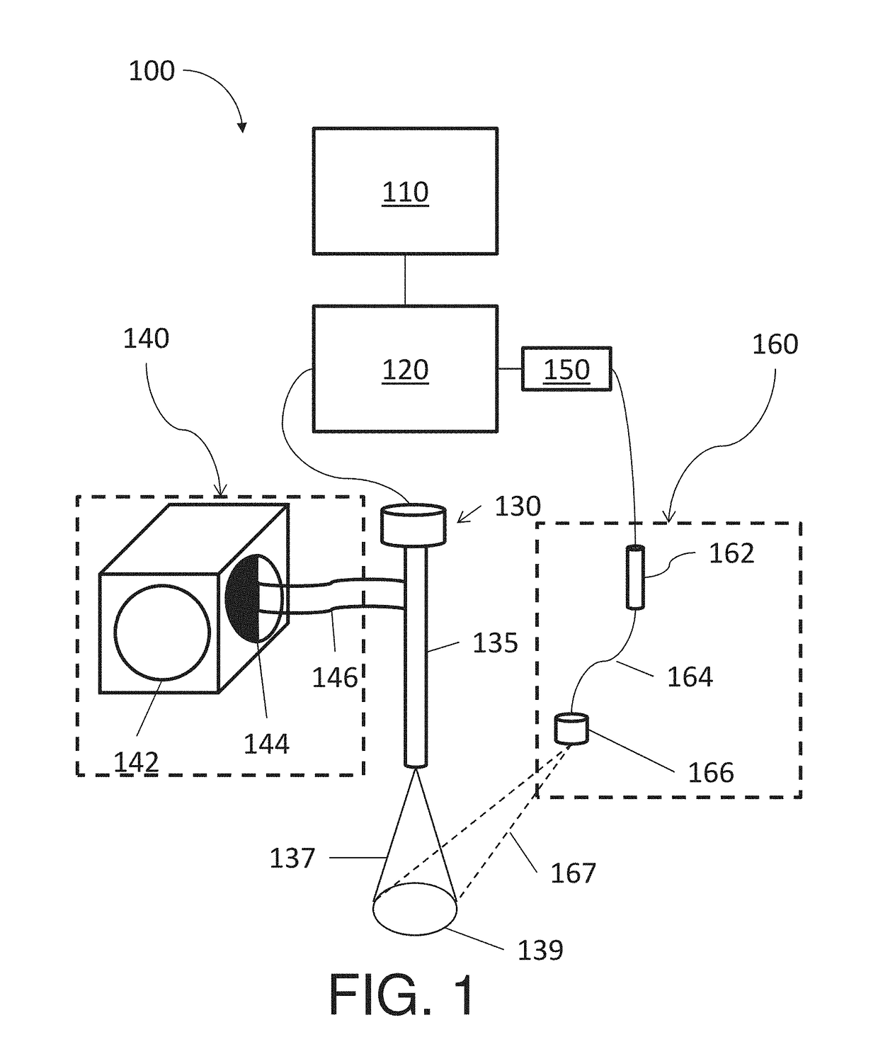
[0021]FIG. 1 shows a laparoscopic system 100 that is capable of imaging subsurface blood flow using laser light and imaging inside of a patient's body using white light. The system 100 includes a display 110, a computing device 120, a laparoscope 130, a white light source 140, a switch 150, and a laser light source 160. The display 110 receives video signals from the computing device 120 and displays the video signal. The display 110 may be any form suitable for displaying medical im...


PUM


Abstract

A laparoscopic apparatus for imaging subsurface blood flow of tissue, the laparoscopic apparatus including a light source emitting white light via a light guide, a laser source emitting laser light via an optical fiber and a laparoscope that alternatively receives reflected laser light emitted from the laser source, and reflected white light emitted from the light source. The apparatus further includes a computing device receives the sensed resulted from the laparoscope and generates laser speckle contrast images or white light images according to an output status of the light source and the laser source, and a display that is operatively associated with the computing device and that displays at least one of the laser speckle contrast images and the white light images, where the laser speckle contrast images show the subsurface blood flow.

Description

CROSS REFERENCE TO RELATED APPLICATION[0001]The present application is a U.S. National Stage application under 35 U.S.C. §371(a) of PCT / CN2014 / 078222 filed May 23, 2014, the entire contents of which are incorporated by reference herein.BACKGROUND[0002]Technical Field[0003]The present disclosure relates to systems for imaging blood flow. More particularly, the present disclosure relates to systems that image subsurface blood flows of tissue with a large field of view.[0004]Discussion of Related Art[0005]Recently, surgeries to internal organs have been greatly increased due to improvements on medical diagnosis, clinicians' operational techniques, medical instruments, and medical systems. Since sufficient blood flow is of fundamental importance in promoting healing after surgery and avoiding leakage of the organ, visually finding and confirming a proper location for surgery is also important. Compared to open surgeries, laparoscopic surgeries leave a relatively small suture on the skin...

Claims


Application Information

Patent Timeline
IPC IPC(8): A61B5/00A61B5/026
CPCA61B5/0084A61B5/6847A61B5/0059A61B5/0261A61B5/026
Inventor LUO, ZHONGCHIWU, XINMIN
Owner TYCO HEALTHCARE GRP LP
Why Eureka
Try Eureka