
System and method for medical diagnostics

Active Publication Date: 2017-10-26
WELCH ALLYN INC
View PDF6 Cites 27 Cited by
  • Summary
  • Abstract
  • Description
  • Claims
  • Application Information

AI Technical Summary

Benefits of technology

The medical diagnostic device described in this patent text can enable a patient to perform a self-examination and collect samples without a clinician's presence. The device is easy to operate and has integrated sample collecting functions to protect the sample collector from contamination. The device also includes an imager to help direct the sampling device to the intended target of interest. The images or video can be stored for future reference or training purposes, and the patient can see the images directly. Overall, this device simplifies patient care and improves the accuracy and efficiency of medical diagnosis.

Problems solved by technology

However, inserting the imaging device can disturb the inserted vaginal speculum and can be uncomfortable for the patient.
The sample collecting device, however, may contact additional surfaces during the process, thus potentially contaminating the sample.
However, many patients experience embarrassment and discomfort at the idea of experiencing such an intimate and potentially uncomfortable examination.
Because of this discomfort, many women elect not to have these examinations performed.
As a result, serious medical conditions, such as cervical cancer, can remain undiagnosed or be diagnosed at a stage that is too late to effectively treat.

Method used


Image

  • System and method for medical diagnostics
  • System and method for medical diagnostics
  • System and method for medical diagnostics

Examples


Embodiment Construction

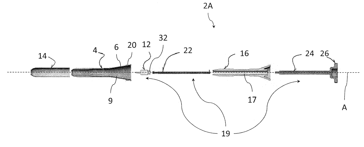
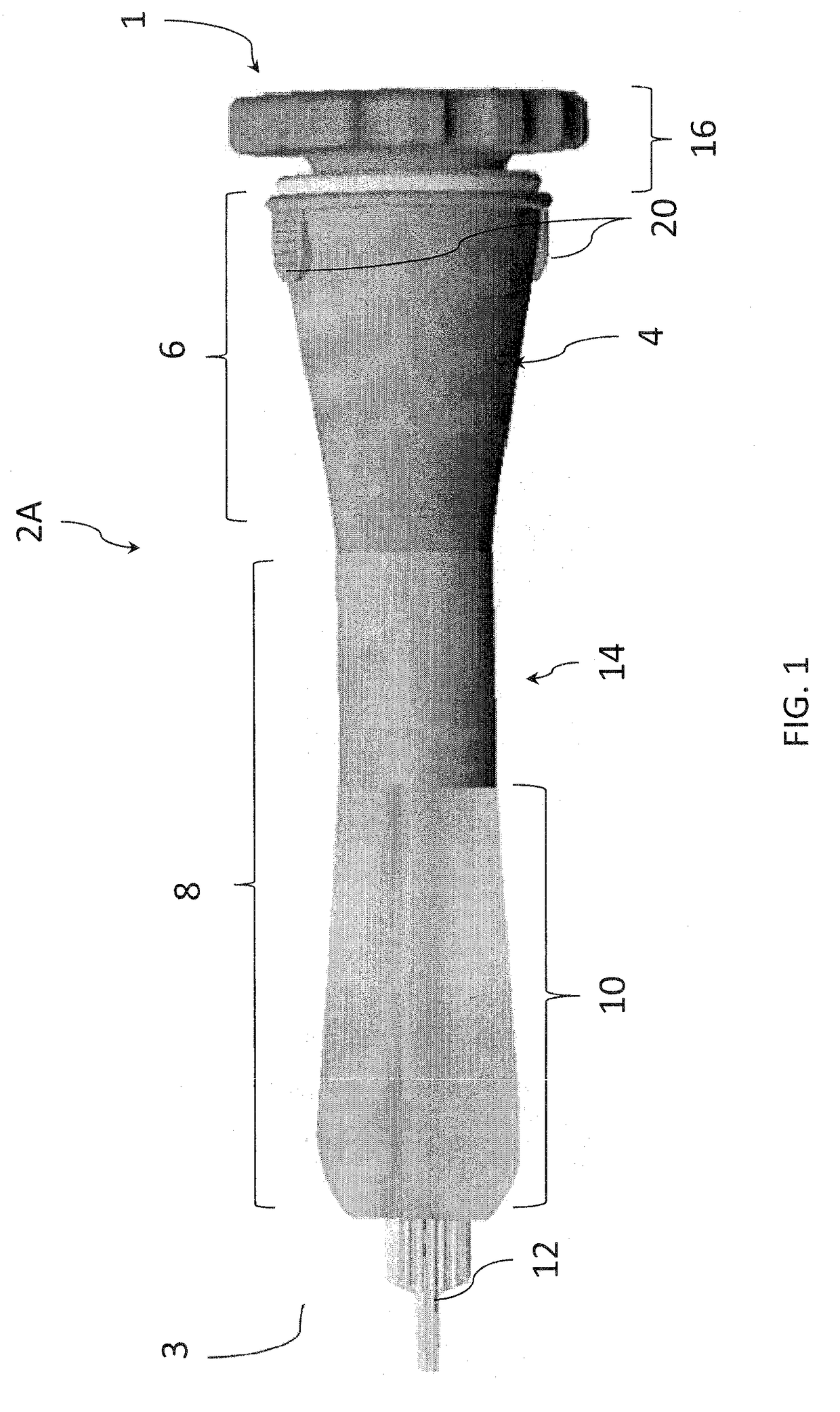
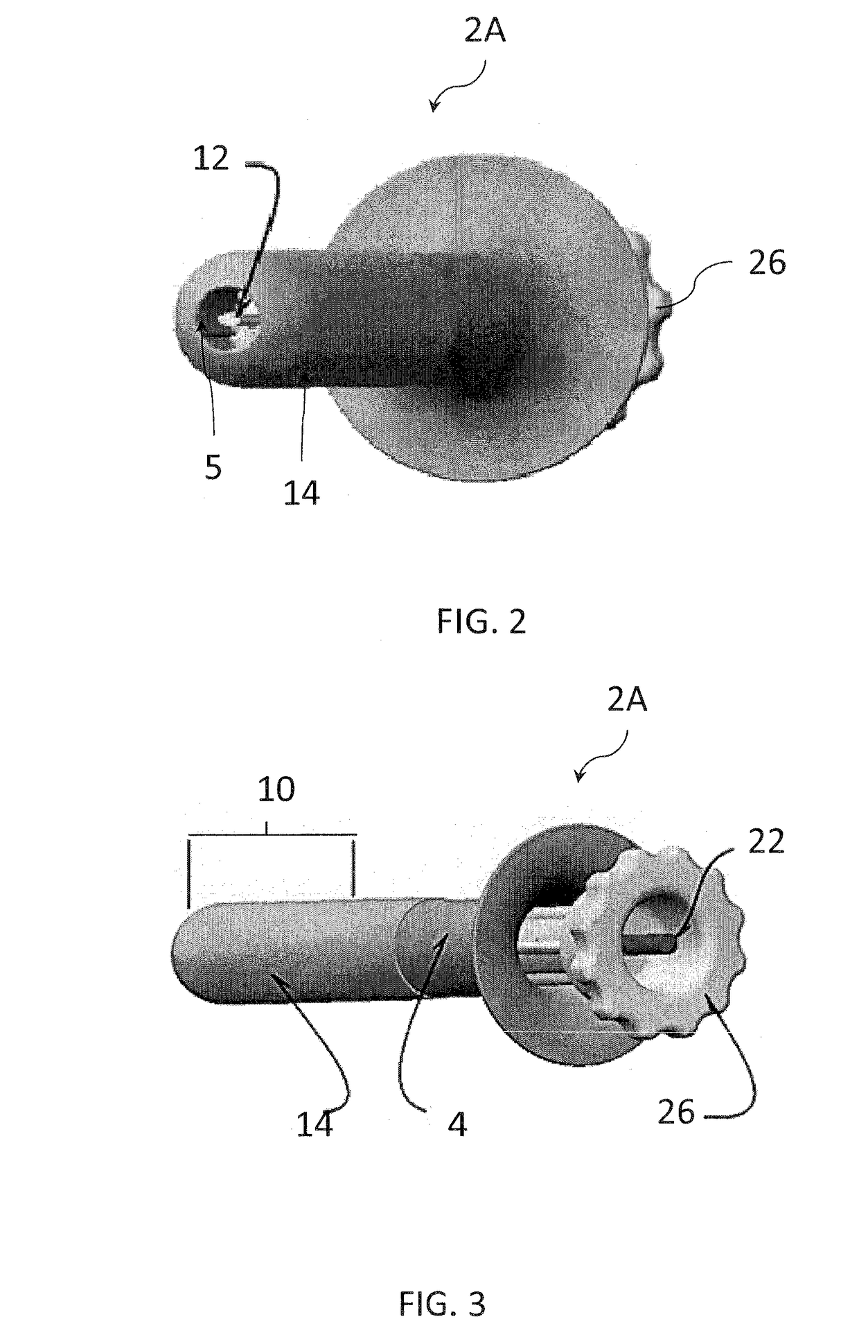
[0043]The following Detailed Description should be read with reference to the accompanying drawings, in which like elements in different drawings are identically numbered for the sake of clarity. The drawings, which are not necessarily to scale, are intended to depict salient features of the design in selected embodiments and are not intended to limit the intended scope of the invention, except where so expressly indicated. The Detailed Description illustrates by way of example, not by way of limitation, the principles of the invention. This Description will clearly enable one skilled in the art to make and use the invention, and describes several embodiments, adaptations, variations, alternatives and uses of the invention, including what is presently believed to be the best mode of carrying out the invention.

[0044]In addition, various terms are used throughout in order to provide a suitable frame of reference with regard to the accompanying drawings such as “lower”, “upper”, “top”,...


PUM


Abstract

A system and a method for a medical diagnostic device are described herein. The medical diagnostic device can include an outer sleeve having a hollow interior and open distal and proximal ends, the outer sleeve being defined by a substantially tubular configuration including a proximal section having a substantially conical shape. A core is configured to be inserted axially within the outer sleeve. The core includes an axial inner cavity. The core is further defined by a proximal section having a conical shape substantially conforming to the proximal section of the outer sleeve. The core retains a diagnostic assembly, that can include at least one of a sample collecting assembly and an imaging assembly, that can be integrally or interchangeably attached and wherein the device can be utilized for self-patient or single patient use.

Description

TECHNICAL FIELD[0001]This application generally relates to the field of medical devices and more specifically to a medical diagnostic device that permits various functionalities, such as imaging and sample collection that can be done interchangeably or in tandem. In at least one version, the medical diagnostic device can be used for body cavity examinations, such as examinations of the cervix.BACKGROUND[0002]There are known medical devices used for examining various anatomical cavities of patients. For example, vaginal specula are used in the diagnostic medical field to examine the cervix of a female patient. Over time, a number of various specula designs have been developed. Typically, a Graves speculum is defined by an upper blade and a lower blade, the latter including a pistol-grip like handle portion. The speculum is inserted in the vagina of a female patient and the upper and lower blades are articulated to facilitate examination of the cervix of the female patient.[0003]After...

Claims


Application Information

Patent Timeline
IPC IPC(8): A61B10/02A61B1/04A61B1/303
CPCA61B10/0291A61B2010/0216A61B1/303A61B1/04
Inventor DE KONING, LEZANNEVIVENZIO, ROBERT L.LIA, RAYMOND A.BENNETT, TRACY
Owner WELCH ALLYN INC
Learn More