Endoscopic examination support device, endoscopic examination support method, and endoscopic examination support program

a technology of endoscope and support device, which is applied in the direction of tomography, diagnostic recording/measuring, applications, etc., can solve the problems of difficult to make the distal end of the endoscope reach the target position, require great effort to thoroughly examine all the routes, and difficult to grasp which position within the tubular structure is represented by the endoscopic image, etc., to achieve efficient examination of the tubular structure, easy to recognize, and easy to recognize

Inactive Publication Date: 2017-11-30
FUJIFILM CORP
View PDF4 Cites 41 Cited by
  • Summary
  • Abstract
  • Description
  • Claims
  • Application Information

AI Technical Summary

Benefits of technology

[0021]According to the present invention, passage position information representing a passage position of an endoscope in a tubular structure is acquired using position information of the endoscope inserted into the tubular structure. In addition, passage propriety information representing a portion in the tubular structure through which the endoscope can be passed and a portion in the tubular structure through which the endoscope cannot be passed is acquired by comparing the diameter of the endoscope with the diameter of the tubular structure at each position. Moreover, a tubular structure image generated from a three-dimensional image is displayed by changing a display state of a portion in the tubular structure image through which the endoscope has been passed and a portion in the tubular structure image through which the endoscope has not been passed using the passage position information and a display state of the portion in the tubular structure image through which the endoscope can be passed and the portion in the tubular structure image through which the endoscope cannot be passed using the passage propriety information. For this reason, it is possible to easily recognize a route through which the endoscope has been passed and a route through which the endoscope has not been passed and to easily recognize the portion in the tubular structure through which the endoscope can be passed and the portion in the tubular structure through which the endoscope cannot be passed, through observing the tubular structure image. Accordingly, it is possible to efficiently examine the tubular structure using the endoscope.

Problems solved by technology

For this reason, it is difficult to grasp which position within the tubular structure is represented by the endoscopic image.
Particularly, a bronchial endoscope has a small diameter and a narrow field, and therefore, it is difficult to make a distal end of the endoscope reach a target position.
In such an examination of all branches, it requires great effort to thoroughly examine all the routes.
In addition, the tubular structure has multiple branches, and therefore, there is also a possibility that an unexamined portion may remain.
Therefore, there is a portion in bronchi which cannot be examined depending on the diameter of an endoscope to be used.
However, it is impossible to recognize which portion of the bronchus an endoscope in use can or cannot pass through even by viewing the bronchial image displayed through the method disclosed in JP2007-83034A.

Method used


Image

  • Endoscopic examination support device, endoscopic examination support method, and endoscopic examination support program
  • Endoscopic examination support device, endoscopic examination support method, and endoscopic examination support program
  • Endoscopic examination support device, endoscopic examination support method, and endoscopic examination support program

Examples


Embodiment Construction

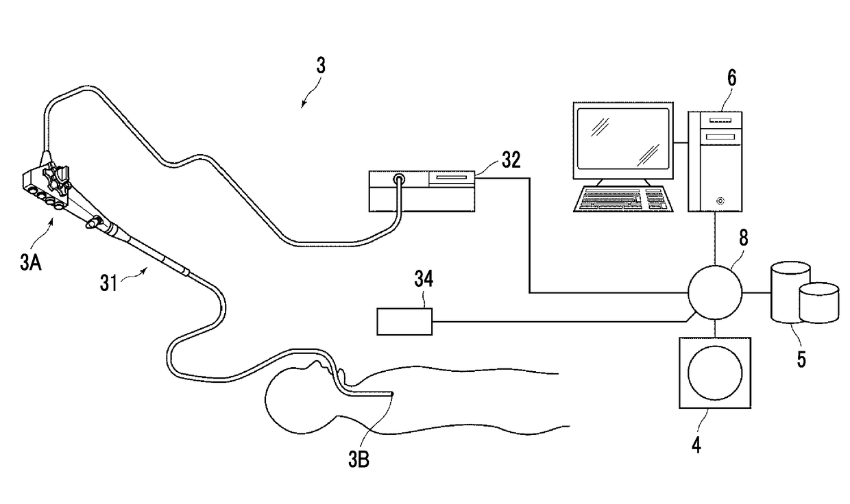
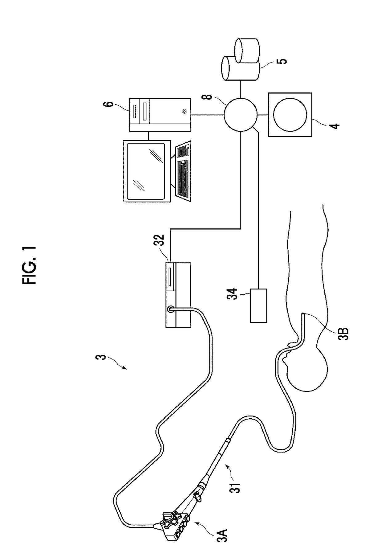
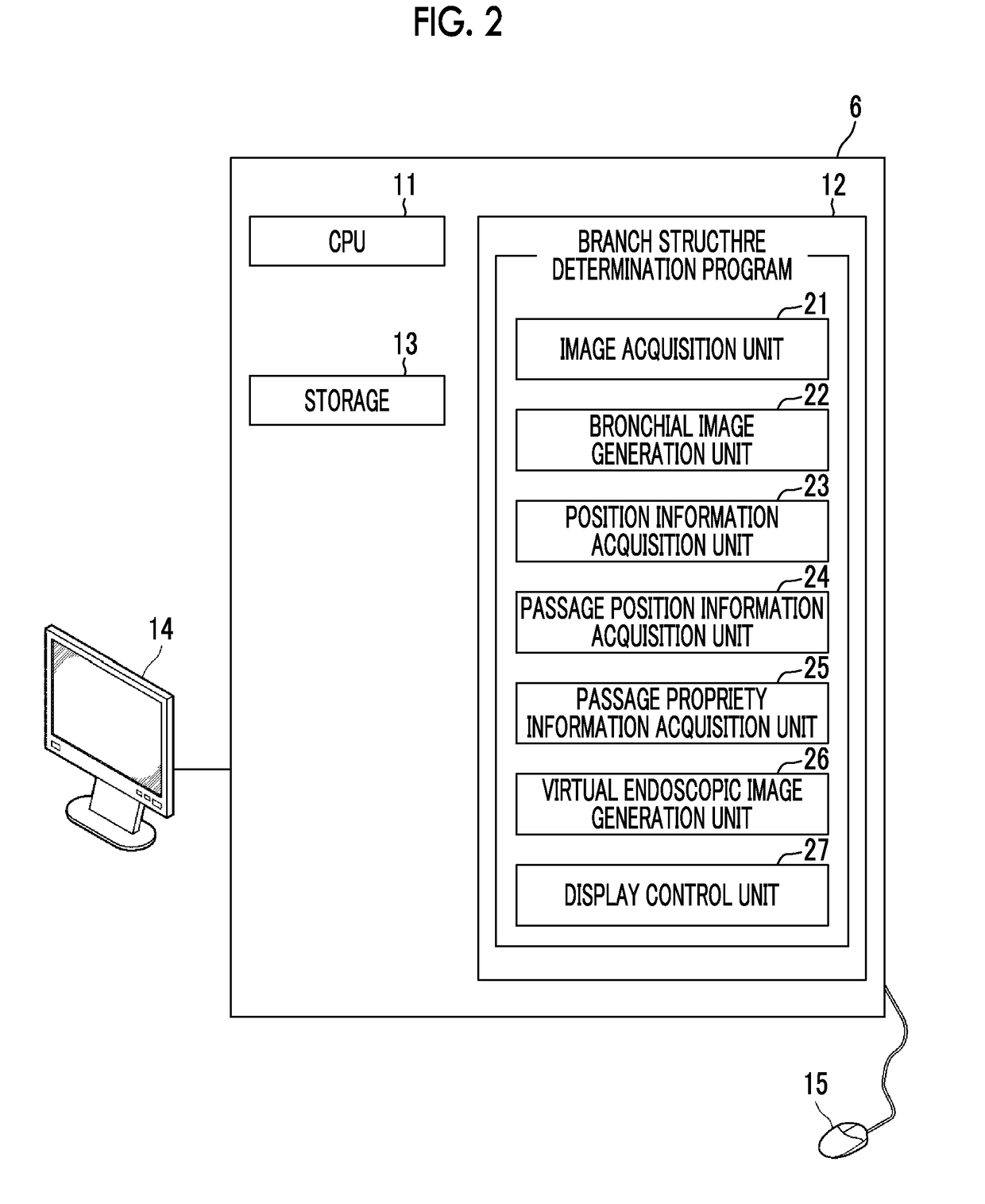
[0030]Hereinafter, an embodiment of the present invention will be described with reference to the drawings. FIG. 1 is a hardware configuration diagram showing an outline of a diagnosis support system to which an endoscopic examination support device according to an embodiment of the present invention is applied. As shown in FIG. 1, an endoscope device 3, a three-dimensional image photographing device 4, an image storage server 5, and an endoscopic examination support device 6 are connected to each other in a communicable state via a network 8 in this system.

[0031]The endoscope device 3 includes an endoscopic scope 31 imaging the inside of a tubular structure of a subject, a processor device 32 generating an image of the inside of the tubular structure based on a signal obtained through imaging, a position detection device 34 detecting the position and the direction of a distal end of the endoscopic scope 31, and the like.

[0032]The endoscopic scope 31 is an endoscopic scope in which ...


PUM


Abstract

A bronchial image generation unit generates a bronchial image and a position information acquisition unit acquires position information of an endoscope in a bronchus. A passage position information acquisition unit acquires passage position information representing a passage position of the endoscope and a passage propriety information acquisition unit acquires passage propriety information representing portions through which the endoscope can be passed and a portion through which the endoscope cannot be passed. A display control unit displays a bronchial image by changing a display state of a portion of the bronchial image through which the endoscope has been passed and a portion of the bronchial image through which the endoscope has not been passed using the passage position information, and changing a display state of portions of the bronchial image through which the endoscope can be passed and cannot be passed using the passage propriety information.

Description

CROSS REFERENCE TO RELATED APPLICATIONS[0001]The present application is a Continuation of PCT International Application No. PCT / JP2016 / 001163 filed on Mar. 3, 2016, which claims priority under 35 U.S.C. §119(a) to Japanese Patent Application No. 2015-062105 filed on Mar. 25, 2015. Each of the above applications is hereby expressly incorporated by reference, in its entirety, into the present application.BACKGROUNDTechnical Field[0002]The present invention relates to an endoscopic examination support device, an endoscopic examination support method, and an endoscopic examination support program for supporting an endoscopic examination of a tubular structure, such as bronchi, which has a branched structure.Description of the Related Art[0003]In recent years, a technique of observing or treating a tubular structure such as the large intestine or bronchi of a patient using an endoscope has been attracting attention. However, an endoscopic image is an image obtained by indicating the insi...

Claims


Application Information

Patent Timeline
Patent Type & Authority Applications(United States)
IPC IPC(8): A61B5/06A61B1/267A61B1/04
CPCA61B5/066A61B1/2676A61B1/04A61B1/0005A61B6/032A61B6/037A61B6/12A61B6/4417A61B6/466A61B6/5217A61B8/0841A61B8/4416A61B8/466A61B8/483A61B8/5223G16H50/30
Inventor YAMADA, KENTA
Owner FUJIFILM CORP
Why Eureka
Try Eureka