T cell-bound cytokine assay for antigen-specific tolerance

a cytokine and antigen-specific technology, applied in the field of t cell-bound cytokine assay for antigen-specific tolerance, can solve the problems of few assays, few assays, and little success in developing in vitro non-animal assays

Inactive Publication Date: 2017-12-14
UNIVERSITY OF PITTSBURGH +2
View PDF2 Cites 1 Cited by
  • Summary
  • Abstract
  • Description
  • Claims
  • Application Information

AI Technical Summary

Benefits of technology

[0005]In the interest of providing a clear and concise summary, the following description references certain exemplary aspects and embodiments. Persons of ordinary skill in the art will, in view of the teachings in this application, readily recognize and appreciate that other aspects, embodiments, configurations, and variations of the technology disclosed herein are possible and that the exemplary aspects and embodiments described in this summary or elsewhere in this application are neither limiting nor exhaustive.

Problems solved by technology

However, drug-free allograft acceptance is rarely encountered in transplant recipients.
However, few assays exist to measure the regulatory portion of an immune response.
In the intervening 15 years, others have tried with little success to develop in vitro non-animal assays to measure and model human DTH responses, but most of these non-animal assays measure antigen non-specific responses using whole donor cells as a source of antigens.

Method used


Image

  • T cell-bound cytokine assay for antigen-specific tolerance
  • T cell-bound cytokine assay for antigen-specific tolerance
  • T cell-bound cytokine assay for antigen-specific tolerance

Examples


example 1

en-Specific Response Following Donor-Specific Transfusion (DST) and Co-Stimulatory Blockade Treatment in CBA-Tolerized Mice

[0050]To initially validate the T-CBC assay, we induced tolerance in C57BL / 6 (“B6”) mice to CBA antigens using a donor-specific transfusion (DST) plus anti-CD154 (CD154 is also known as CD40 Ligand) tolerization method. The term “CBA antigens” refers to a lysate of spleen cells obtained from a mouse of the CBA strain and prepared by sonication. Following sonication and centrifugation at 10,000×g, the liquid supernatant is used as the source of CBA antigens. CD154 (aka CD40 Ligand) is a membrane glycoprotein and differentiation antigen transiently expressed on the surface of activated T cells. Through the binding of CD154 to CD40 on antigen presenting cells (APC) including B cells, monocytes / macrophages, and dendritic cells, it serves a crucial function in T cell-APC cognate interaction. CD154-interaction with CD40 transduces signals for T-dependent B cell activa...

example 2

Quantifying Treg Cells Specific for Self-Antigens or Donor Alloantigens

[0055]Materials and Methods:

[0056]Single cell suspensions of spleen, lymph node cells or peripheral blood mononuclear cells were cultured overnight in DMEM+10% FBS in the presence of a specific test antigen or crude cell lysate. When measuring alloantigen-specific induction of tolerance, the antigen preparation was a supernatant obtained from a sonicate of 10×106 donor spleen, lymph node cells or peripheral blood mononuclear cells, centrifuged at 10,000×g for 14 minutes at 4° C. with phenylmethylsulfonyl fluoride (PMSF) and protease inhibitor iodoacetamide (IAA) at a concentration of 4×10⁷ cell equivalents per mL. To measure allospecific Treg, no intact donor cells are used. Both nanovesicles and soluble protein are present in these crude antigen preps. See e.g., Bracamonte-Baran and Burlingham, Biomed. J. 2015, 38(1):39-51.

[0057]When detecting self-antigen-specific Treg cells, the antigen preparations can contai...

example 3

face Staining

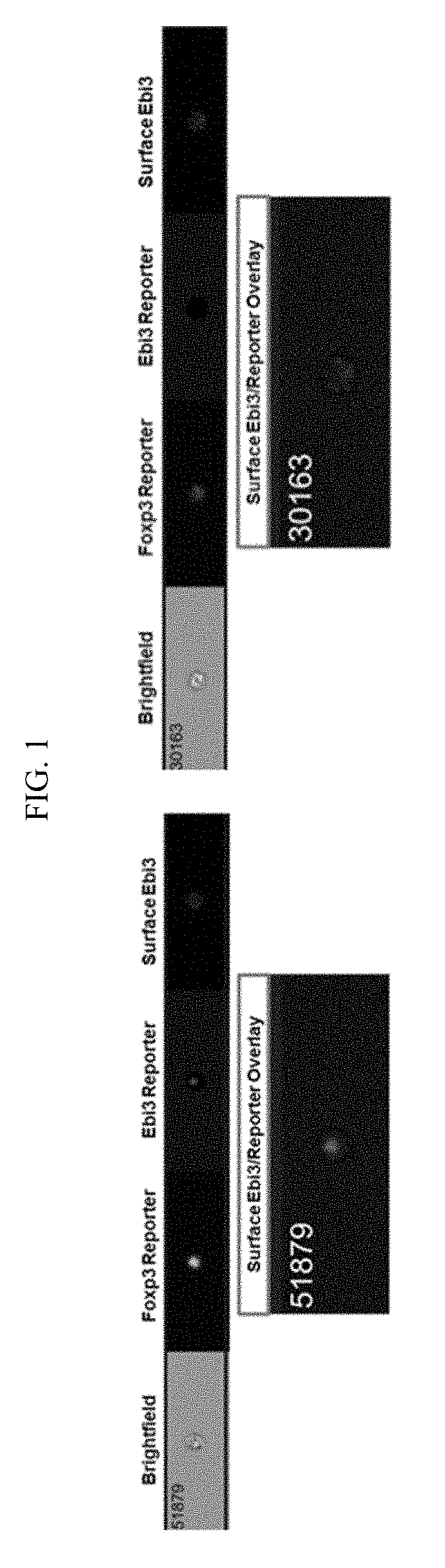
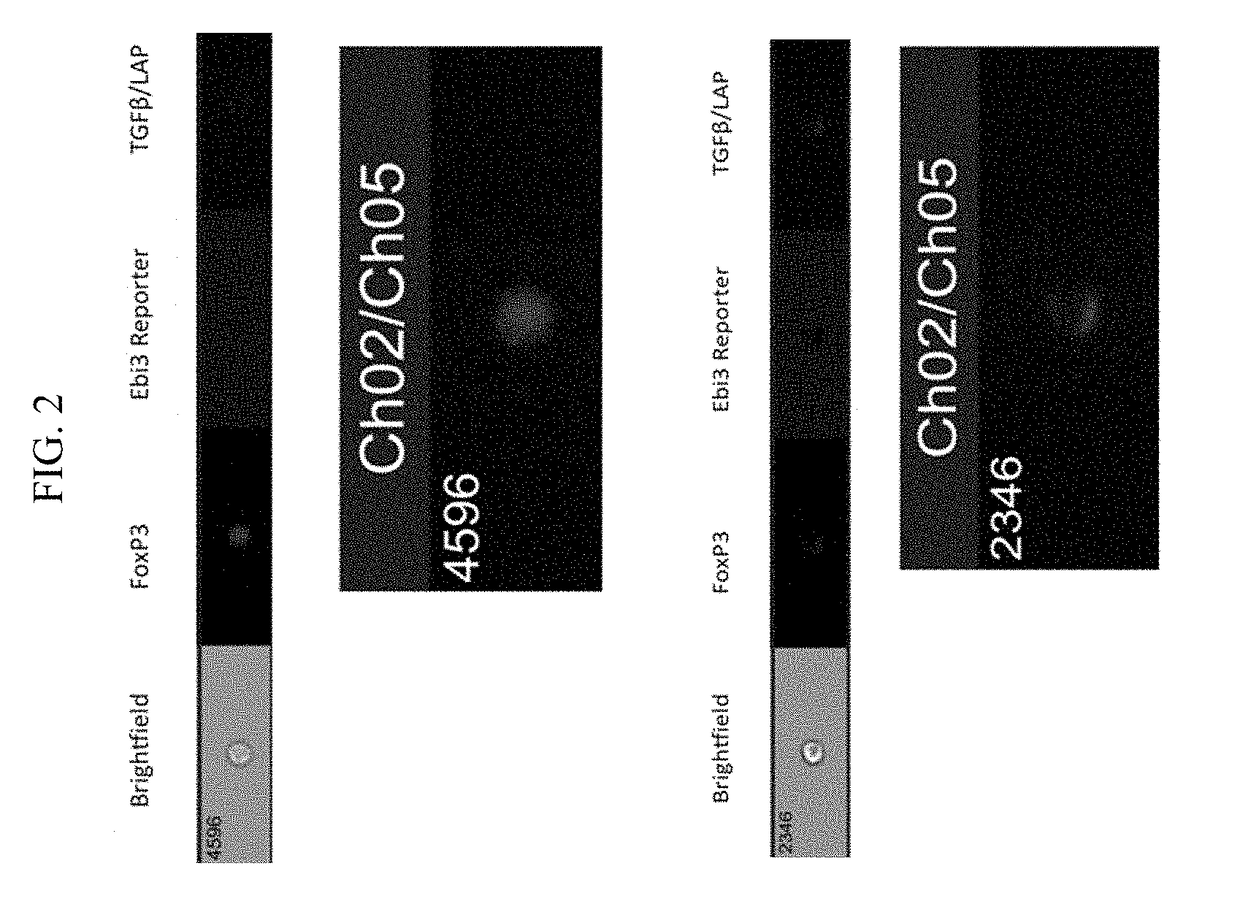
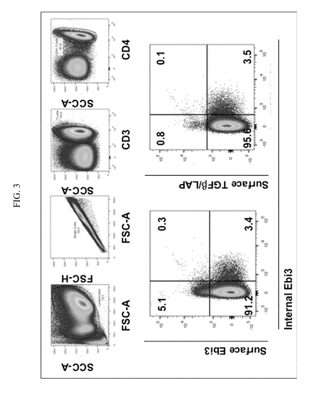
[0068]Single cell suspensions were prepared from spleen, lymph nodes, or other tissues of interest. Staining for live and dead cells was performed prior to surface staining with IL-35 antibody. LIVE / DEAD® fixable dead cell stain Aqua (Invitrogen) was used at a dilution of 1:700 in phosphate buffered saline. Alternatively, scatter gates can be used to exclude apoptotic cells and doublets. After washing off the live / dead stain, we prepared a staining cocktail for surface markers for staining different lymphocyte populations based on the experimental needs. A Foxp3 reporter mouse was used for Treg analysis. A staining cocktail composition for detecting surface expression of IL-35 on Treg / Teff cells is listed below:

[0069]NMS (10%) of staining buffer (FACS) volume

[0070]CD4 (at 1:500 dilution)

[0071]CD25 (at 1:500 dilution)

[0072]CD45RB (at 1:500 dilution)

[0073]Ebi3 Biotin-linked antibody (V1.4F5.29 clone described by Collison et al, Nature Immunol., 2010). The stock concentrat...


PUM

PropertyMeasurementUnit
sizeaaaaaaaaaa
concentrationaaaaaaaaaa

Abstract

In vitro methods for detecting and measuring an antigen-specific regulatory T cell response are described. In particular, there is provided, for example, a method of detecting a change in surface expression of particular T cell markers in T cells obtained from the subject as a way to detect induced immune suppression in response to exposure to a particular antigen or plurality of antigens.

Description

CROSS-REFERENCE TO RELATED APPLICATIONS[0001]This application claims the benefit of U.S. Provisional Application No. 62 / 348,653, filed Jun. 10, 2016, which is hereby incorporated by reference in its entirety for all purposes.STATEMENT REGARDING FEDERALLY SPONSORED RESEARCH OR DEVELOPMENT[0002]This invention was made with government support under AI066219 awarded by the National Institutes of Health. The government has certain rights in the invention.BACKGROUND[0003]Establishment of tolerance to a well-functioning transplant with limited specific inductive therapy is a major goal of organ transplantation. Such tolerance manifests as drug-free allograft acceptance, meaning that a transplant recipient exhibits tolerance to alloantigens (antigens present in members of the same species and used by the immune system to distinguish self from non-self) derived from the transplanted cells, tissue, or organ. However, drug-free allograft acceptance is rarely encountered in transplant recipient...

Claims


Application Information

Patent Timeline
Patent Type & Authority Applications(United States)
IPC IPC(8): G01N33/50G01N33/74G01N33/68G01N33/564
CPCG01N33/5091G01N33/564G01N33/6869G01N2800/245G01N2333/54G01N2333/495G01N33/74
Inventor BURLINGHAM, WILLIAM JAMESVIGNALI, DARIO ALBERTO
Owner UNIVERSITY OF PITTSBURGH
Why Eureka
Try Eureka