Measuring breast density using breast computed technology

a breast density and computed technology, applied in the field of breast imaging, can solve the problems of deterring some women from regular mammograms, uncomfortable compression, limitations of mammography, etc., and achieve the effect of improving the ability to perform biopsies

Active Publication Date: 2018-01-11
RGT UNIV OF CALIFORNIA
View PDF2 Cites 14 Cited by
  • Summary
  • Abstract
  • Description
  • Claims
  • Application Information

AI Technical Summary

Benefits of technology

[0009]The present invention provides a set of tools that improves the ability to perform biopsies when using a breast CT machine. The invention allows the spatial coordinates of the suspicious lesions to be extracted directly from the breast CT images and these spatial coordinates to be used to guide a biopsy without repositioning or re-registration of the patient.
[0010]The invention first provides a multi-image display of breast CT data in any of several different formats: axial slices, coronal, sagittal and axial projections, volume rendered and / or virtual or actual compression mammograms. Each image may have a cursor that tracks with cursors in other images following a common position within the breast. The cursors allow accurate determination of the coordinates of the suspicious lesion as identified by a variety of methods including computer analysis of image / volume data and operator inspection of the images. As part of this process, an iterative matching system makes it possible to superimpose matching cursors, not only on the CT generated images, but on a standard mammogram taken earlier with a standard mammography machine.
[0011]Once the location of the suspicious lesion is identified, a robotic biopsy system incorporated into the CT system may provide a biopsy without the need to re-position the patient. Geometric correction of the CT machine using a specially designed phantom and / or the ability to image portions of the robot, before and during the insertion of the biopsy needle (using periodic updates of the breast image), allows an accurate biopsy on the same machine. The breast may be stabilized during this process with a support, for example, a suction collar having apertures for admitting a biopsy needle.

Problems solved by technology

Despite the important role that mammography plays in breast cancer screening, it is well recognized that mammography has limitations, particularly in women with dense breasts.
Further, even with compression of the breast, minor overlap of normal breast anatomy can sometimes obscure suspicious areas—especially in women with breast implants or very dense breasts.
The compression can be uncomfortable and may deter some women from regular mammograms.
Nevertheless, standard whole-body CT is impractical for imaging the breasts because the axial scan used in such machines would deposit significant radiation in the torso and lungs near the breast.
One drawback to such breast CT machines, in instances where a suspicious lesion is found and a biopsy is required, is that compression of the breast in a standard mammography fixture and possibly re-imaging of the breast may be required to provide the necessary guidance for the biopsy procedure.

Method used


Image

  • Measuring breast density using breast computed technology
  • Measuring breast density using breast computed technology
  • Measuring breast density using breast computed technology

Examples


Embodiment Construction

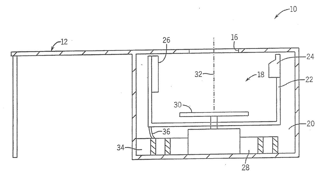
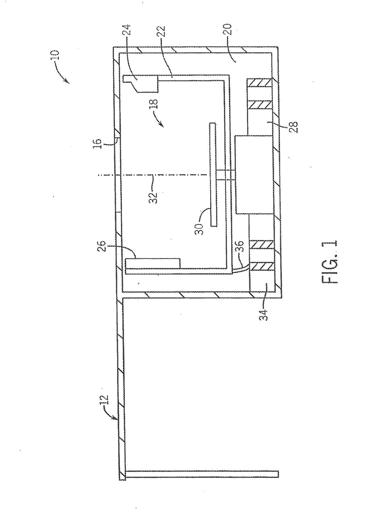
[0068]Referring more specifically to the drawings for illustrative purposes, the present invention is embodied in the apparatus generally shown in FIG. 1 through FIG. 37. It will be appreciated that the apparatus may vary as to configuration and as to details of the parts, and that the method may vary as to the specific steps and sequence, without departing from the basic concepts as disclosed herein.

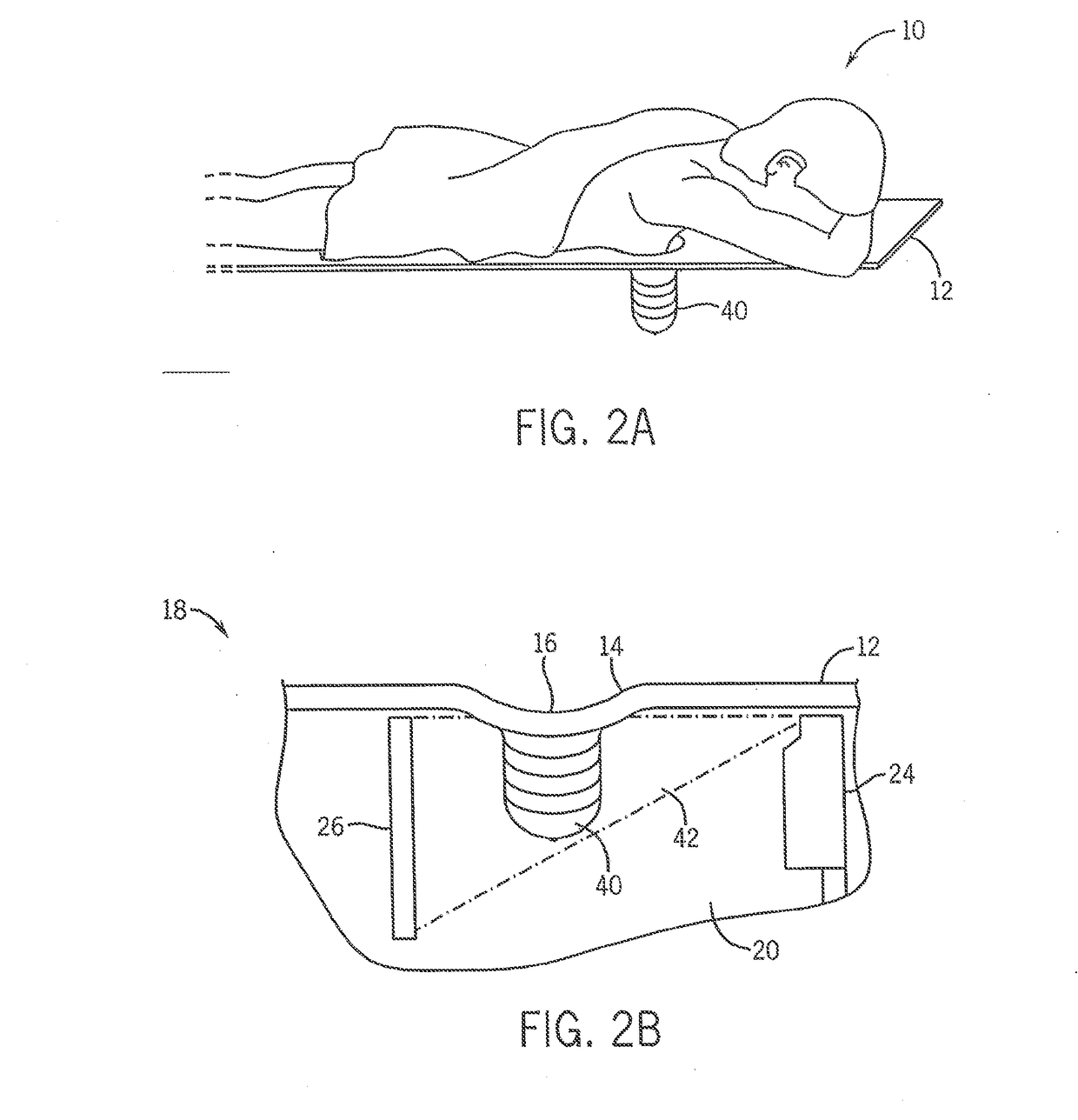
[0069]1. CT Imaging of Pendant Breast.

[0070]Referring to FIG. 1, a breast CT scanner 10 tailored specifically for breast cancer screening is shown. The scanner has a padded table 12 with a circular opening 16 through which the patient may individually position the breast to be screened. A CT scanning mechanism 18 is positioned in chamber 20 under the table 12. The scanning mechanism 18 comprises an x-ray emitter 24 and detector 26 supported on opposing sides of a rotating gantry 22. A centrally located motor 28 is mechanically coupled to the gantry 22 so that the gantry 22 is rotated in...


PUM


Abstract

A device and methods for performing a simulated CT biopsy on a region of interest on a patient. The device comprises a gantry (22) configured to mount an x-ray emitter (24) and CT detector (26) on opposing sides of the gantry, a motor (28) rotatably coupled to the gantry such that the gantry rotates horizontally about the region of interest, and a high resolution x-ray detector (172) positioned adjacent the CT detector in between the CT detector and the x-ray emitter.

Description

CROSS-REFERENCE TO RELATED APPLICATIONS[0001]This application is based on U.S. Provisional Application 60 / 677,704 filed May 3, 2005, hereby incorporated by reference.STATEMENT REGARDING FEDERALLY-SPONSORED RESEARCH OR DEVELOPMENT[0002]This invention was made with Government support under Grant Nos. EB002138 and EB89260, awarded by the National Institute of Health. The government has certain rights in this invention.BACKGROUND OF THE INVENTION1. Field of the Invention[0003]This invention pertains generally to imaging of a patient's breast, and more particularly to improved methods and apparatus for breast diagnosis, evaluation and therapy through CT imaging.2. Description of Related Art[0004]X-ray mammography is used as the primary breast cancer screening test worldwide, and recent reports have suggested that mammography has been responsible for a sizeable reduction in breast cancer mortality. In standard mammography, the breast is generally compressed between two radiolucent plates ...

Claims


Application Information

Patent Timeline
Patent Type & Authority Applications(United States)
IPC IPC(8): A61B6/00A61B6/03A61B6/04A61B8/00A61B8/08A61B6/12
CPCA61B6/03A61B6/405A61B6/4417A61B6/4028A61B8/4416A61B6/4423A61B6/5235A61B6/502A61B6/0435A61B6/584A61B6/037A61B6/12A61B8/0825A61B6/482A61N2005/1061A61B10/0041A61B6/469A61B6/4452A61B6/032A61B6/488A61B6/06G16H50/70
Inventor BOONE, JOHN M.YELLEN-NELSON, THOMAS R.
Owner RGT UNIV OF CALIFORNIA
Why Eureka
Try Eureka