Methods of selecting and isolating cancer stem cells

a cancer stem cell and stem cell technology, applied in the field of cancer stem cell selection and isolating, can solve the problems of phenotypical heterogeneity and relatively unstable cscs, and achieve the effect of lowering the fluorescence level

Active Publication Date: 2018-01-11
RGT UNIV OF CALIFORNIA
View PDF1 Cites 3 Cited by
  • Summary
  • Abstract
  • Description
  • Claims
  • Application Information

AI Technical Summary

Benefits of technology

[0031]In another aspect, provided herein is a method of isolating leukemia stem cells of B-cell type acute lymphoblastic leukemia (B-cell ALL) from a sample (e.g., a biological sample or a cell culture sample). The sample can be a B-cell ALL cell line or a sample obtained from a patient with B-cell ALL. The method includes: (a) incubating the sample with a fluorescent glucose analog under suitable conditions, (b) measuring or detecting the level of fluorescence in the sample using, for example, flow cytometry; and (c) isolating (e.g., separating or purifying) the leukemia stem cells having a lower level of fluorescence compared to the non-leukemia stem cells in the sample. Optionally, the isolated leukemia stem cells can be confirmed to be stem cells of B-cell ALL by detecting a higher or lower expression level of at least one leukemia stem cell marker of Table 3 in an isolated leukemia stem cell compared to a non-leukemia stem cell. In some embodiments, a higher expression level of at least one leukemia stem cell marker selected from the group consisting of leukemia stem cell marker Nos. 1-9 of Table 3 indicates that the isolated leukemia stem cells are B-cell ALL stem cells. In other embodiments, a lower expression level of at least one leukemia stem cell marker selected from the group consisting of leukemia stem cell marker Nos. 10-80 of Table 3 indicates that the isolated leukemia stem cells are B-cell ALL stem cells. In yet other embodiments, a higher expression level of at least one leukemia stem cell marker selected from the group consisting of leukemia stem cell marker Nos. 1-9 of Table 3 in combination with a lower expression level of at least one leukemia stem cell marker selected from the group consisting of leukemia stem cell marker Nos. 10-80 of Table 3 indicates that the isolated leukemia stem cells are B-cell ALL stem cells.
[0032]In another aspect, provided herein is a method of isolating leukemia stem cells of T-cell type acute lymphoblastic leukemia (T-cell ALL) from a sample (e.g., a biological sample or a cell culture sample). The sample can be a T-cell ALL cell line or a sample obtained from a patient with T-cell ALL. The method includes: (a) incubating the sample with a fluorescent glucose analog under suitable conditions, (b) measuring or detecting the level of fluorescence in the sample using, for example, flow cytometry; and (c) isolating (e.g., separating or purifying) the leukemia stem cells having a lower level of fluorescence compared to the non-leukemia stem cells in the sample. Optionally, the isolated leukemia stem cells can be confirmed to be stem cells of T-cell ALL by detecting a higher or lower expression level of at least one leukemia stem cell marker of Table 4 in an isolated leukemia stem cell compared to a non-leukemia stem cell. In some embodiments, a higher expression level of at least one leukemia stem cell marker selected from the group consisting of leukemia stem cell marker Nos. 1-19 of Table 4 indicates that the isolated leukemia stem cells are T-cell ALL stem cells. In other embodiments, a lower expression level of at least one leukemia stem cell marker selected from the group consisting of leukemia stem cell marker Nos. 20-105 of Table 4 indicates that the isolated leukemia stem cells are T-cell ALL stem cells. In yet other embodiments, a higher expression level of at least one leukemia stem cell marker selected from the group consisting of leukemia stem cell marker Nos. 1-19 of Table 4 in combination with a lower expression level of at least one leukemia stem cell marker selected from the group consisting of leukemia stem cell marker Nos. 20-105 of Table 4 indicates that the isolated leukemia stem cells are T-cell ALL stem cells.

Problems solved by technology

Challenges remain for isolating CSCs including LSCs because CSCs are phenotypically heterogeneous and are relatively unstable.

Method used


Image

  • Methods of selecting and isolating cancer stem cells
  • Methods of selecting and isolating cancer stem cells
  • Methods of selecting and isolating cancer stem cells

Examples


example 1

Stem Cells (LSCs) in Acute Lymphoblastic Leukemia (ALL): Unveiling Hierarchical Structure at Single Cell Resolution

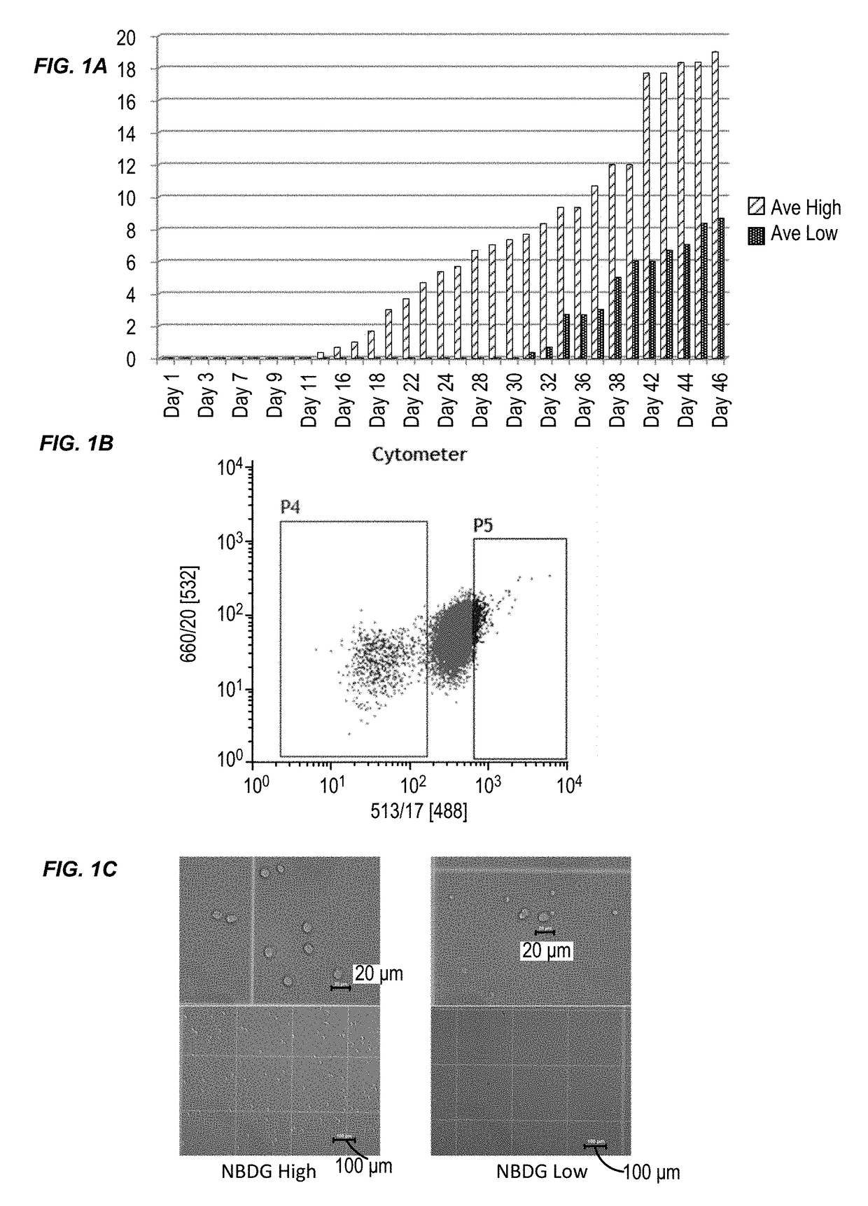
[0132]This example illustrates a novel method to isolate ALL LSCs based on their cellular metabolic activity. This example also shows that these isolated LSCs have in vivo leukemia-initiating capability (LIC). This example also describes a series of primary ALL xenograft mouse models generated from ALL patient samples and NOD / SCID / IL2Rγ− / − (NSG) mice.

[0133]Leukemia cells harvested from several generations of these mice were used in this study. LSCs and non-LSCs were isolated from 4 different B-cell type ALL samples and transplanted separately into healthy NSG mice. Cell numbers used varied between 5,000; 10,000; and 50,000 per mouse, and the number of the animals varied between three and eight per group. All the animals transplanted with LSCs developed leukemia between 5-14 weeks, whereas those transplanted with non-LSCs did not develop the disease within the same timef...

example 2

Stem Cells (LSCs) in B-Cell Acute Lymphoblastic Leukemia (B-Cell ALL)

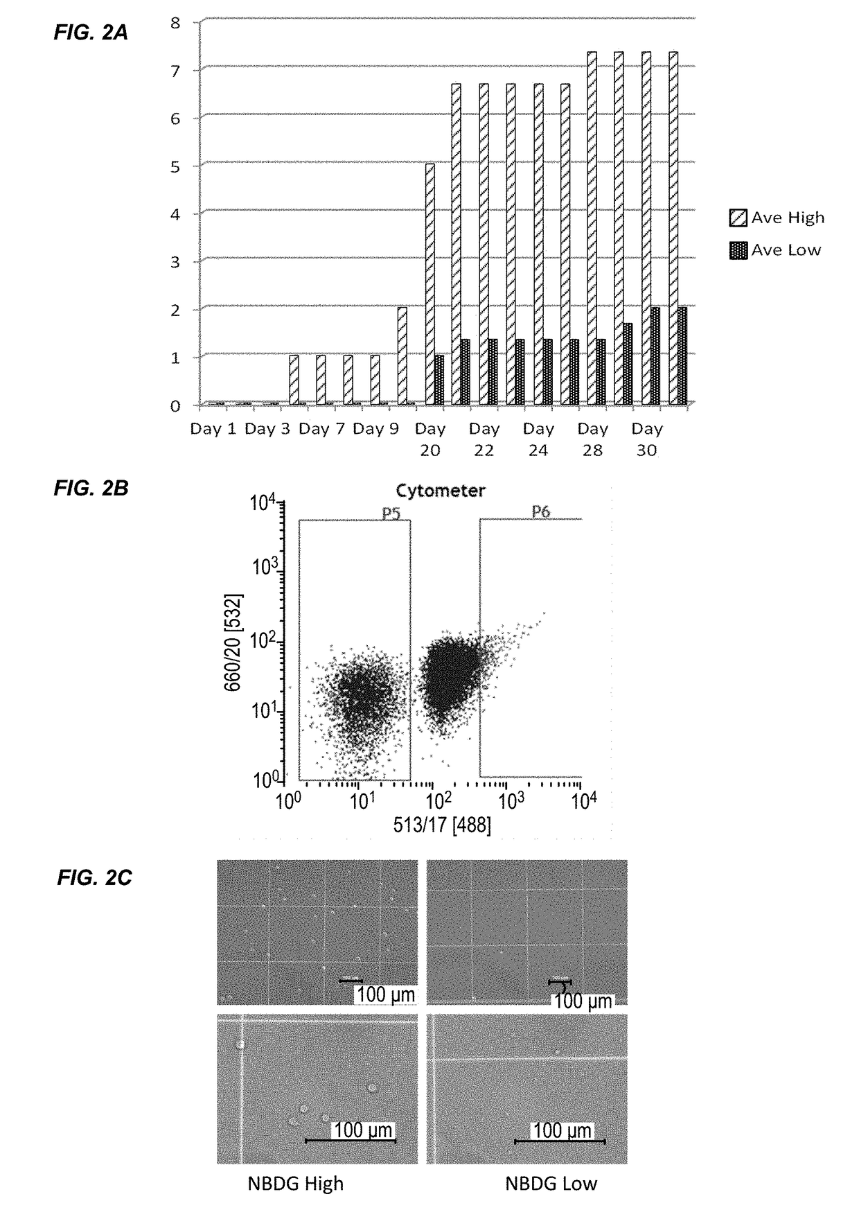
[0178]This example describes the results of a genome-wide microarray gene expression profiling of RNA isolated from the JM1 cell line. The experimental procedures are described in Example 1. Briefly, JM1 cells were treated with NBDG and FACS sorted to isolate two cell populations: a low NBDG group and a high NBDG group. The cells of each group were subject to transcriptome analysis using a gene expression microarray (SurePrint® G3 Human Gene Expression 8×60K v2 Microarray, Agilent Technologies). The data is provided in Table 3. There was an average fold change of ≧1.5 between the groups (i.e., low NBDG vs. high NBDG) with paired t-test results of p≦0.05. 80 genes exhibited a differential expression level between B-cell ALL stem cells and non-B-cell ALL stem cells. The expression level difference for each of the 80 genes was statistically significant.

example 3

Stem Cells (LSCs) in T-Cell Acute Lymphoblastic Leukemia (T-Cell ALL)

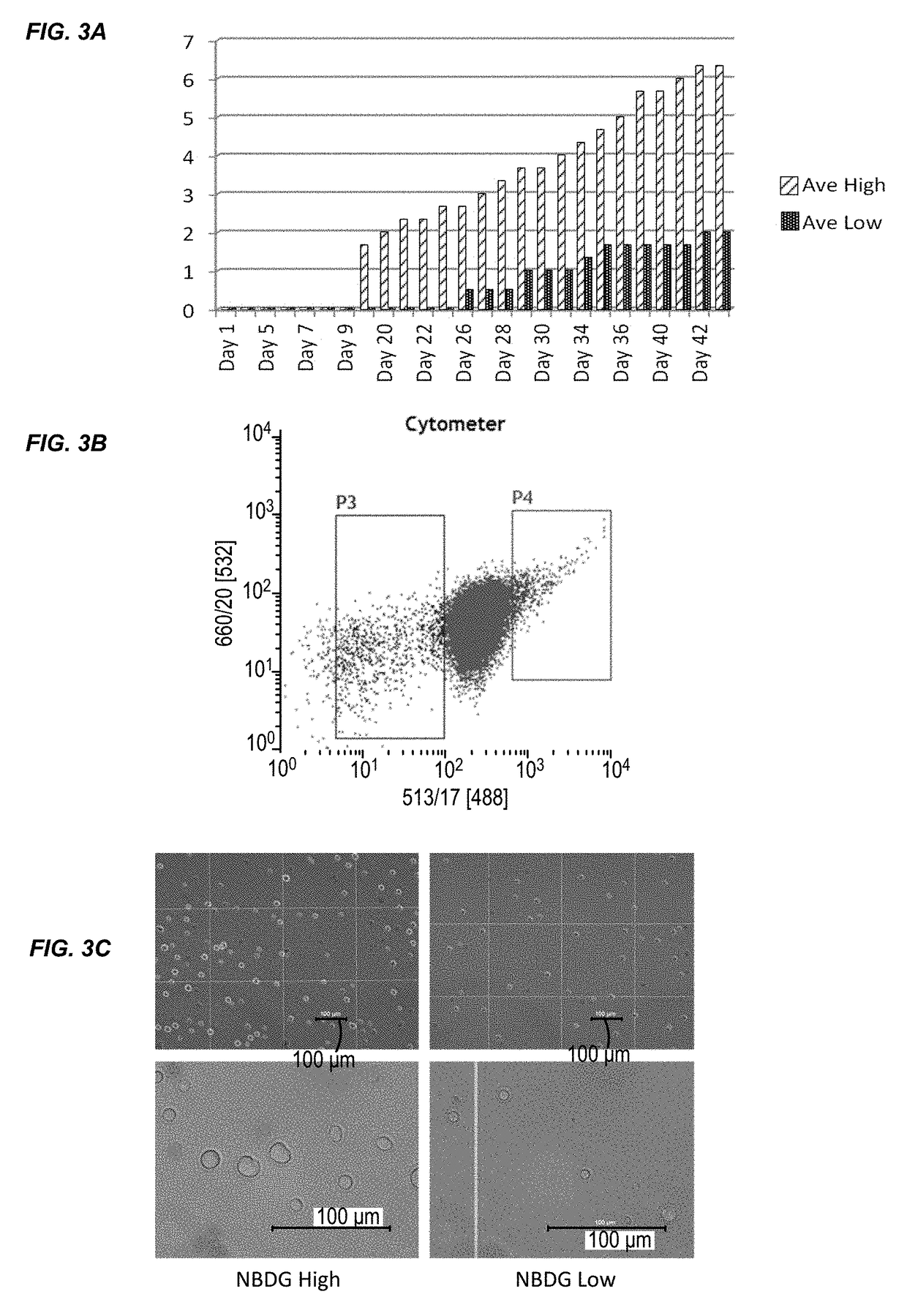
[0179]This example describes the results of a genome-wide microarray gene expression profiling of RNA isolated from the Molt4 cell line. The experimental procedures are described in Example 1. Briefly, Molt4 cells were treated with NBDG and FACS sorted to isolate two cell populations: a low NBDG group and a high NBDG group. The cells of each group were subject to transcriptome analysis using a gene expression microarray (SurePrint® G3 Human Gene Expression 8×60K v2 Microarray, Agilent Technologies). The data is provided in Table 4. There was an average fold change of ≧1.5 between the groups (i.e., low NBDG vs. high NBDG) with paired t-test results of p≦0.05. 105 genes exhibited a differential expression level between T-cell ALL stem cells and non-T-cell ALL stem cells. The expression level difference for each of the 105 genes was statistically significant.


PUM

PropertyMeasurementUnit
pHaaaaaaaaaa
temperatureaaaaaaaaaa
sizeaaaaaaaaaa

Abstract

Provided herein are methods for selecting/identifying and/or isolating cancer stem cells from a biological sample or a cell culture sample using a fluorescent glucose analog. Also provided herein are methods for selecting/identifying and/or isolating leukemia stem cells and subpopulations thereof. The present invention is based, in part, on the discovery that cancer stem cells can be selected/identified based upon a lower level of fluorescence of the fluorescent glucose analog compared to non-cancer stem cells and that specific genes are differentially expressed in leukemia stem cells compared to non-leukemia stem cells.

Description

CROSS-REFERENCES TO RELATED APPLICATIONS[0001]This application is a continuation of International Patent Application No. PCT / US2015 / 059269, filed Nov. 5, 2015, which claims priority to U.S. Provisional Application No. 62 / 075,751, filed Nov. 5, 2014, which applications are incorporated herein by reference in their entirety for all purposes.BACKGROUND OF THE INVENTION[0002]Cancer develops in a hierarchical pattern originating from cancer stem cells (CSCs) that self-renew and give rise to more differentiated cancer cells that are unable to initiate the disease. Cancer stem cells have been identified in several types of cancer, including acute myeloid leukemia (AML), breast cancer, head and neck cancer, glioma, lung cancer, prostate cancer, mesenchymal neoplasm, melanoma, and colon cancer to name a few. Methods of selecting CSCs include using a combination of cell surface markers such as, EpCAM, CD166, CD144, CD133, and CD44.[0003]Cancer stem cells are key to the progression of cancer, ...

Claims


Application Information

Patent Timeline
Patent Type & Authority Applications(United States)
IPC IPC(8): G01N33/574
CPCG01N33/57426G01N33/574G01N33/57407G01N33/5005G01N33/582
Inventor SATAKE, NORIKONITIN, NITIN
Owner RGT UNIV OF CALIFORNIA
Why Eureka
Try Eureka