Methods and systems for enhancing microangiography image quality

a microangiography and image quality technology, applied in image enhancement, instruments, applications, etc., can solve the problems of difficult to obtain detailed information of pathology and significant vision loss, and achieve the effect of enhancing the quality of flow images

Active Publication Date: 2018-02-15
UNIV OF WASHINGTON
View PDF0 Cites 2 Cited by
  • Summary
  • Abstract
  • Description
  • Claims
  • Application Information

AI Technical Summary

Benefits of technology

[0008]In accordance with the present invention, a system and a method are defined for enhancing quality of a flow image of a sample of a subject. In one embodiment, the computer-implemented method may comprise acquiring a first flow image from a plurality of first OMAG scans of the sample. The sample comprises a biological tissue having fluid flow therethrough. The method includes acquiring a structure image from a second OMAG scan of the sample, plotting, onto a graph, data based on pixel intensity values from the flow image and pixel intensity values from the structure image and differentiating the data, based on location on the graph, into a first data group representing static structure signals and a second data group representing flow signals. The method then includes suppressing pixels in the flow image corresponding to the first data group. The graph may be a two-dimensional (2D) graph or a three-dimensional (3D) graph. The fluid flow may be a blood flow, and the method may further comprise quantifying blood perfusion within the biological tissue in the image.

Problems solved by technology

Retinal pathologies can cause alteration in the retinal vascular network and affect photoreceptor function, which may lead to a significant loss of vision.
However, due to its relatively low spatial resolution and lack of depth information, it is difficult to obtain detailed information of the pathology, such as the location of affected vessels and the specific morphological changes in the capillary network.

Method used


Image

  • Methods and systems for enhancing microangiography image quality
  • Methods and systems for enhancing microangiography image quality
  • Methods and systems for enhancing microangiography image quality

Examples


Embodiment Construction

[0064]In the following detailed description, reference is made to the accompanying figures, which form a part thereof. In the figures, similar symbols typically identify similar components, unless context dictates otherwise. The illustrative embodiments described in the detailed description, figures, and claims are not meant to be limiting. Other embodiments may be utilized, and other changes may be made, without departing from the spirit or scope of the subject matter presented herein. It will be readily understood that the aspects of the present disclosure, as generally described herein, and illustrated in the figures, can be arranged, substituted, combined, separated, and designed in a wide variety of different configurations, all of which are explicitly contemplated herein.

I. Overview

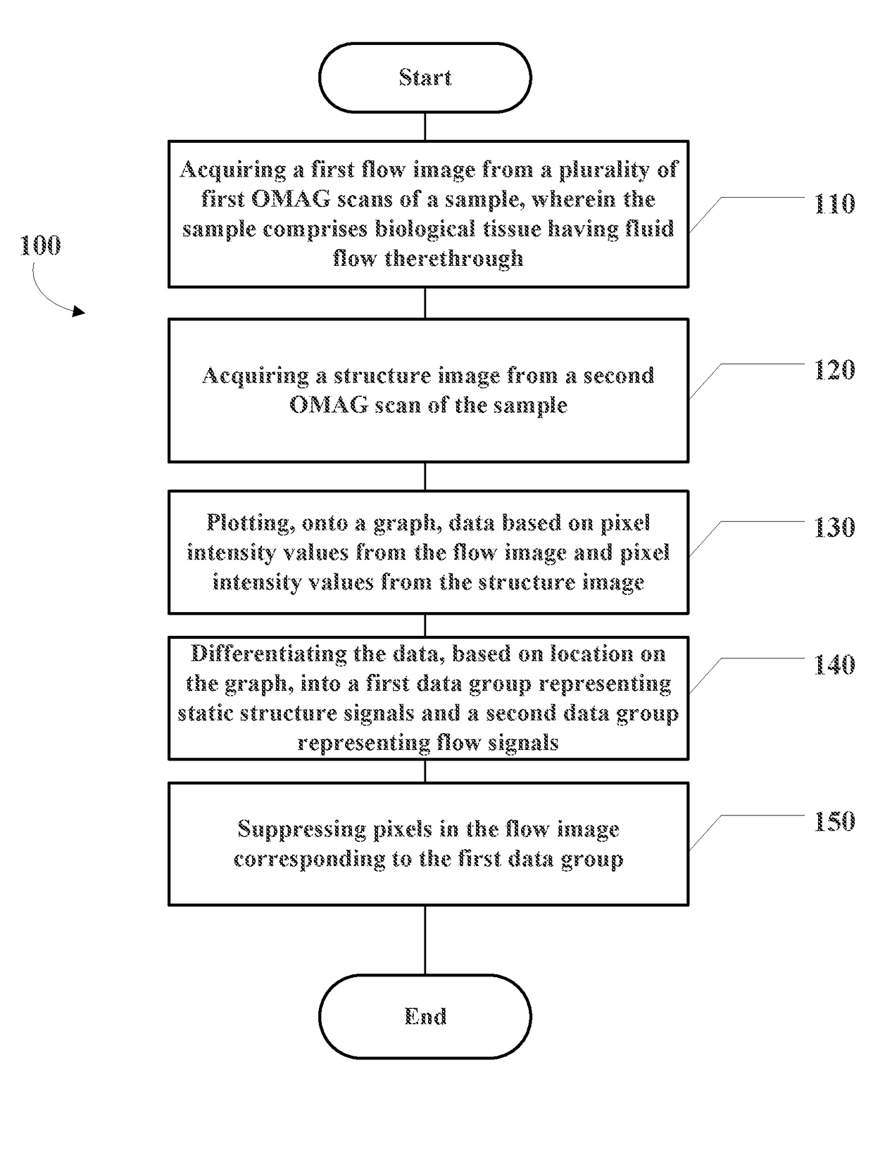
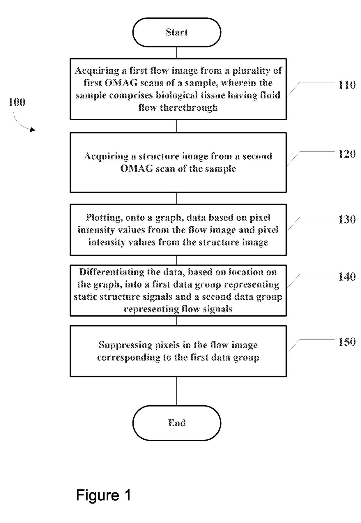
[0065]Angiography methods, such as OMAG, provide for the visualization of functional blood vessels noninvasively and with exceptional sensitivity. Yet such methods are prone to capturing static back...


PUM


Abstract

Systems and methods for enhancing quality of a flow image of a sample of a subject are provided. The method comprises acquiring a first flow image from a plurality of first OMAG scans of the sample, and acquiring a structure image from a second OMAG scan of the sample. Data based on pixel intensity values from the flow image and pixel intensity values from the structure image are then plotted onto a graph and from the graph, may be differentiated into a first data group representing static structure signals and a second data group representing flow signals. The method then includes suppressing pixels in the flow image corresponding to the first data group. The flow signal may also be multiplied by a weighting factor to suppress artifacts in the image.

Description

CROSS-REFERENCE TO RELATED APPLICATIONS[0001]This application claims priority to U.S. Provisional Patent Application Ser. No. 62 / 129,410 filed on Mar. 6, 2015, which is hereby incorporated by reference in its entirety.STATEMENT REGARDING FEDERALLY SPONSORED RESEARCH[0002]This invention was made with U.S. government support under Grant No. R01 EY024158, awarded by the National Eye Institute. The government has certain rights in the invention.BACKGROUND[0003]Retinal microcirculation is important in maintaining normal visual functions, providing oxygen and nutrients to active retinal tissue, and removing metabolites and waste from the tissue. Retinal pathologies can cause alteration in the retinal vascular network and affect photoreceptor function, which may lead to a significant loss of vision. Visualizing such alterations in retinal vasculature is thus desirable.[0004]Fluorescein angiography (FA) is a minimally invasive method used for the clinical investigation and diagnosis of the ...

Claims


Application Information

Patent Timeline
Patent Type & Authority Applications(United States)
IPC IPC(8): A61B3/10A61B5/00A61B5/026G06T5/00
CPCA61B3/102G06T5/005A61B5/0066A61B5/0261A61B5/489A61B3/14A61B2576/02G06T2207/20072A61B3/0025A61B3/1241G06T2207/10101G06T2207/20076G16H30/40
Inventor WANG, RUIKANG K.ZHANG, ANQIZHANG, QINQIN
Owner UNIV OF WASHINGTON
Why Eureka
Try Eureka