Device and method for non-invasive recording of the erg and vep response of an eye

a non-invasive recording and eye technology, applied in the field of ophthalmology, can solve the problems of examining a large number of patients very quickly, and achieve the effects of reducing the invasiveness of the electrode in contact, and reducing the risk of contamination

Active Publication Date: 2018-04-26
C S O SRL
View PDF2 Cites 0 Cited by
  • Summary
  • Abstract
  • Description
  • Claims
  • Application Information

AI Technical Summary

Benefits of technology

[0007]The device and the method according to the invention are adapted for implementing an improved technique that provides for recording the ERG and VEP through the eyelid and therefore with the eye closed, eliminating the invasiveness of the electrodes in contact with the eye surface. Concerning this, it should be noted how according to current reference standards for medical devices, the eye surface is considered an orifice or stroma of the human body. On the other hand, the eyelid is considered as the epidermal surface, so that simple contact with it, indeed according to the invention, achieves a totally non-invasive condition, contrary to the prior art.
[0008]By keeping the patient under examination with eyes closed during the examination, the intrinsic measurement uncertainty linked, in the prior art with open eye tests, to the natural tendency for the patient to blink during the test is avoided. The device according to the invention is also more suitable, for obvious reasons, for carrying out examinations on paediatric patients. A further advantage of the technique according to the invention derives from the reduction of the risks of contamination. The electrodes rested on the outside of the eyelids indeed do not normally come into contact with organic fluids and therefore their cleaning, even where it is judged suitable for absolute safety and hygiene, can be carried out with maximum ease and speed. This also leads to the possibility of examining a large number of patients very quickly.

Problems solved by technology

This also leads to the possibility of examining a large number of patients very quickly.

Method used


Image

  • Device and method for non-invasive recording of the erg and vep response of an eye
  • Device and method for non-invasive recording of the erg and vep response of an eye
  • Device and method for non-invasive recording of the erg and vep response of an eye

Examples


Embodiment Construction

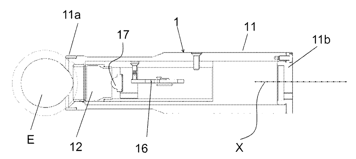
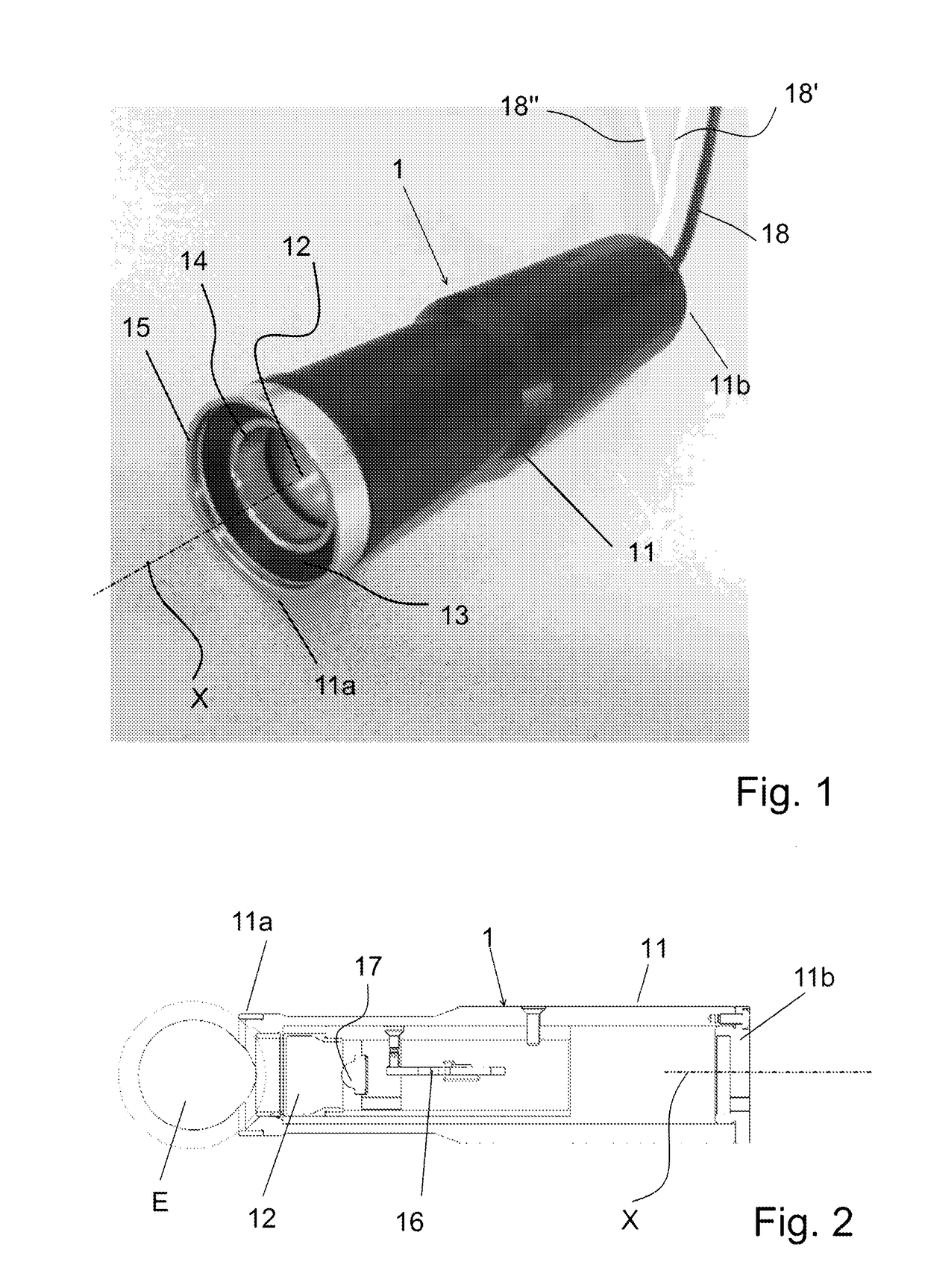
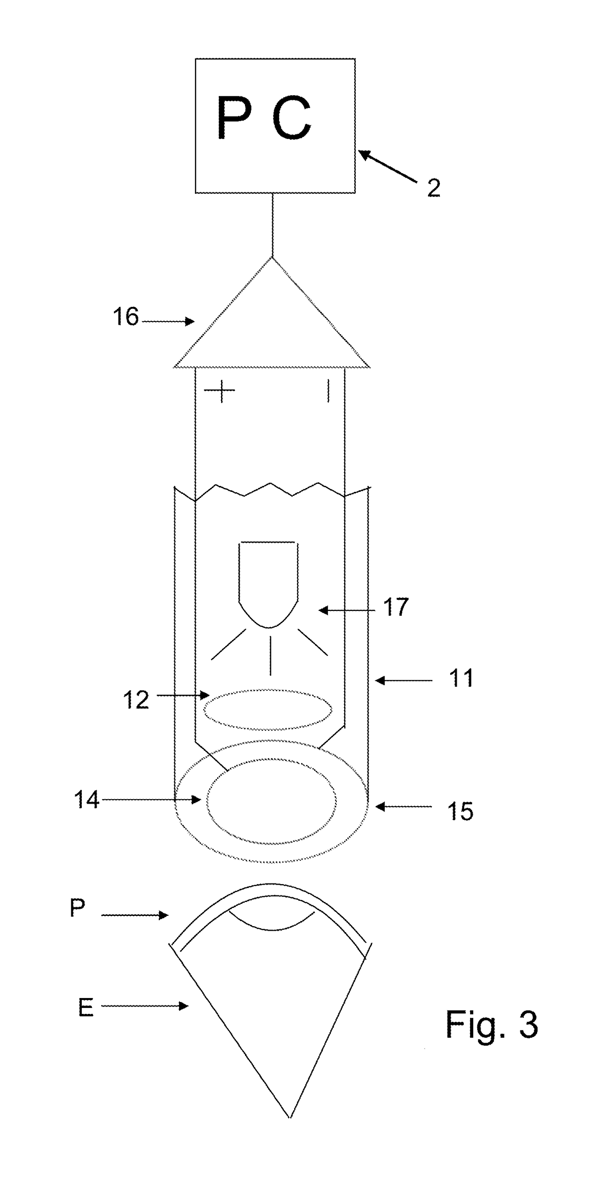
[0013]With reference to said figures, a device according to the invention comprises a handpiece 1 adapted for being gripped by a medical operator. For this purpose it has a tubular case 11, preferably ergonomically shaped and of tubular structure of substantial axial symmetry about an axis X and of diameter substantially corresponding to the average or standard eye socket of an adult patient (possibly handles of different sizes can be provided for better adaptation of the handpiece to different patients, in particular with respect to paediatric patients).

[0014]Along the aforementioned axis two ends of the case 11, and therefore of the handpiece 1, are defined. A first end 11a or front end is that intended to make contact with an eyelid P of a patient's eye E; opposite the first end, a second end or rear end 11b is defined, at which the case 11 is blind and it is possible there to arrange for the connection of one or more wires 18, 18′, 18″ for the transmission of power and signal, w...


PUM


Abstract

The present invention refers to the field of ophthalmology, and in particular to that of devices and methods for supporting the diagnosis of important eye pathologies such as Age-related Macular Degeneration (AMD), Diabetic Retinopathy (DR), anomalies and dysfunctions of the retina and of sight in general such as degeneration of the retinal structure of the optical nerve and of the visual cortex. More specifically, the invention concerns a new device and method for recording the electroretinogram (so-called ERG) of an eye, i.e. the bioelectric response of the retina induced by a light stimulus, through the eyelid.

Description

FIELD OF THE INVENTION[0001]The present invention refers to the field of ophthalmology, and in particular to that of devices and methods for supporting the diagnosis of important eye pathologies such as Age-related Macular Degeneration (AMD), Diabetic Retinopathy (DR), anomalies and dysfunctions of the retina and of sight in general such as degeneration of the retinal structure of the optical nerve and of the visual cortex. More specifically, the invention concerns a new device and method for recording the electroretinogram (so-called ERG) and the visual evoked potential (so-called VEP) of an eye, i.e. the bioelectric response of the retina induced by a light stimulus, through the eyelid in a non-invasive manner.BACKGROUND OF THE INVENTION[0002]The recording of the bioelectric responses generated by the retina as a consequence of a visual stimulus perceived by the patient is a remarkably useful tool in order to support the medical operator in diagnostic activity. The aforementioned ...

Claims


Application Information

Patent Timeline
Patent Type & Authority Applications(United States)
IPC IPC(8): A61B5/0496A61B5/00A61B3/12
CPCA61B5/0496A61B5/7228A61B3/12A61F9/007A61B3/113A61B5/04842A61B5/378A61B5/398
Inventor BAGLINI, CLAUDIO
Owner C S O SRL
Why Eureka
Try Eureka