Tissue microarray analysis

Active Publication Date: 2018-08-30
KONINKLJIJKE PHILIPS NV
View PDF2 Cites 5 Cited by
  • Summary
  • Abstract
  • Description
  • Claims
  • Application Information

AI Technical Summary

Benefits of technology

[0051]The images of cores corresponding to the same tissue block of origin can be combined in a single image and registered to the reference image. This can be images from different stains. The virtual staining outside the area of the core can be a combined staining from different images to represent a combination of information. As an example one can combine a cell membrane staining for instance for human epidermal growth factor receptor 2 (HER2) with a nuclear staining, for instance for estrogen receptors (ER) and optionally combine that with a Fluorescence in Situ Hybridization (FISH) s

Problems solved by technology

The validation of these biomarkers using the standard histopathological techniques may be time-consuming and labor intensi

Method used


Image

  • Tissue microarray analysis
  • Tissue microarray analysis
  • Tissue microarray analysis

Examples


Embodiment Construction

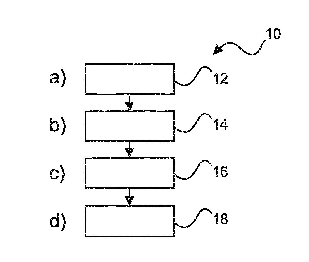
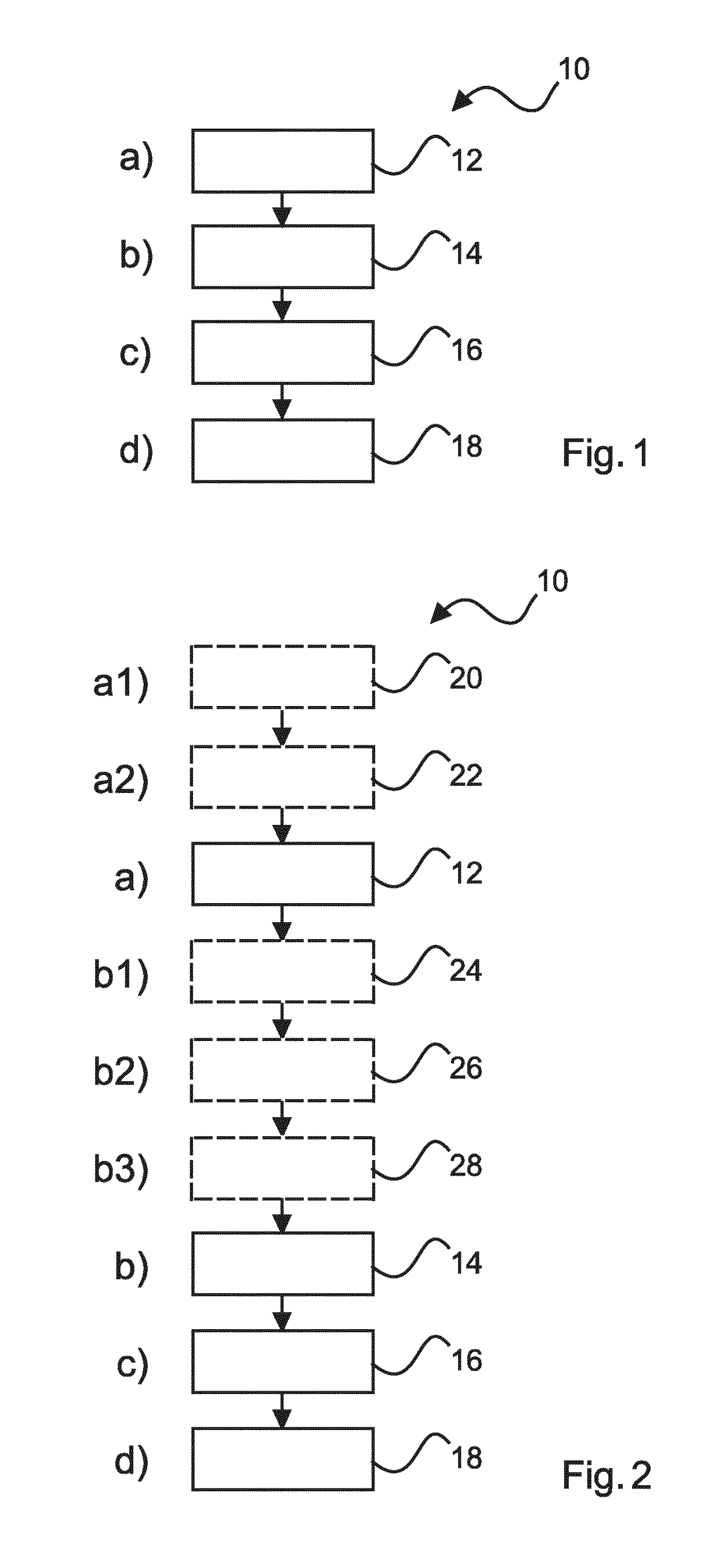
[0067]FIG. 1 shows basic steps of a method 10 for tissue examination. The method comprises the following steps:

[0068]In a first step 12, also referred to as step a), a reference image of a reference slice obtained from a tissue sample block is provided.

[0069]In a second step 14, also referred to as step b), a microarray image of a microarray slice comprising at least one tissue core obtained from at least the tissue sample block is provided.

[0070]In a third step 16, also referred to as step c), tissue core images of the at least one tissue core are registered with the reference image based on a spatial arrangement of the respective tissue core within the tissue sample block.

[0071]In a fourth step 18, also referred to as step d), the registered tissue core images are provided in combination with the reference image for analyzing purposes.

[0072]In step a), the reference image may be previously stored in an image management system, which allows for retrieval either locally or remotely....


PUM


Abstract

The present invention relates to digital pathology. In order to facilitate analyzing a tissue microarray, an apparatus is provided for tissue examination. The apparatus comprises a data input (102), a tissue microarray analyzing unit (104), and an output (106). The data input is configured to receive a reference image of a reference slice obtained from a tissue sample block; and to receive a microarray image of a microarray slice comprising at least one tissue core obtained from at least the tissue sample block. The tissue microarray analyzing unit is configured to register tissue core images of at least one tissue core with the reference image based on a spatial arrangement of the respective tissue core within the tissue sample block. The output is configured to provide a registered result obtained from the tissue microarray analyzing unit for further analyzing purposes.

Description

FIELD OF THE INVENTION[0001]The present invention relates to the field of digital pathology, and in particular to an apparatus, to a system, and to a method for tissue examination. Furthermore, the invention relates to a computer program element as well as to a computer-readable medium.BACKGROUND OF THE INVENTION[0002]Pathology diagnostic investigation of patient material (e.g. tissue and cells) is the basis of many treatment decisions, e.g. in oncology. In standard anatomical pathology, diagnosis is made on the basis of cell morphology and staining characteristics. The investigation of the pathogenesis and progression of diseases, such as cancer, may require the use of multiple biomarkers for staining. The validation of these biomarkers using the standard histopathological techniques may be time-consuming and labor intensive.[0003]In recent years, tissue microarray has been proposed to overcome these problems. Tissue microarrays are paraffin blocks produced by extracting tissue cor...

Claims


Application Information

Patent Timeline
IPC IPC(8): G06K9/00G06T7/00G06T7/33G06K9/20G06V20/20
CPCG06K9/0014G06K9/00147G06T7/0014G06T7/337G06K9/2063G06T2207/30024G06T2207/30072G06T2207/20212G06F3/147G06T2207/10056G06K2209/05G06V20/695G06V20/20G06V2201/04G06F18/00G06V20/698G06V2201/03
Inventor WIMBERGER-FRIEDL, REINHOLDPIERIK, ANKE
Owner KONINKLJIJKE PHILIPS NV
Why Eureka
Try Eureka