Oligonucleotide-based probes for detection of circulating tumor cell nucleases

Inactive Publication Date: 2018-10-04
UNIV OF IOWA RES FOUND
0 Cites 1 Cited by

AI-Extracted Technical Summary

Problems solved by technology

MBC can be managed, but cannot be cured.
CTCs constitute seeds for subsequent growth of additional tumors (metastasis) in other areas of the body, a primary cause of death for those with...

Benefits of technology

[0006]Accordingly, in certain embodiments, the present invention provides a substrate probe for detecting a circulating tumor cell (CTC) nuclease comprising an oligonucleotide of 2-30 nucleotides in length, a fluorophore operably linked to the oligonucleotide, and a quencher operably linked to the oligonucleotide, wherein the oligonucleotide c...

Abstract

The present invention relates to a rapid detection of circulating tumor cell (CTC)-associated nuclease activity with chemically modified nuclease substrate probes and compositions useful in detection assays.

Application Domain

Microbiological testing/measurement

Technology Topic

OligonucleotideNuclease +7

Image

  • Oligonucleotide-based probes for detection of circulating tumor cell nucleases
  • Oligonucleotide-based probes for detection of circulating tumor cell nucleases
  • Oligonucleotide-based probes for detection of circulating tumor cell nucleases

Examples

  • Experimental program(3)

Example

Example 1
Rapid and Sensitive Detection of Circulating Tumor Cells with Nuclease-Activated Oligonucleotide Probes
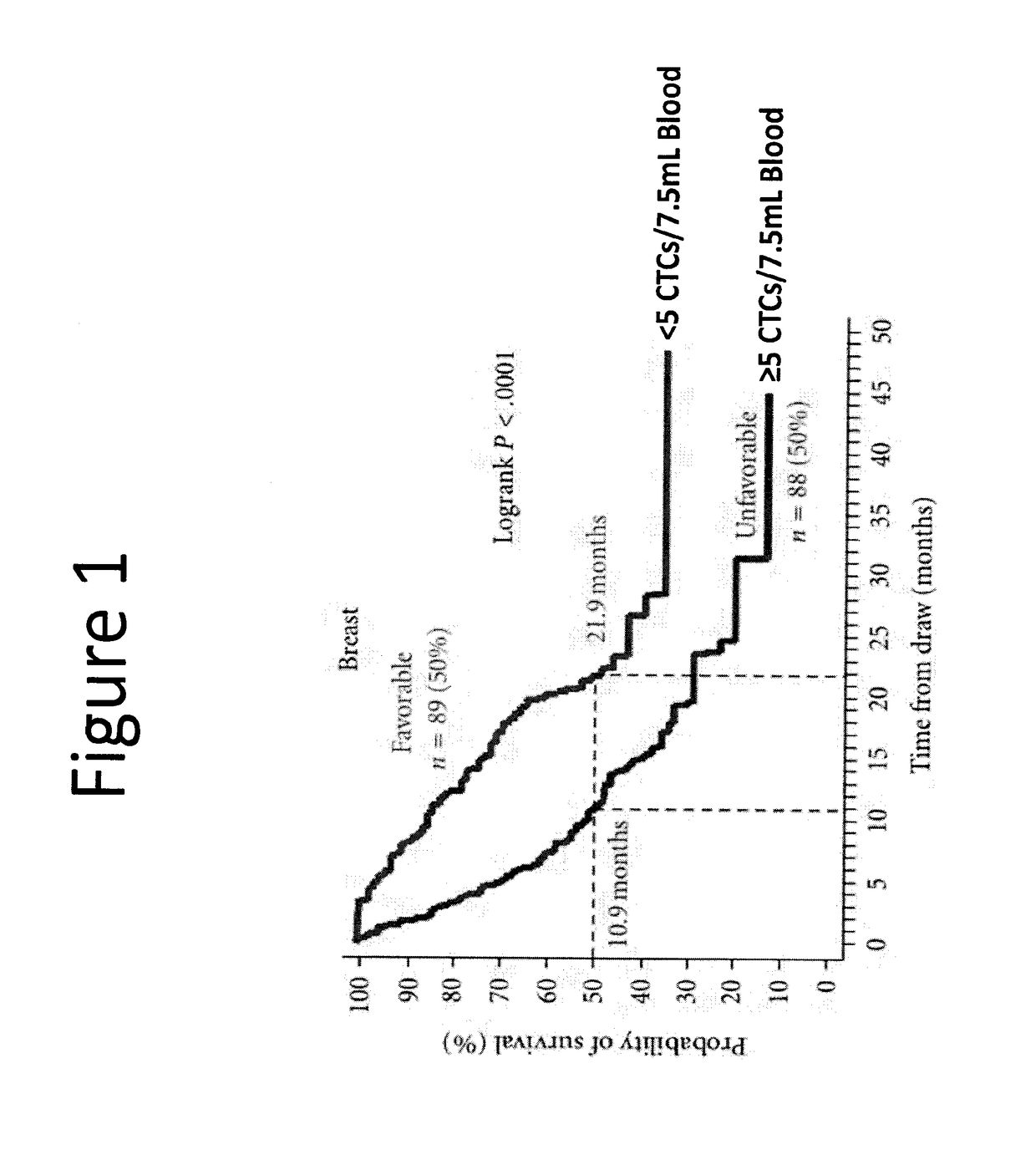
[0142]Metastatic breast cancer is the second leading cause of female cancer deaths in the United States. Despite substantial progress in its treatment, metastatic breast cancer remains incurable. Early identification of breast cancer patients at greatest risk of developing metastatic disease is thus an important goal that would enable oncologists to aggressively treat these patients while the cancer is still vulnerable. In addition, this would spare patients who do not need or would not benefit from further treatments from having to endure the harmful side-effects of chemotherapeutic drug regimens. Circulating tumor cells (CTCs) are rare cancer cells found in the blood circulation of cancer patients that provide a non-invasively accessible cancer cell specimen (liquid biopsy) from patients. The number of circulating tumor cells (CTCs) in cancer patients has recently been shown to be a valuable diagnostic indicator of the state of metastatic breast cancer. In particular, patients with few or no CTCs were found to have a better overall prognosis compared to patients with high numbers of CTCs.
[0143]Despite the implications of CTCs as diagnostics for advanced breast cancer treatment, a critical challenge for adopting CTC-based diagnostic tests has been the development of methods with sufficient sensitivity to reliably detect the small number of CTCs that are present in the circulation. Furthermore, current tests for CTC detection are expensive, have high false positives and negatives, have high background noise, are time consuming and require a significant level of expertise to conduct. To overcome the limitations of current CTC detection assays and develop more sensitive, rapid and cost effective CTC detection methods, we explored the potential of detecting CTCs by measuring their nuclease activity with nuclease-activated probes (Hernandez F J, et al., Noninvasive imaging of staphylococcus aureus infections with a nuclease-activated probe. Nat Med. 2014; 20:301-306; Hernandez F J, et al., Degradation of nuclease-stabilized RNA oligonucleotides in mycoplasma-contaminated cell culture media. Nucleic Acid Ther. 2012; 22:58-68).
[0144]Data is presented toward the development of a rapid and highly-sensitive CTC detection assay based on nuclease-activated oligonucloetide probes that are selective digested (activated by target nucleases expressed in breast cancer cells. It was confirmed that these probes were not activated by serum nucleases or nucleases from a lymphoblastic cell line (e.g., K-562). Furthermore, we present extensive data towards the optimization of activity and sensitivity of these probes in cell lysates from various breast cancer cell lines and in blood from breast cancer patients. In conclusion, this work describes a robust assay for detection of breast cancer CTCs that is straightforward to implement in most clinical diagnostic labs.
[0145]Chemically Modified Oligonucleotides
[0146]Artificial RNA reagents such as siRNAs and aptamers often must be chemically modified for optimal effectiveness in environments that include nucleases. Synthetic RNA that is exposed to cells or tissues must be protected from nuclease degradation in order to carry out its intended function in most cases. Common approaches for avoiding nuclease degradation include nanoparticle encapsulation which insulates the RNA from exposure to nucleases and chemical modification to render it resistant to degradation. Modification of RNA by substituting O-methyl or fluoro groups for the hydroxyl at the 2′-position of the ribose can greatly enhance its stability in the presence of extracellular mammalian nucleases.
[0147]These modifications are widely employed in the development of siRNAs and RNA aptamers for both research and therapeutic applications. siRNAs can be modified with 2′-O-methyl substitutions in both sense and antisense strands without loss of silencing potency, but only a subset of nucleotides are typically modified with 2′-O-methyls as over-modification of the siRNA can reduce or eliminate its silencing ability. siRNAs with 2′-fluoro modified pyrimidines have also been reported to retain silencing activity in vitro as well as in vivo.
[0148]Substrates probes were synthesized with chemical modifications indicated in figure legends, flanked by a FAM (5′-modification) and a pair of fluorescence quenchers, “ZEN” and “Iowa Black” (3′-modifications).
[0149]Oligonucleotide Probe Synthesis and Purification
[0150]Oligonucleotide probes were synthesized and purified. Briefly, all the FAM-labeled probes were synthesized using standard solid phase phosphoramidite chemistry, followed by high performance liquid chromatography (HPLC) purification. For the Cy5.5-labeled probes, the sequences were first synthesized with ZEN and Iowa Black quenchers or inverted dT on the 3′-ends and amine on the 5′-ends using the standard solid phase phosphoramidite chemistry, and purified with HPLC. These purified sequences were then set to react with Cy5.5 NHS ester (GE Healthcare, Piscataway, N.J.) to chemically conjugate the Cy5.5 label on the sequences. The Cy5.5-labeled probes were further purified with a second HPLC purification. All probe identities were confirmed by electron spray ionization mass spectrometer (ESI-MS) using an Oligo HTCS system (Novatia LLC, Princeton, N.J.). The measured molecular weights are within 1.5 Daltons of the expected molecular weights. The purity of the probes was assessed with HPLC analysis and is typically greater than 90%. Quantitation of the probes was achieved by calculating from their UV absorption data and their nearest-neighbor-model-based extinction coefficients at 260 nm. Extinction coefficients of 2′-O-methyl-nucleotides and 2′-fluoro-nucleotides are assumed to be the same as that of RNA.
[0151]Breast Cancer Cell Lysates have High Nuclease Activity Against Probes
[0152]Cells from breast cancer cell lines or normal breast cell line were washed, lysed, and dialyzed against buffer containing 1 mM DTT, 1% Triton X-100, 50 mM Tris pH9, 150 mM NaCl, and 10 mM MgCl2. Additionally, the buffer contains complete ULTRA protease inhibitor (Roche, product number 05892791001), at a concentration 1 tablet per 10 mL of buffer. The lysates were incubated with 50 pmol of probe, and fluorescence was measured after 1.5 hours. It was found that the breast cancer cell lysates did exhibit high nuclease activity against the probes. (FIG. 4).
[0153]Breast Cancer Cells Secrete Nucleases
[0154]Next, it was examined whether breast cancer cells secrete nucleases. Breast cancer cell lines and normal breast cell lines ere incubated with serum free media overnight. The media was collected and the cell debris was spun down. The media was dialyzed against PBS+/+ with protease inhibitors, and the supernatants were incubated with 50 pmol of probe. Fluorescence was measured after 1.5 hours. It was found that the breast cancer cells did secrete nucleases. (FIG. 5).
[0155]Incubation Conditions
[0156]Incubation conditions were evaluated and optimized. MDA231, Human PBMC cell lysates, or human serum were dialyzed with Tris-buffer ranging from pH 7 to pH 10. A pH of 9 was found to be optimum (FIG. 6).
[0157]It was also evaluated whether the concentration of Ca2+ and/or Mg2+ affected the nuclease activity. MDA231 cell lysates were dialyzed with buffer containing various concentrations of Ca2+ or Mg2+, and nuclease activity against DNA probe was measured. It was found that the concentration of Ca2+ was not important, but that a concentration of 10 mM of Mg2+ was optimal (FIG. 7).
[0158]Controls
[0159]Both MCF10a (a mammary epithelia cell line) and K562 (hematopoietic cell line) cells were tested as possible controls. It was found that the K562 cells were better controls (FIG. 8).
[0160]Sensitivity
[0161]It was determined what is the lowest number of breast cancer cells detectable with these probes, and how much probe is optimal. Kinetics of nuclease degradation of different amounts of double stranded DNA (Oligo 1) probe (100 pmol, 25 pmol, 12.5 pmol, 6.25 pmol) by lysates of 100, 30, or 10 SKBr3 cells, or lysis buffer alone. Probes were digested 2.5 hours at 37 degrees C., with measurements taken every 15 minutes. It was determined that as few as 10 SKBr3 cells were detectable, and that using about 6.25 pmol of probe was effective (FIG. 9). It was determined that the present probes were effective for detecting several breast cancer subtypes (FIGS. 10 and 11).
[0162]Detection in Blood Samples
[0163]It was investigated whether breast cancer cell could be detected in blood using the present method. Briefly, 105, 104, or 103 SKBr3 cells were spiked into 100 μL of human blood. The cells were pelleted cells and the plasma was removed. The cells were lysed with optimized buffer (described above) and tested for nuclease activity. It was possible to detect breast cancer cell could be detected in blood (FIGS. 12 and 13).

Example

Example 2
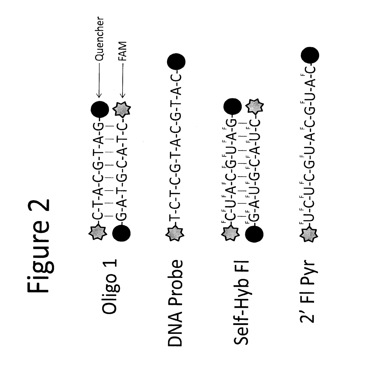
[0164]The sensitivity of the assay in blood is low due to background activity from blood cells (FIG. 14), which further confirmed the need for capturing/enriching CTCs from blood. CTCs can be enriched from blood using size exclusion filters. Two commercially available filters are the ISET filter from Rarecells and the filter from ScreenCell. The filter from ScreenCell was more effective at reducing the background signal from blood compared to the ISET filter (FIG. 15A). The variability in the background signal from blood derived from several healthy donors was examined (FIGS. 15 B and C). Fixed amounts of breast cancer cells were spiked in blood and process the mixture with the ScreenCell filters (FIG. 16A). It was possible to robustly detect 200 cancer cells spiked into 1 mL of blood, which was a 500 fold improvement in sensitivity over no filtration. The probes were validated in blood from patients with stage IV breast cancer. The probe used in this example was the dsDNA probe (FIG. 16B). The blood from patients and healthy donors was processed using the ScreenCell filter. These data clearly show a statistically significance between blood from patience with stage IV and healthy donor blood showing that the nuclease activated probes can successfully identify CTCs in patient samples. FIG. 17 shows the variability in probe activity from draw to draw. This variability could be due to the changes in potential response to treatment or disease progression. FIG. 18 shows that it was possible to rule out that the nuclease activity observed in the patient samples was due to higher amounts of blood cells present in the blood of these patients. Together, these data confirm that the nuclease activated robes can be used to detect CTC-nuclease activity in blood from patients with stage IV breast cancer.

Example

Example 3
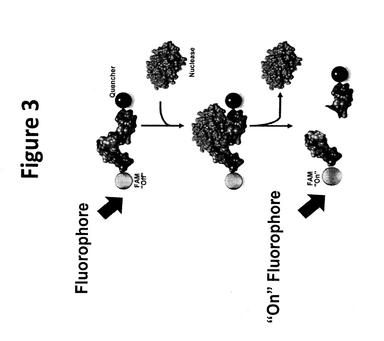
[0165]Experiments were performed to evaluate nuclease activity in supernatants from cancer cell lines. Secreted nuclease active was measured in contrast to intracellular nuclease activity. Experiments were performed to demonstrate that the ssDNA and 2′F-ssRNA nuclease activated probes were activated by secreted nucleases from breast cancer cells (FIGS. 19A-D and 20A-C). The data show that the probes can be used to detect nucleases that are secreted from CTCs in blood of breast cancer patients.
[0166]Although the foregoing specification and examples fully disclose and enable the present invention, they are not intended to limit the scope of the invention, which is defined by the claims appended hereto.
[0167]All publications, patents and patent applications are incorporated herein by reference. While in the foregoing specification this invention has been described in relation to certain embodiments thereof, and many details have been set forth for purposes of illustration, it will be apparent to those skilled in the art that the invention is susceptible to additional embodiments and that certain of the details described herein may be varied considerably without departing from the basic principles of the invention.
[0168]The use of the terms “a” and “an” and “the” and similar referents in the context of describing the invention are to be construed to cover both the singular and the plural, unless otherwise indicated herein or clearly contradicted by context. The terms “comprising,”“having,”“including,” and “containing” are to be construed as open-ended terms (i.e., meaning “including, but not limited to”) unless otherwise noted. Recitation of ranges of values herein are merely intended to serve as a shorthand method of referring individually to each separate value falling within the range, unless otherwise indicated herein, and each separate value is incorporated into the specification as if it were individually recited herein. All methods described herein can be performed in any suitable order unless otherwise indicated herein or otherwise clearly contradicted by context. The use of any and all examples, or exemplary language (e.g., “such as”) provided herein, is intended merely to better illuminate the invention and does not pose a limitation on the scope of the invention unless otherwise claimed. No language in the specification should be construed as indicating any non-claimed element as essential to the practice of the invention.
[0169]Embodiments of this invention are described herein, including the best mode known to the inventors for carrying out the invention. Variations of those embodiments may become apparent to those of ordinary skill in the art upon reading the foregoing description. The inventors expect skilled artisans to employ such variations as appropriate, and the inventors intend for the invention to be practiced otherwise than as specifically described herein. Accordingly, this invention includes all modifications and equivalents of the subject matter recited in the claims appended hereto as permitted by applicable law. Moreover, any combination of the above-described elements in all possible variations thereof is encompassed by the invention unless otherwise indicated herein or otherwise clearly contradicted by context.
<160> NUMBER OF SEQ ID NOS: 4
<210> SEQ ID NO: 1
<211> LENGTH: 8
<212> TYPE: DNA
<213> ORGANISM: Artificial Sequence
<223> OTHER INFORMATION: Description of Artificial Sequence Synthetic
probe
<400> SEQENCE: 1
ctacgtag 8
<210> SEQ ID NO: 2
<211> LENGTH: 12
<212> TYPE: DNA
<213> ORGANISM: Artificial Sequence
<223> OTHER INFORMATION: Description of Artificial Sequence Synthetic
probe
<400> SEQENCE: 2
tctcgtacgt ac 12
<210> SEQ ID NO: 3
<211> LENGTH: 8
<212> TYPE: RNA
<213> ORGANISM: Artificial Sequence
<223> OTHER INFORMATION: Description of Artificial Sequence Synthetic
probe
<400> SEQENCE: 3
cuacguag 8
<210> SEQ ID NO: 4
<211> LENGTH: 12

PUM

PropertyMeasurementUnit
Area6600000.0m ** 2
Nanoscale particle size494.0nm
Nanoscale particle size495.0nm

Description & Claims & Application Information

We can also present the details of the Description, Claims and Application information to help users get a comprehensive understanding of the technical details of the patent, such as background art, summary of invention, brief description of drawings, description of embodiments, and other original content. On the other hand, users can also determine the specific scope of protection of the technology through the list of claims; as well as understand the changes in the life cycle of the technology with the presentation of the patent timeline. Login to view more.
Why Eureka
Try Eureka