Spectral imaging for measurement of nuclear pathology features in cancer cells prepared for in situ analysis

a nuclear pathology and in situ analysis technology, applied in cell components, instruments, batteries, etc., can solve the problems of inability to directly associate objective measures, increase the chance of false negative results associated with cytological staining methods or false positives, and limit the usefulness of combining such measurements in a meaningful way. , to achieve the effect of increasing the statistical confidence of identification

Active Publication Date: 2018-10-11
VENTANA MEDICAL SYST INC +1
View PDF0 Cites 1 Cited by
  • Summary
  • Abstract
  • Description
  • Claims
  • Application Information

AI Technical Summary

Benefits of technology

[0031]d. comparing the combined in-situ molecular and morphometric data obtained from the test cell sample of step (c) with the library data in step (b) and providing increased statistical confidence of identification of the test cell sample as a cancer or pre-cancer cell sample.

Problems solved by technology

Further, current methods usually require a single staining method at a time, and if more than one staining method is performed, it is usually not on the same exact cells.
This adds to the chance of either false negative results associated with cytological staining methods or false positive results associated with immunogenic or activity-based staining methods.
The inability to directly associate objective measures of morphology with particular genetic rearrangements when separate slides are used has limited usefulness of combining such measurements in a meaningful way.
While each year over 300,000 men are diagnosed with prostate cancer in the U.S. alone, the currently available tests are notoriously inaccurate and subjective.
These methodologies cannot differentiate between benign disease and cancer.
The failure to differentiate can result, for example, in exposure of patients with benign disease to treatments that are unnecessary and have side effects (e.g., impotence and incontinence).
Unfortunately, these techniques generally have only marginal predictive value.
Moreover, it is estimated that PSA testing misses 20%-30% of all individuals with cancer.

Method used


Image

  • Spectral imaging for measurement of nuclear pathology features in cancer cells prepared for in situ analysis
  • Spectral imaging for measurement of nuclear pathology features in cancer cells prepared for in situ analysis
  • Spectral imaging for measurement of nuclear pathology features in cancer cells prepared for in situ analysis

Examples


examples

Correlative Value to Nuclear Morphology and ERG Rearrangement for Prostate Cancer Cells

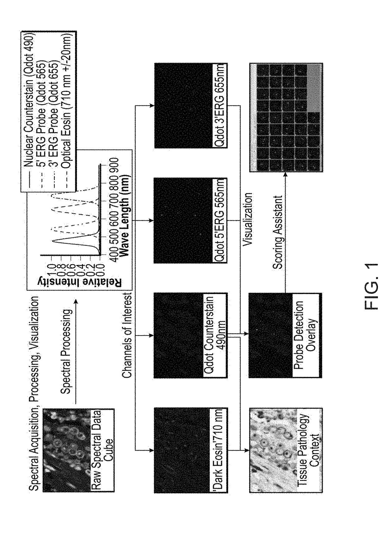
[0074]Quantitative technologies have been advanced and applied in this study to permit extraction of morphometric data from tissue prepared for fluorescent in-situ molecular analysis of multiplexed probes. A highly characterized spectral imaging approach is used to produce high resolution (wavelength resolution, spatial resolution and intensity resolution) data (FIG. 1). FIG. 1 depicts the steps of the present technology where raw data acquired through quantitative spectral imaging is de-composited on the basis of wavelength signal distribution from the nuclear stain and probe detection. This produces a quantifiable image representing the true relative distribution of label on the tissue section. The signal to noise ratio of such images is very high, in part due to the ability to separate the true signal from contaminating signals constitutive to the tissue.

[0075]These data are subsequently proces...


PUM

PropertyMeasurementUnit
wavelengthaaaaaaaaaa
depth of fieldaaaaaaaaaa
nuclear morphologyaaaaaaaaaa

Abstract

In general, the presently disclosed technology relates to identification of cancer subtypes. More specifically, the technology relates to methods for determining molecular drivers of cancer and / or progression using a multivariate image data and statistical analysis of in-situ molecular markers and morphological characteristics in the same cells of a biological sample suspected of b cancer. This analysis takes place after a single acquisition that obtains the molecular and anatomic morphology data in parallel. The analysis compares specific morphological and molecular markers to known samples exhibiting particular genetic drivers of the cancer. This method provides statistical information that allows for an increased confidence in the identification of specific molecular drivers of the cancer.

Description

FIELD OF THE INVENTION[0001]In general, the disclosed technology relates to identification of cancer subtypes. More specifically, the disclosed technology relates to methods for determining molecular drivers of cancer and / or progression using a multivariate image data and statistical analysis of in-situ molecular markers and morphological characteristics in the same cells of a tissue sample of a cancer. This analysis takes place after a single acquisition that obtains the molecular and anatomic morphology data in parallel. The analysis compares specific morphological and molecular markers to known samples exhibiting particular genetic drivers of the cancer. This method provides statistical information that allows for an increased confidence in the identification of specific molecular drivers of the cancer.BACKGROUND OF THE INVENTION[0002]Pathological prognostic assays are used to provide information to help guide and develop treatment regimes and predict outcomes for a myriad of can...

Claims


Application Information

Patent Timeline
Patent Type & Authority Applications(United States)
IPC IPC(8): C12Q1/6886H01M2/34H01H35/14G06F19/18G16B20/00G16B20/20
CPCH01M2/345H01H35/146H01M2/34H01M2/347H01M2/348H01M2220/20C12Q1/6886G06F19/18H01M50/579H01M50/581H01M50/578G16B20/00G16B20/20G01N33/57407C12Q1/6841C12Q2600/156Y02E60/10H01M50/572
Inventor DEMICHELIS, FRANCESCAGARSHA, KARLMILLER, PHILLIP C.NAGLE, RAY B.OTTER, MICHAELPESTANO, GARY ANTHONYRUBIN, MARK
Owner VENTANA MEDICAL SYST INC
Why Eureka
Try Eureka