
Ultrasound imaging apparatus and ultrasound imaging method for inspecting a volume of a subject

a technology of ultrasound imaging and ultrasound, applied in applications, ultrasonic/sonic/infrasonic image/data processing, ultrasonic/sonic/infrasonic diagnostics, etc., can solve the problem of reducing the usability of three-dimensional ultrasound systems for precise quantification analysis or size measurements, reducing spatial resolution, etc. problem, to achieve the effect of low time consumption, high quality and precise measuremen

Pending Publication Date: 2019-01-17
KONINKLJIJKE PHILIPS NV
View PDF13 Cites 10 Cited by
  • Summary
  • Abstract
  • Description
  • Claims
  • Application Information

AI Technical Summary

Benefits of technology

The present invention aims to provide an ultrasound imaging apparatus that can produce better quality images with minimal effort. A corresponding ultrasound imaging method is also provided.

Problems solved by technology

The three-dimensional ultrasound systems have due to the huge amount of measurement data in the field of view a reduced spatial resolution and a reduced image quality in real-time so that the three-dimensional ultrasound systems have a limited usability for precise quantification analysis or size measurements.

Method used


Image

  • Ultrasound imaging apparatus and ultrasound imaging method for inspecting a volume of a subject
  • Ultrasound imaging apparatus and ultrasound imaging method for inspecting a volume of a subject
  • Ultrasound imaging apparatus and ultrasound imaging method for inspecting a volume of a subject

Examples


Embodiment Construction

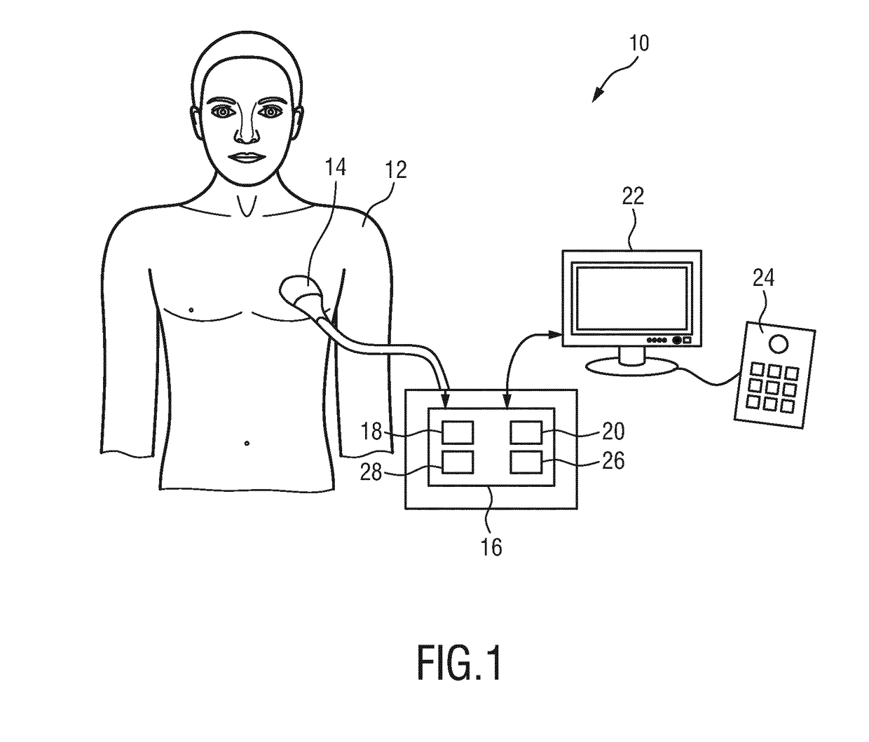
[0042]FIG. 1 shows a schematic illustration of an ultrasound imaging apparatus generally denoted by 10. The ultrasound imaging apparatus 10 is applied to inspect a volume of an anatomical site, in particular an anatomical site of a patient 12. The ultrasound imaging apparatus 10 comprises an ultrasound probe 14 having at least one transducer array including a multitude of transducer elements for transmitting and receiving ultrasound waves. The transducer elements are preferably arranged in a 2D array for providing multi-dimensional image data, in particular three-dimensional ultrasound image data and bi-plane image data. Bi-plane image data can be acquired by sweeping two intersecting 2D image planes. Generally in bi-pane imaging the two 2D planes are orthogonal to an emitting surface of the array and can intersect under a different angle.

[0043]The ultrasound imaging apparatus 10 comprises in general a control unit 16 connected to the ultrasound probe 14 for controlling the ultrasou...


PUM


Abstract

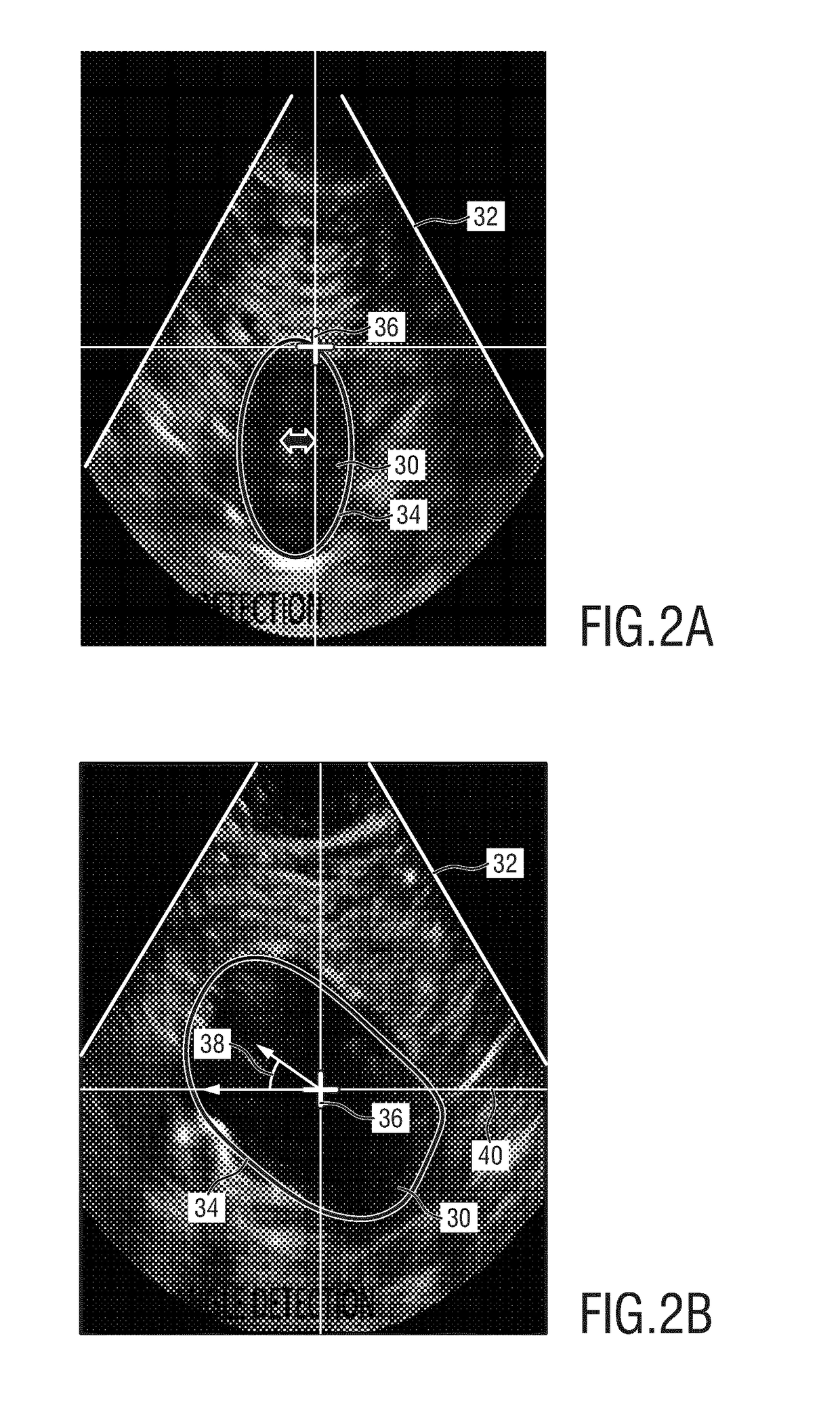
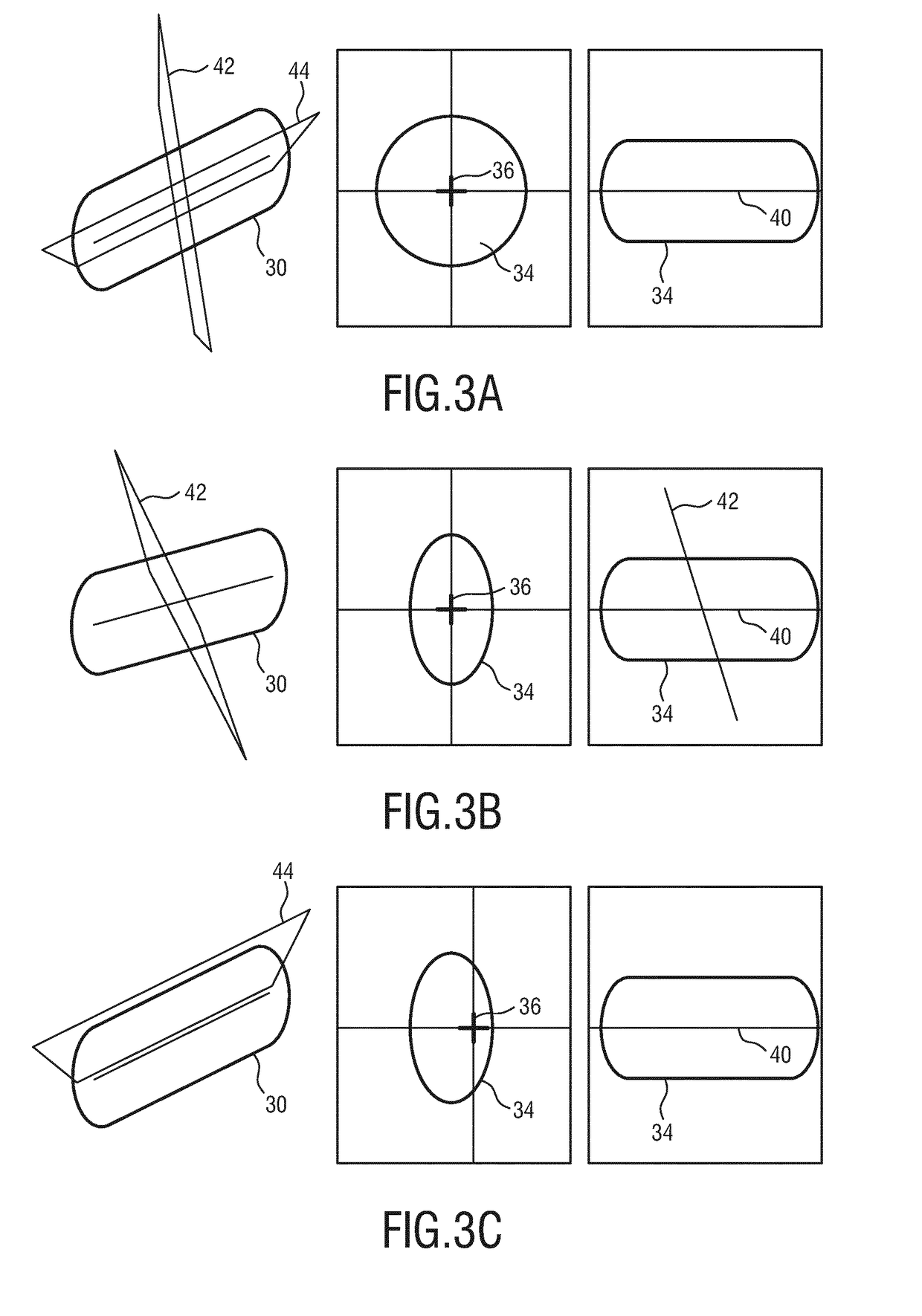
An ultrasound imaging apparatus (10) for inspecting a volume of a subject (12) is disclosed. The ultrasound imaging apparatus comprises an ultrasound probe (14) including a plurality of ultrasound transducer elements for acquiring three-dimensional ultrasound data in a field of view (32), and for providing three-dimensional and two-dimensional ultrasound image data in the field of view. An image processing unit (18) is coupled to the ultrasound probe for receiving the two-dimensional ultrasound image data in an image plane (42, 44) and for determining an anatomical feature (30) in the two-dimensional ultrasound image data. An evaluation unit (20) is provided for evaluating the two-dimensional ultrasound image data and for determining a quality parameter based on a positional relation of the anatomical feature with respect to the image plane, and an alignment unit (28) is provided for aligning or indicating an alignment of the image plane bases on the positional relation and the quality parameter.

Description

FIELD OF THE INVENTION[0001]The present invention relates to an ultrasound imaging apparatus for inspecting a volume of a subject. The present invention further relates to an ultrasound imaging method for inspecting a volume of a subject. The present invention in particular relates to real-time user guidance and optimization of image quality during ultrasound inspection of a volume of a subject in order to provide a size measurement of an anatomical object of the subject based on ultrasound data.BACKGROUND OF THE INVENTION[0002]In the field of medical imaging systems three-dimensional ultrasound measurement units are well-known for quantification analysis and size measurements such as diameters or volumes of anatomical objects of a patient. A corresponding ultrasound apparatus is e.g. known from WO 2014 / 097090 A1.[0003]The three-dimensional ultrasound systems have due to the huge amount of measurement data in the field of view a reduced spatial resolution and a reduced image quality...

Claims


Application Information

Patent Timeline
IPC IPC(8): A61B8/08A61B8/00
CPCA61B8/483A61B8/5223A61B8/4245A61B8/5269A61B8/523G16H50/30
Inventor ROUET, LAURENCECOLLET-BILLON, ANTOINEENTREKIN, ROBERT RANDALL
Owner KONINKLJIJKE PHILIPS NV
Learn More